Search results (275 results)
-
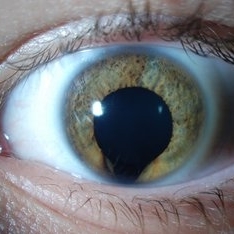
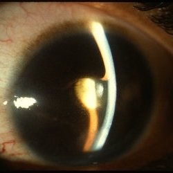
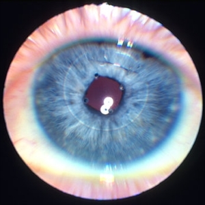
 Busacca nodules
May 2 2013 by Henry J. Kaplan, MD
Typical Busacca iris stromal nodules in sarcoid uveitis; notice the ps formation.
Condition/keywords: busacca nodulaes, granulomatous uveitis, iris nodules, sarcoid bussaca iris nodules
-
---thumb.jpg/image-square;max$300,300.ImageHandler) Roth Spot
Roth Spot
Feb 27 2013 by Henry J. Kaplan, MD
Roth spots due to subacute bacterial endocardiris in a patient with the diagnosis of AIDS .
Condition/keywords: AIDS, subacute bacterial endocardiris, white centered retinal hemorrhage (Roth Spot)
-
 Keyhole Pupil Coloboma
Jul 13 2013 by Jason S. Calhoun
14-year-old male presents with decreased vision in the left eye. Dx with iris and retinal coloboma in the left eye. Patient VA was 20/20, right eye, 20/100 left eye with pinhole improvement 20\50. Patient was fitted for SCL in the left eye.
Photographer: Jason S. Calhoun, Department of Ophthalmology, Mayo Clinic Jacksonville, Florida
Imaging device: TOPCON D-90 SL NIKON CAMERA
Condition/keywords: deformed pupil
-
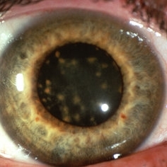
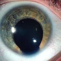
---thumb.jpg/image-square;max$300,300.ImageHandler) Iris Nodules (Lisch)
Iris Nodules (Lisch)
Dec 27 2013 by David Callanan, MD
57-year-old female patient with iris nodules.
Condition/keywords: iris nodules
-
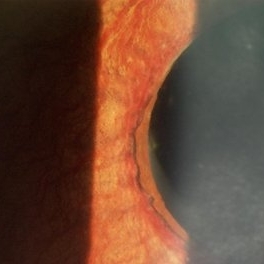
---thumb.jpg/image-square;max$300,300.ImageHandler) Inferior Sector Iris Atrophy With Depigmentation
Inferior Sector Iris Atrophy With Depigmentation
Aug 1 2013 by From the Collections of Thomas M. Aaberg, MD and Thomas M. Aaberg Jr., MD
Inferior sector iris atrophy with depigmentation.
Condition/keywords: depigmentation, inferior sector iris atrophy
-
 Iris Bombe with Rubeosis
Oct 8 2012 by Jeffrey G. Gross, MD, FASRS
Iris bombe, with rubeosis.
Condition/keywords: iris bombe, rubeosis
-
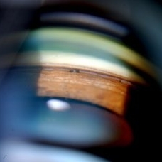
 Gonioscopy: Pigment Dispersion Glaucoma
Jul 8 2013 by Jason S. Calhoun
Patient with no family history of glaucoma, came in with elevated IOP. During gonioscopy exam. brown pigment overlying the trabecular meshwork. Also, trans-illumination defects on the iris.
Photographer: Jason S. Calhoun, Department of Ophthalmology, Mayo Clinic Jacksonville, Florida
Condition/keywords: gonioscopy, pigment dispersion syndrome of iris
-
---thumb.JPG/image-square;max$300,300.ImageHandler) Retinal Coloboma
Retinal Coloboma
Jul 8 2013 by Jason S. Calhoun
14-year-old male with decreased vision in the left eye. Dx with iris and retinal coloboma in the left eye. Patient VA was 20/20, right eye, 20/100 left eye with pinhole improvement 20\50. Patient was fitted for SCL in the left eye.
Photographer: Jason S. Calhoun, Department of Ophthalmology, Mayo Clinic Jacksonville, Florida
Condition/keywords: chorioretinal coloboma
-
 Schaffer's Sign
Dec 23 2019 by Hashim Ali Khan, OD, FAAO
Brown iris pigment in vitreous of a pseudophakic eye without retinal detachment or breaks/ holes in retina.
Condition/keywords: detached vitreous, Schaffer's sign, vitreous pigment
-
 Siderosis
May 2 2013 by Henry J. Kaplan, MD
Iron deposition in the iris epithelium and sphincter and on lens epithelium in the same patient ; #2.
Condition/keywords: siderosis
-
 Iris Nevus
Apr 11 2016 by Kathy Karsten, COT
Anterior segment photo of an iris nevus with a peaked pupil observed over time for growth.
Photographer: KATHY KARSTEN
Imaging device: TOPCON 50-DX
Condition/keywords: iris, nevus
-
---thumb.JPG/image-square;max$300,300.ImageHandler) TID (Trans Illumination Defect)
TID (Trans Illumination Defect)
Jul 8 2013 by Jason S. Calhoun
74-year-old patient who VA 20/70 OD, 20/50 OS. Complaints of blurred vision. Iris atrophy, both eyes. ERM, right eye, Patient to have cataract surgery to improve distance vision.
Photographer: Jason S. Calhoun, Department of Ophthalmology, Mayo Clinic Jacksonville, Florida
Condition/keywords: translucency of iris
-
 Koeppe nodules
May 2 2013 by Henry J. Kaplan, MD
Granulomatous Koeppe nodules at the pupillary margin.
Condition/keywords: iris nodules, Koeppe nodules
-
 Iris Bombe
-
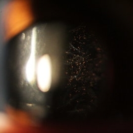
 Gonioscopy; Scattered Peripheral Anterior Synechiae
Jul 8 2013 by Jason S. Calhoun
Patient came in for evaluation for glaucoma. Patient also has a history of uveitis. Last flare up was back in 1990. Patient's VA was 20/30, Right eye and 20/40-1, Left eye. Slit Lamp Gonioscopy reveals iris bow with scattered PAS around the angles of the anterior chamber. You can also see pigmentation in the trabecular meshwork. Patient will follow up in 3 months.
Photographer: Jason S. Calhoun, Department of Ophthalmology, Mayo Clinic Jacksonville, Florida
Condition/keywords: gonioscopy, goniosynechiae
-
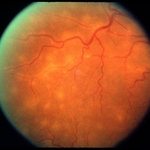 Tuberculosis Panuveitis
Feb 25 2013 by Henry J. Kaplan, MD
Peripheral vascular sheathing and multiple choroidiris foci in a patient with tuberculosis panuveitis.
Condition/keywords: panuveitis, tuberculosis
-
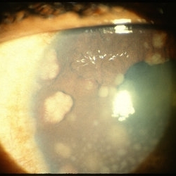 Sarcoid Bussaca Iris Nodules
Oct 11 2012 by Jeffrey G. Gross, MD, FASRS
Sarcoid bussaca iris nodules.
Condition/keywords: autoimmunity, sarcoid bussaca iris nodules, sarcoidosis
-
---thumb.jpg/image-square;max$300,300.ImageHandler) Iris Nodules (Lisch)
Iris Nodules (Lisch)
Dec 27 2013 by David Callanan, MD
57-year-old female patient with iris nodules.
Condition/keywords: iris nodules
-
 Iris clip IOL
Jan 11 2013 by Alex P. Hunyor, MD
Iris fixated IOL - note stainless steel suture.
Condition/keywords: intraocular lens (IOL), iris clip intraocular lens
-
 Russell bodies Observed in a Patient with Fuchs' Heterochromic Iridocyclitis
Nov 1 2016 by PAVEL FLORES-MORENO
Anterior chamber shows in iris surface: small, refractile iris crystals.
Photographer: Pavel Flores-Moreno
Imaging device: Anterior chamber camera
Condition/keywords: Fuchs, Russell bodies
-
 Iris Coloboma
-
 X-linked ocular albinism slide 1
Oct 22 2012 by Ronald C. Gentile, MD
Anterior segment photo revealed lightly pigmented iris and yellow-white lashes. His complexion was much lighter then both his parents and unaffected siblings.
Photographer: The New York Eye & Ear Infirmary Department of Medical Imaging
Condition/keywords: Nettleship-Falls ocular albinism, ocular albinism
-
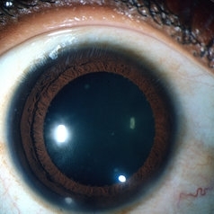
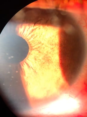
 Advanced Stage of Neovascular Glaucoma
Mar 21 2013 by Yusuke Oshima, MD, PhD
An 82-year-old man with a advanced stage of neovascular glaucoma. A slit-lamp photograph illustrates iris ectropion with prominent iris neovascularization.
Photographer: Yusuke Takada, Osaka University Graduate School of Medicine
Condition/keywords: neovascular glaucoma
-
---thumb.jpg/image-square;max$300,300.ImageHandler) Siderosis
Siderosis
May 4 2013 by Henry J. Kaplan, MD
Iris heterochromia due to siderosis as a result of retained metallic intraocular foreign body; notice the dilated pupil in the involved site. #1
Condition/keywords: siderosis
 Loading…