Search results (275 results)
-
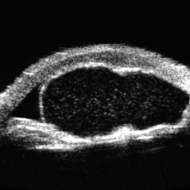
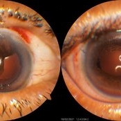
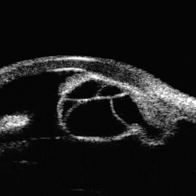 Idiopathic Iris Cyst
Aug 13 2025 by Virginia Gebhart
74 year old female with idiopathic iris cyst encroaching visual axis. Lesion is cystic with baseline internal reflectivity, unequivocal growth since first exam in 2022. Pt remains asymptomatic, will continue to observe.
Photographer: Virginia Gebhart, Retina Consultants of Carolina
Imaging device: Ellex Eye Cubed
Condition/keywords: cyst, cystic lesion, idiopathic cysts, ultrasound biomicroscopy
-
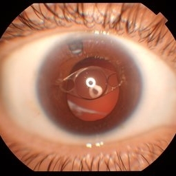 Anterior Iris Claw Artisan Lens
May 14 2025 by Moazzam Parvez
Anterior segment image of a 40 year old gentleman with a anteriorly placed iris claw lens post retinal detachment surgery.
Photographer: Dr Moazzam Parvez, Netralayam , Kolkata
Imaging device: Topcon DC-4
Condition/keywords: Anteriorly placed iris claw lens
-
Eye of the Hurricane
Apr 9 2025 by Gustavo Uriel Fonseca Aguirre
Ultrasound biomicroscopy of a post-operative eye (status post trabeculectomy and phacoemulsification) reveals a patent ostium on the right side, along with an intraocular lens in position. A hyphema is observed displaying small convection currents, creating a circular motion pattern due to the temperature gradient between the iris and cornea. Notably, the blood flow can be seen circulating toward the trabeculectomy site.
Condition/keywords: hyphema, trabeculectomy
-
 Eye of the Hurricane
Apr 8 2025 by Gustavo Uriel Fonseca Aguirre
Ultrasound biomicroscopy of a post-operative eye (status post trabeculectomy and phacoemulsification) reveals a patent ostium on the right side, along with an intraocular lens in position. A hyphema is observed displaying small convection currents, creating a circular motion pattern due to the temperature gradient between the iris and cornea. Notably, the blood flow can be seen circulating toward the trabeculectomy site.
Photographer: Gustavo U. Fonseca Aguirre, Hospital Conde de Valenciana, Ciudad de México
Condition/keywords: Hyphema, trabeculectomy
-
 Iris Discoloration
Apr 1 2025 by Korey Starkey
4 year-old patient sent for genetic testing to rule out possibility of Waardenburg syndrome with Hirschsprung disease. Left eye iris has no discoloration present, vision in both eyes is 20/40.
Photographer: Korey Starkey
Imaging device: Topcon
Condition/keywords: external, external photography, iris, topcon
-
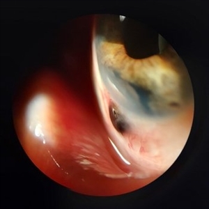
 Ciliary Body Metastasis
Mar 26 2025 by Virginia Gebhart
54 year old female referred for iris mass. UBM shows large solid mass originating in the ciliary body and eroding into the anterior chamber under the iris epithelium. Recent CT scans revealed multiple bilateral pulmonary and hepatic nodules. Pt has been scheduled for PET scan and liver biopsy by radiation oncologist.
Photographer: Virginia Gebhart, Retina Consultants of Carolina
Imaging device: Samsung Galaxy
Condition/keywords: choroidal metastasis, ciliary body mass, metastatic cancer
-
 Dislocated Lens
Jan 30 2025 by Kimberly Wakester
Fundus photograph of a 37-year-old man with an anteriorly dislocated lens in the left eye. The natural lens has displaced anteriorly in the AC secondary to trauma to the eye. There is also a Macular hole present with vitreous hemorrhage. Patient was recommended to proceed with lensectomy, iris repair and MH repair in the left eye.
Photographer: Kimberly Wakester, COA
Imaging device: Topcon TRC-50DX
Condition/keywords: dislocated lens, iridodialysis
-
 Macular Hole
Jan 30 2025 by Kimberly Wakester
Fundus photograph of a 37-year-old man with an anteriorly dislocated lens in the left eye. The natural lens has displaced anteriorly in the AC secondary to trauma to the eye. There is also a Macular hole present with vitreous hemorrhage. Patient was recommended to proceed with lensectomy, iris repair and MH repair in the left eye.
Photographer: Kimberly Wakester, COA
Imaging device: Optos California
Condition/keywords: dislocated lens, macular hole, vitreous hemorrhage
-
 Iris Nevus
Jan 28 2025 by Korey Starkey
Slit-lamp image of an 89-year-old patient with an iris nevus. Nevus appeared stable on exam, will continue to monitor.
Photographer: Korey Starkey
Imaging device: Slit lamp camera
Condition/keywords: ectropion uveae, iris nevus, slit lamp photo
-
 Iris Melanoma
Jan 28 2025 by Korey Starkey
Slit-lamp image of 90-year-old patient with iris melanoma and new hemorrhage affecting the right eye. Patient re-presented after nearly 1 year, now seeking treatment. Given iris location of tumor, multiple clock hours of iris involved, and increase in size of the known malignant transformation; safest approach was enucleation.
Photographer: Korey Starkey
Imaging device: Slit lamp camera
Condition/keywords: anterior chamber, hemorrhage, iris melanoma, slit lamp photo
-
 Sectoral Ocular Melanocytosis
Jan 17 2025 by Virginia Gebhart
67 year old female with congenital sectoral ocular melanocytosis. Pigmentation on nasal sclera and nasal iris of right eye, as well as deep pigmentation nasally of fundus. Will continue close observation
Photographer: Virginia Gebhart
Imaging device: Topcon 50DX/Samsung Galaxy
Condition/keywords: choroidal melanocytosis, heterochromia, ocular melanocytosis, Oculodermal Melanocytosis
-
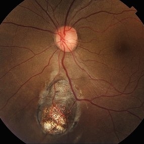
 Unilateral Coloboma Involving Disc and Macula
Dec 27 2024 by Tejaswita Verma
Fundus image of a 15 years old male presenting with unilaterally diminished vision since childhood in RE with CF3mt vision and inferior iris coloboma and retinochoroidal coloboma with nystagmus and cataract.
Photographer: DR. TEJASWITA VERMA
Imaging device: MIRANTE
Condition/keywords: chorioretinal coloboma, iridofundal coloboma
-
 Coloboma in a Unicameral Eye
Dec 20 2024 by Virginia Gebhart
59 year old female with choroidal coloboma extending into iris. Pt had PC IOL placed in 2016, removed in Aug due to suspected UGH syndrome. Lens haptics were oriented vertically causing haptic to chafe iris superiorly. Most likely etiology was loss of inferior zonules from coloboma. Pt remains aphakic.
Photographer: Virginia Gebhart, Retina Consultants of Carolina
Imaging device: Optos California
Condition/keywords: choroidal coloboma, coloboma
-
 NVI
NVI
Oct 24 2024 by Korey Starkey
Iris FA of a 74 year old male with neovascularization of the iris. Noted mild activity of NVI at the superior pupillary margin, recommending observation at time of visit.
Photographer: Korey Starkey
Imaging device: Heidelberg Spectralis
Condition/keywords: FA, Heidelburg Spectralis, Iris, iris fluorescein angiogram, neovascularization of iris (NVI), smokestack
-
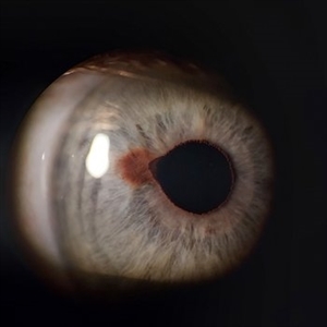
 Suspicious Lesion 18 Years s/p Iris Resection
Oct 15 2024 by Virginia Gebhart
85 year old female with small pigmented lesion present s/p sectoral iridectomy in 2006. Lesion is suspicious for recurrence of melanoma after 18 years. Stable compared to previous exam in March 2024, unclear if this is a new lesion or has been present for an extended time. Will monitor closely.
Photographer: Virginia Gebhart, Retina Consultants of Carolina
Imaging device: Samsung Galaxy
Condition/keywords: iris melanoma, melanoma
-
 New Iris Melanoma
Oct 10 2024 by Virginia Gebhart
56 year old male with new amelanotic melanoma emanating from the ciliary body through the posterior iris epithelium. CT scan showed no evidence of metastatic disease. Pt scheduled for radioactive plaque and tumor biopsy
Photographer: Virginia Gebhart, Retina Consultants of Carolina
Imaging device: Samsung Galaxy
Condition/keywords: amelanotic melanoma, iris melanoma
-
 Retinal Colomoba
Jul 21 2024 by César Adrián Gómez Valdivia, MD
Retinal Coloboma found in a female 41 year old patient. Iris, Lens, Ciliary Body, Zonules, Choroid and Retina were involved.
Photographer: Erika Paulina Ornelas Cazares
Imaging device: TOPCON TRC-50DX
Condition/keywords: coloboma
-
 Scleral Ectasia Post Radiation for Iris Melanoma
Jul 5 2024 by Zach Seim
Slit-Lamp Photograph of a 52 year old male with Scleral Ectasia post radiation for Iris Melanoma.
Photographer: Zach Seim
Imaging device: Slit Lamp Photography on Samsung Galaxy 7
Condition/keywords: Iris, iris melanoma, scleral ectasia, slit lamp photo, slit lamp photography
-
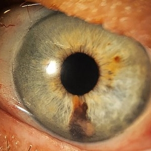
 Iris Nevus
Jul 3 2024 by Zach Seim
Slit Lamp Photograph of an 88 year old man with an Iris Nevus. Patient presented with DCC 20/60+1. Plan to monitor.
Photographer: Zach Seim
Imaging device: Slit Lamp photography with Samsung Galaxy 7
Condition/keywords: iris, iris nevus, nevus, right eye, slit lamp photo, slit lamp photography
-
 Iris Melanoma
Feb 1 2024 by Virginia Gebhart
90 year old female with elevated pigmented lesion, amelanotic portion extending toward the angle, questionable vascularity on UBM.
Photographer: Virginia Gebhart
Imaging device: Samsung Galaxy Z Flip
Condition/keywords: iris lesion, iris melanoma
-
 Tilted IOL
Dec 7 2023 by Virginia Gebhart
61 year old female with inferiorly tilted IOL. UBM shows haptic rubbing against the iris causing transillumination defect from 5 to 6 o'clock.
Photographer: Virginia Gebhart
Condition/keywords: lens dislocation, transillumination
-
 Idiopathic Iris Cyst
Oct 25 2023 by Virginia Gebhart
UBM of recurring idiopathic iris cyst in 72 year old female
Photographer: Virginia Gebhart
Imaging device: Ellex Eye Cubed
Condition/keywords: anterior chamber, cyst, immersion ultrasound, iris
-
 Iridodialysis Repair
Jul 11 2023 by Maneesh M Bapaye, MD, MBA
A 68 year old female patient was referred with large iridodialysis in superotemporal and temporal quadrant in right eye, as seen in left side image in the panel. She underwent iridodialysis repair using 9-0 prolene suture on single armed needle to suture the iris to iris root in hangback fashion. The patient had satisfactory anatomical outcome as seen in right side image in the panel.
Photographer: Dr.Maneesh Bapaye
Condition/keywords: Intraoperative iridodialysis
-
 Fundus Coloboma
Feb 22 2023 by Zach Seim
An ultra-widefield fundus image of a 25 year old male with Fundus Coloboma, as well as Iris Coloboma affecting both eyes. Patient's vision at the time of the image was 20/100-2. Discussed genetic testing as patient reports that he has a child with coloboma and patient agrees. There is a possibility of this finding being syndromic given cornea has small WTW and possibly microphthalmia. The patient has old tractional exudation at edge (abutting fovea). Recommended observation without treatment.
Photographer: Zach Seim
Imaging device: Optos California
Condition/keywords: coloboma, coloboma of optic disc, fundus photograph, Optos, scanning laser ophthalmoscope, ultra-wide field imaging
-
 Iris Coloboma
Feb 22 2023 by Zach Seim
An external image of a 25 year old male with Iris Coloboma, as well as Fundus Coloboma affecting both eyes. Patient's vision at the time of the image was 20/80. Discussed genetic testing as patient reports that he has a child with coloboma and patient agrees. There is a possibility of this finding being syndromic given cornea has small WTW and possibly microphthalmia. Recommended observation without treatment.
Photographer: Zach Seim
Imaging device: Topcon 50DX
Condition/keywords: coloboma, iris, left eye, Topcon
 Loading…