Search results (275 results)
-
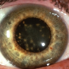
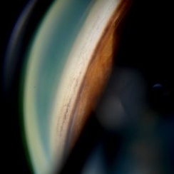
 New Iris Melanoma
Oct 10 2024 by Virginia Gebhart
56 year old male with new amelanotic melanoma emanating from the ciliary body through the posterior iris epithelium. CT scan showed no evidence of metastatic disease. Pt scheduled for radioactive plaque and tumor biopsy
Photographer: Virginia Gebhart, Retina Consultants of Carolina
Imaging device: Samsung Galaxy
Condition/keywords: amelanotic melanoma, iris melanoma
-
 Iris Pigmented Lesion
Apr 27 2018 by Mark Lazcano
Gonio photograph of 20-year-old male with pigmented iris lesion consistent with melanocytoma
Photographer: mark Lazcano,University of Miami , Bascom Palmer Eye Institute
Imaging device: gonio Prism
Condition/keywords: pigmented lesion
-
 Iris
Iris
Apr 29 2019 by Stephanie Moolman
Multi-color images after Yag PI of iris.
Photographer: Stephanie Moolman, Dr Marissa Willemse, Pretoria, South Africa
Imaging device: Heidelberg Spectralis
Condition/keywords: glaucoma, iris, multicolor, NdYAG laser, peripheral iridotomy
-
 Iris Racemose Hemangioma
Jan 1 2023 by Maxwell J Wingelaar, MD
Fluorescein Angiogram of 66 year old female presented with an iris racemose hemangioma
Photographer: Ken Huff
Condition/keywords: Racemose hemangioma
-
 Wrinkled Anterior Capsule 40X zoom
Feb 18 2023 by Ahmed Abbas Hashmi, OD
Imprint of Iris Pigmentation on Anterior Lens Surface with wrinkled anterior capsule
Photographer: Ahmed Abbas Hashmi
Condition/keywords: lens opacity
-
 Cat Eye Syndrome
Feb 11 2020 by Sophia El Hamichi, MD
A 3-year-old female with cat eye syndrome including iris, chorioretinal and optic nerve colobomas. Note the CNV temporally to the optic nerve coloboma (blue arrows)
Photographer: Giselle De Oliveira, Bascom Palmer Eye Institute, Miami
Imaging device: RetCam
Condition/keywords: cat eye syndrome, chorioretinal coloboma, choroidal neovascularization (CNV), coloboma, coloboma of optic disc, optic nerve coloboma
-
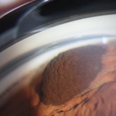
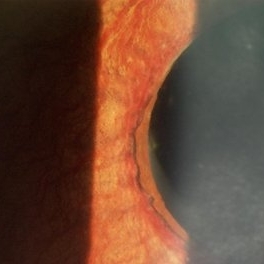
 Ciliary Body Metastasis
Mar 26 2025 by Virginia Gebhart
54 year old female referred for iris mass. UBM shows large solid mass originating in the ciliary body and eroding into the anterior chamber under the iris epithelium. Recent CT scans revealed multiple bilateral pulmonary and hepatic nodules. Pt has been scheduled for PET scan and liver biopsy by radiation oncologist.
Photographer: Virginia Gebhart, Retina Consultants of Carolina
Imaging device: Samsung Galaxy
Condition/keywords: choroidal metastasis, ciliary body mass, metastatic cancer
-
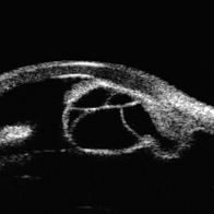
Eye of the Hurricane
Apr 9 2025 by Gustavo Uriel Fonseca Aguirre
Ultrasound biomicroscopy of a post-operative eye (status post trabeculectomy and phacoemulsification) reveals a patent ostium on the right side, along with an intraocular lens in position. A hyphema is observed displaying small convection currents, creating a circular motion pattern due to the temperature gradient between the iris and cornea. Notably, the blood flow can be seen circulating toward the trabeculectomy site.
Condition/keywords: hyphema, trabeculectomy
-
 Eye of the Hurricane
Apr 8 2025 by Gustavo Uriel Fonseca Aguirre
Ultrasound biomicroscopy of a post-operative eye (status post trabeculectomy and phacoemulsification) reveals a patent ostium on the right side, along with an intraocular lens in position. A hyphema is observed displaying small convection currents, creating a circular motion pattern due to the temperature gradient between the iris and cornea. Notably, the blood flow can be seen circulating toward the trabeculectomy site.
Photographer: Gustavo U. Fonseca Aguirre, Hospital Conde de Valenciana, Ciudad de México
Condition/keywords: Hyphema, trabeculectomy
-
 Idiopathic Iris Cyst
Aug 13 2025 by Virginia Gebhart
74 year old female with idiopathic iris cyst encroaching visual axis. Lesion is cystic with baseline internal reflectivity, unequivocal growth since first exam in 2022. Pt remains asymptomatic, will continue to observe.
Photographer: Virginia Gebhart, Retina Consultants of Carolina
Imaging device: Ellex Eye Cubed
Condition/keywords: cyst, cystic lesion, idiopathic cysts, ultrasound biomicroscopy
-
---thumb.jpg/image-square;max$300,300.ImageHandler) Iris Nodules (Lisch)
Iris Nodules (Lisch)
Dec 27 2013 by David Callanan, MD
57-year-old female patient with iris nodules.
Condition/keywords: iris nodules
-
 NVI
NVI
Oct 24 2024 by Korey Starkey
Iris FA of a 74 year old male with neovascularization of the iris. Noted mild activity of NVI at the superior pupillary margin, recommending observation at time of visit.
Photographer: Korey Starkey
Imaging device: Heidelberg Spectralis
Condition/keywords: FA, Heidelburg Spectralis, Iris, iris fluorescein angiogram, neovascularization of iris (NVI), smokestack
-
 Siderosis
May 2 2013 by Henry J. Kaplan, MD
Iron deposition in the iris epithelium and sphincter and on lens epithelium in the same patient ; #2.
Condition/keywords: siderosis
-
 Waardenberg Syndrome
Jan 21 2021 by Jamin S. Brown, MD
9-year-old African American female, fellow eye color blue.
Photographer: Stefanie Palmer CRA, Retina Vitreous Surgeons of CNY
Condition/keywords: anterior segment, iris, waardenburg syndrome
-
 4 Point Scleral Fixation Akreos AO60 With Gore Tex Suture
May 21 2021 by Jesus Lozano, MD
Anterior segment photo of a 54-year-old man after 4 point scleral fixation Akreos AO60 with Gore Tex suture plus PPV who had a severe traumatic iris defect and was aphakic after ocular trauma.
Photographer: Luigi Zinn, Hadassah Medical Center, Jerusalem.
Condition/keywords: aphakia, cornea rupture, lens, penetrating trauma
-
 Advanced Stage of Neovascular Glaucoma
Mar 21 2013 by Yusuke Oshima, MD, PhD
An 82-year-old man with a advanced stage of neovascular glaucoma. A slit-lamp photograph illustrates iris ectropion with prominent iris neovascularization.
Photographer: Yusuke Takada, Osaka University Graduate School of Medicine
Condition/keywords: neovascular glaucoma
-
 Artificial Fellow Iris Comparison
Dec 13 2019 by Eliot Bergandine
Iris implant on patient in late 60's.
Photographer: Eliot Bergandine
Imaging device: Canon CR-2 AF
Condition/keywords: artificial, implant, iris
-
 Artificial Iris Implant
Dec 13 2019 by Eliot Bergandine
Iris implant on patient in late 60's.
Photographer: Eliot Bergandine
Imaging device: Canon CR-2 AF
Condition/keywords: artificial, implant, iris
-
 Artificial Iris Implant Close Up
Dec 13 2019 by Eliot Bergandine
Iris implant on patient in late 60's.
Photographer: Eliot Bergandine
Imaging device: Canon CR-2 AF
Condition/keywords: artificial, implant, iris
-
 Chorioretinal Coloboma with Retinal Detachment
Dec 5 2020 by Niloofar Piri, MD
14-year-old female with 1q21.1 microdeletion syndrome and behavioral, intellectual, and systemic abnormalities, including congenital microcornea, iris coloboma, and chorioretinal and optic nerve coloboma presented with decreased vision. Right eye fundus taken with RetCam shows coloboma with retinal detachment. (Left eye showed white cataract with funnel RD on B-scan).
Photographer: Niloofar Piri MD, Douglas Snyder MD
Condition/keywords: chorioretinal coloboma, optic nerve coloboma
-
 Disseminated Chorioretinitis With Unknown Etiology
Apr 5 2018 by Kim Barrett
Ultra-wide field fluorescein angiogram of a 31-year-old female with intermittent pain in her left eye. Her condition has been managed in Liberia until recently when she moved to the United States. She suffers from multiple modalities including central retinal artery occlusion, posterior synechiae of the iris, interstitial keratitis, disseminated chorioretinitis, as well as HIV. An infectious cause is high on the differential in light of her HIV status. DDx: hypertensive crisis, an embolism (? IV drug use), coagulopathy, trauma, infectious. Blood work was normal. Her current vision is 20/30 right eye and 20/400 left eye.
Photographer: Kim Barrett, COA
Imaging device: Optos
Condition/keywords: central retinal artery occlusion (CRAO), chorioretinal scar, ciliary artery sparring, disseminated chorioretinitis, HIV, left eye, optic atrophy, staining
-
 Fundus Coloboma
Feb 22 2023 by Zach Seim
An ultra-widefield fundus image of a 25 year old male with Fundus Coloboma, as well as Iris Coloboma affecting both eyes. Patient's vision at the time of the image was 20/100-2. Discussed genetic testing as patient reports that he has a child with coloboma and patient agrees. There is a possibility of this finding being syndromic given cornea has small WTW and possibly microphthalmia. The patient has old tractional exudation at edge (abutting fovea). Recommended observation without treatment.
Photographer: Zach Seim
Imaging device: Optos California
Condition/keywords: coloboma, coloboma of optic disc, fundus photograph, Optos, scanning laser ophthalmoscope, ultra-wide field imaging
-
 Gonioscopy: Pigment Dispersion Glaucoma
Jul 8 2013 by Jason S. Calhoun
Patient with no family history of glaucoma, came in with elevated IOP. During gonioscopy exam. brown pigment overlying the trabecular meshwork. Also, trans-illumination defects on the iris.
Photographer: Jason S. Calhoun, Department of Ophthalmology, Mayo Clinic Jacksonville, Florida
Condition/keywords: gonioscopy, pigment dispersion syndrome of iris
-
 Gonioscopy; Scattered Peripheral Anterior Synechiae
Jul 8 2013 by Jason S. Calhoun
Patient came in for evaluation for glaucoma. Patient also has a history of uveitis. Last flare up was back in 1990. Patient's VA was 20/30, right eye and 20/40-1, left eye. Slit Lamp gonioscopy reveals iris bow with scattered PAS around the angles of the anterior chamber. You can also see pigmentation in the trabecular meshwork. Patient will follow up in 3 months.
Photographer: Jason S. Calhoun, Department of Ophthalmology, Mayo Clinic Jacksonville, Florida
Condition/keywords: gonioscopy, goniosynechiae
-
 Iris Bombe
Sep 15 2020 by Sophia El Hamichi, MD
A 23-year-old female with Coats' disease OS.
Photographer: Belinda Rodriguez, Murray Ocular Oncology and Retina, Miami
Condition/keywords: Coats' disease, iris bombe, silicone oil
 Loading…