Search results (52 results)
-
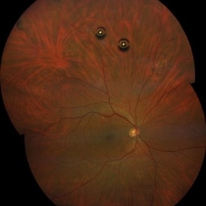
 Ozurdex implant
Aug 23 2012 by Daniel A. Adelberg, MD, FASRS
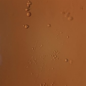
Anterior Segment photograph of a 50 year old with Uveitis and Cystoid Macular Edema status post Intravitreal injection of an Ozurdex dexamethasone implant
Photographer: Robert Ramsey, Southwestern Eye Center, Mesa Arizona
Condition/keywords: Ozurdex implant
-
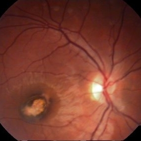
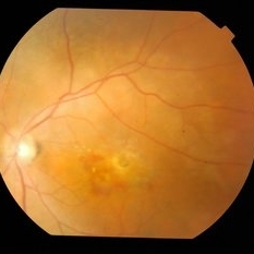
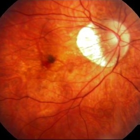 Myopic Choroidal Neovascular Membrane
Mar 25 2013 by Ratimir Lazic, MD, PhD
Color fundus photography of a 33-year-old female. In macular area subretinal hemorrhage can be seen. Area of atrophy temporal from PNO. Myopic changes of posterior pole and mid periphery can be noticed. The patient has been treated with 2 consecutive ranibizumab intravitreal injections. BCVA at baseline was 0,05 (Snellen lines) and 0,3 (Snellen lines) 2 months after.
Photographer: Marko Lukic, MD
Imaging device: Zeis Visucam Lite 2
Condition/keywords: high myopia, myopic choroidal neovascularization (CNV), ranibizumab
-
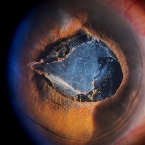
 cRORA
cRORA
Aug 5 2020 by Dhaivat Shah
A 54-year-old healthy male presented to us with a decreased vision in right eye since past 8 years. The patient gave a history of bleed in right eye before 8 years for which some intravitreal injection was given; post which there no major visual improvement. No details or documentation was available regarding the same. His BCVA in the right eye was 5/60. Fundus examination revealed a sharply demarcated hypopigmented patch over the macula with mild posterior excavation suggestive of macular scar. OCT image shows foveal thinning with loss of Retinal pigment epithelium and outer retinal layers (RORA). There are 2 types of RORAs, complete and incomplete. Complete RORA and incomplete RORA are entities defined by various imaging modalities describing atrophy of the retinal pigment epithelial and the outer retinal layers. OCT imaging defines incomplete RORA (iRORA) as a region of signal hyper transmission into the choroid and a corresponding zone of attenuation ordisruption of the RPE (<250um) and evidence of overlying photoreceptor degeneration (<250um). There should not be any RPE tear associated with it. OCT imaging describes complete RORA (cRORA) based on 4 inclusion criteria. These include, area of hypertransmission of more than 250um, zone of attenuation or disruption of the RPE of more than 250um in diameter, evidence of overlying photoreceptor degeneration and absence of scrolled RPE or other signs of an RPE tear. Other modalities used to define these include fundus autoflourescence(FAF), near infrared reflectance(NIR) and color fundus photograph(CFP). On CFP, it shows a sharply demarcated hypopigmented of >250um size with better visibility of choroidal vessels. FAF shows a hypo autoflourescent patch with sharply demarcated borders of size >250um, the colour of which is similar to that of the optic nerve head or retinal blood vessels excluding any pigmentation or artefact. On NIR, it shows a hyperreflective area with sharply demarcated borders of >250um size excluding any artefact. RORA can be seen in conditions like geographical atrophy in ARMD, central areolar choroidal dystrophy, atrophy secondary to anti-VEGF treatment. References: 1. Sadda SR, Guymer R, Holz FG, et al. Consensus Definition for Atrophy Associated with Age-Related Macular Degeneration on OCT: Classification of Atrophy Report 3 [published correction appears in Ophthalmology. 2019 Jan;126(1):177]. Ophthalmology. 2018;125(4):537-548. 2. Guymer RH, Rosenfeld PJ, Curcio CA, et al. Incomplete Retinal Pigment Epithelial and Outer Retinal Atrophy in Age-Related Macular Degeneration: Classification of Atrophy Meeting Report 4. Ophthalmology. 2020;127(3):394-409. 3. Eng VA, Rayess N, Nguyen HV, Leng T. Complete RPE and outer retinal atrophy in patients receiving anti-VEGF treatment for neovascular age-related macular degeneration. PLoS One. 2020;15(5):e0232353.
Photographer: Miss Anjum Zafar Khan
Imaging device: Choithram Netralaya
Condition/keywords: macular scar, outer retina, retinal pigment epithelium
-
 Funnel Retinal Detachment
Feb 2 2015 by Matt Poe, COA
The patient presented with total vision loss for >2months. Patient had history of exudative ARMD with intravitreal injections. No surgical intervention was done due to the long standing detachment and patient health.
Photographer: Matt Poe, COA. Northwest Arkansas Retina Associates, Springdale, AR.
Condition/keywords: retinal defect
-
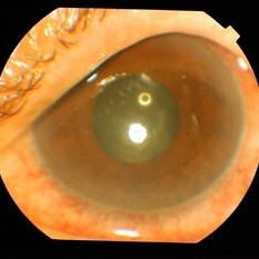
 Intravitreal Injection
Apr 12 2017 by John T. Thompson, MD
Intravitreal injection of anti-VEGF drug.
Imaging device: Cell phone with macro lens
Condition/keywords: intravitreal injection
-
 RPE tear in an 82-Year-Old Woman
Dec 7 2015 by Roy Schwartz, MD
An RPE tear in an 82-year-old woman, found incidentally after a year of visual deterioration. She hasn't visited an ophthalmologist since then. She has never underwent an intravitreal injection.
Photographer: Galit Yair Pur
Condition/keywords: retinal pigment epithelium (RPE) tear
-
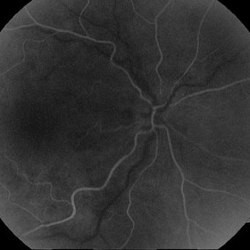
 Central Retinal Vein Occlusion
Feb 28 2013 by Theodore Leng, MD, MS, FASRS
OCT scan 4 weeks after a single intravitreal injection of aflibercept.
Imaging device: Zeiss Cirrus HD-OCT
Condition/keywords: aflibercept, central retinal vein occlusion (CRVO), cystoid macular edema (CME), EYLEA
-
---thumb.JPG/image-square;max$300,300.ImageHandler) spontaneous submacular hemorrhage
spontaneous submacular hemorrhage
Nov 3 2012 by Mallika Goyal, MD
Displacement of spontaneous submacular hemorrhage from subfoveal location 24 hours after intravitreal injection of SF6 gas and prone positioning. No macular abnormality was found.
Photographer: Mallika Goyal, MD
Condition/keywords: spontaneous submacular hemorrhage
-
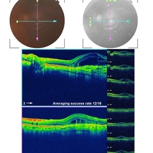 Resistant Wet AMD Responsive on Aflibercept
Mar 12 2014 by Ratimir Lazic, MD, PhD
An OCT image of a 77-year-old female with wet AMD poorly responsive to previously administered 15 monthly consecutive anti-VEGF intravitreal injections. The image was taken on the day of administration of the intravitreal aflibercept.
Photographer: Ivan Boras
Imaging device: Topcon 3D OCT
Condition/keywords: anti-VEGF, optical coherence tomography (OCT), wet age-related macular degeneration (wet AMD)
-
 Intravitreal Air Bubbles after anti-VEGF Injection
Jul 24 2020 by Darin R. Goldman, MD
Widefield fundus photograph of a 56-year-old male patient immediately following intravitreal anti-VEGF injection. Two air bubbles are visible within the mid-vitreous cavity.
Photographer: Alberto Velez, Retina Group of Florida
Imaging device: Zeiss Clarus
Condition/keywords: anti-VEGF, intravitreal, intravitreal injection
-
 Post Injection Candida Endophthalmitis
Jul 15 2014 by Mallika Goyal, MD
Left eye of a 75-year-old gentleman shows conjunctival congestion, chemosis, vitreous exudates 4 weeks following an intravitreal injection of avastin+triamcinolone acetonide combination for wet AMD non-responding to anti-VEGF monotherapy as well as PDT+Anti-VEGF combination therapy. Vitreous RNA-DNA chip analysis revealed candida species.
Photographer: Mallika Goyal, MD, Apollo Health City, Jubilee Hills, Hyderabad-500033
Condition/keywords: post injection candida endophthalmitis
-
 CNV due to Toxoplasmosis
Apr 6 2014 by Ratimir Lazic, MD, PhD
A color fundus image of a 7-year-old boy. Pigmented chorioretinal scar sorrounded by subretinal hemorrhage can be seen. VA is 0,2 by Snellen lines. The image presents the baseline clinical picture. The antiVEGF intravitreal injection, under general anesthesia, was administered.
Photographer: Marko Vlasic, University Eye Clinic Svjetlost
Imaging device: Zeis Visucam Lite 2
Condition/keywords: choroidal neovascularization (CNV), subretinal hemorrhage, toxoplasmosis
-
 Post Injection Candida Endophthalmitis
Jul 15 2014 by Mallika Goyal, MD
Left eye of a 75-year-old gentleman shows conjunctival congestion, chemosis, vitreous exudates 4 weeks following an intravitreal injection of avastin+triamcinolone acetonide combination for wet AMD non-responding to anti-VEGF monotherapy as well as PDT+Anti-VEGF combination therapy. Vitreous RNA-DNA chip analysis revealed candida species.
Photographer: Mallika Goyal, MD, Apollo Health City, Jubilee Hills, Hyderabad-500033
Condition/keywords: post injection candida endophthalmitis
-
 Inflammatory pupillary membrane in patient with endophthalmitis
Jan 28 2023 by Kingston Rodolfo Ureña-Wong, MD, Opht, MSc
Anterior segment photography of a 54-year-old woman with post phacoemulsification endophthalmitis. She did not improve after first intravitreal antibiotics injection and develop an inflammatory pupillary membrane. After two vitrectomies, and a complete three intravitreal injections scheme, we decided to remove the intraocular lens and capsules.
Photographer: Marco Antonio Rubio-Atonal,UNAM, Asociación para evitar la ceguera en México
Imaging device: Zeiss Clarus 700
Condition/keywords: endophthalmitis, pupillary membranes
-
 Methotrexate Bubble following Intravitreal Injection for PVR
Sep 21 2022 by Zach Seim
Ultra-widefield fundus photograph of an 81 year old female with a Methotrexate bubble following an Intravitreal Injection for Proliferative Vitreoretinopathy. Patient has been presenting to the office for two week interval Methotrexate injections in her left eye. The image was taken prior to her eighth injection which revealed a residual Methotrexate bubble in her inferior retinal image. This patient was seeing "lots" of floaters, as well as having visual acuity of cc20/400 cc20/200 PH.
Photographer: Zach Seim
Imaging device: OPTOS California
Condition/keywords: bubble, fundus photograph, fundus photography, intravitreal injection, left eye, methotrexate, nasal retina, Optos, proliferative vitreoretinopathy (PVR), pseudocolor, ultra-wide field imaging
-
 Silicone Oil Droplets in the Vitreous
Sep 25 2019 by Gustavo Barreto de Melo, MD, PhD, FASRS
A 65-year-old female presented with an acute decrease in VA in the left eye 48 hours after an intravitreal injection of an antiangiogenic drug. Slit-lamp examination showed AC cells (4+) and vitritis. No pain or hyperemia. Multiple silicone oil droplets were seen in the anterior vitreous.
Photographer: Celso Dias, Hospital de Olhos de Sergipe
Condition/keywords: silicone oil
-
 Post Injection Candida Endophthalmitis
Jul 15 2014 by Mallika Goyal, MD
Left eye of a 75-year-old gentleman shows status prior to intravitreal injection of avastin+triamcinolone acetonide combination for wet AMD non-responding to anti-VEGF monotherapy as well as PDT+Anti-VEGF combination therapy.
Photographer: Mallika Goyal, MD, Apollo Health City, Jubilee Hills, Hyderabad-500033
Condition/keywords: post injection candida endophthalmitis
-
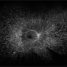
 Central Retinal Vein Occlusion - After 3 Consecutive Ranibizumab Injections
Jan 26 2013 by Ratimir Lazic, MD, PhD
FAG image(arterial phase) of a 67-year-old female after 3 consecutive ranibizumab intravitreal injections due to CME caused by CRVO.
Photographer: Marko Lukic, MD
Imaging device: Zeis Visucam Lite 2
Condition/keywords: central retinal vein occlusion (CRVO)
-
 Management of Submacular Hemorrhage
Mar 22 2020 by Anfisa Ayalon, MD
Intraoperative images were taken during the management of submacular hemorrhage in age-related macular degeneration. The goal of the surgery was the physical displacement of SMH out of the fovea using expansile gas. The image from the left was done during ILM peeling. Note the massive collection of subretinal blood. The image from the right was done after submacular injection of t-PA, bevacizumab and filtered air. Intravitreal injection of 20% SF6 completed the surgery. The visual acuity improved after the surgery from HM to 1/60.
Photographer: Anfisa Ayalon, MD., Meir Medical Center, Kfar Saba, Israel.
Condition/keywords: spontaneous submacular hemorrhage, submacular hemorrhage, vitreomacular surgery, wet age-related macular degeneration (wet AMD)
-
 Proliferative Diabetic Retinopathy
Nov 13 2019 by Olivia Rainey
Ultra-wide field fluorescein angiogram at 29 seconds of a 52-year-old male with proliferative diabetic retinopathy affecting his right eye. Patient is receiving Eylea intravitreal injections and has had panretinal photocoagulation in the past. Patient's vision tested 20/40 and with pinholes to 20/30.
Photographer: Olivia Rainey
Imaging device: Optos California
Condition/keywords: diabetes, diabetic macular edema, early phase, FA early phase, fluorescein angiogram (FA), intravitreal injection, ischemia, pan-retinal photocoagulation (PRP), proliferative diabetic retinopathy (PDR)
-
 Post Injection Candida Endophthalmitis
Jul 15 2014 by Mallika Goyal, MD
Left eye of a 75-year-old gentleman shows status prior to intravitreal injection of avastin+triamcinolone acetonide combination for wet AMD non-responding to anti-VEGF monotherapy as well as PDT+Anti-VEGF combination therapy.
Photographer: Mallika Goyal, MD, Apollo Health City, Jubilee Hills, Hyderabad-500033
Condition/keywords: post injection candida endophthalmitis
-
 Retinopathy of Prematurity Stage 4A Treated with Laser and Bevacizumab Intravitreal Injection
Jan 6 2020 by Sophia El Hamichi, MD
A 41-week-old girl, with a gestational age of 23 weeks and 4 days presenting with stage 4 A of ROP OU. She was treated with laser and intravitreal injections of bevacizumab.
Photographer: Abby Orcutt-Hayes, Murray Ocular Oncology and Retina
Condition/keywords: intravitreal bevacizumab, laser photocoagulation, retinopathy of prematurity stage 4a
-
 Ischemic BRVO OD Color
Apr 12 2018 by Aaron P. Appiah, MD
68-years-old, African American female with history of diabetes mellitus and ischemic BRVO OD, S/P Eylea intravitreal injections for CSME -resolved.
Imaging device: Optos California
Condition/keywords: branch retinal vein occlusion (BRVO)
-
 Montage Photo: Retinopathy of Prematurity Stage 4A Treated with Laser and Bevacizumab Intravitreal Injection
Jan 6 2020 by Sophia El Hamichi, MD
A 41-week-old girl, with a gestational age of 23 weeks and 4 days presenting with stage 4 A of ROP OU. She was treated with laser and intravitreal injections of bevacizumab.
Photographer: Abby Orcutt-Hayes, Murray Ocular Oncology and Retina
Condition/keywords: intravitreal bevacizumab, laser photocoagulation, montage, retinopathy of prematurity stage 4a
 Loading…