Search results (73 results)
-
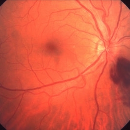
 Lyme Disease
Feb 13 2013 by From the Collections of Thomas M. Aaberg, MD and Thomas M. Aaberg Jr., MD
Papilledema, intra-retinal hemorrhage, periopticneuritis.
Condition/keywords: intraretinal hemorrhage, Lyme disease, periopticneuritis
-
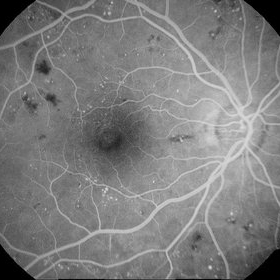
 Inferonasal Branch Retinal Vein Occlusion
Aug 23 2012 by Gerardo Garcia-Aguirre, MD
Fundus of a 55-year-old male showing intraretinal hemorrhages in the inferonasal quadrant.
Photographer: Noemí Hernández, Asociación para Evitar la Ceguera en México
Condition/keywords: branch retinal vein occlusion (BRVO), intraretinal hemorrhage
-
---thumb.jpg/image-square;max$300,300.ImageHandler) Peripapillary Atrophy
Peripapillary Atrophy
Feb 13 2013 by From the Collections of Thomas M. Aaberg, MD and Thomas M. Aaberg Jr., MD
Papilledema, intra-retinal hemorrhage, periopticneuritis.
Condition/keywords: intraretinal hemorrhage, papilledema, periopticneuritis, peripapillary atrophy
-
---thumb.jpg/image-square;max$300,300.ImageHandler) Fibrovascular Proliferation
Fibrovascular Proliferation
Feb 13 2013 by From the Collections of Thomas M. Aaberg, MD and Thomas M. Aaberg Jr., MD
Neovascularization, fibrous proliferation, intraretinal hemorrhage.
Condition/keywords: fibrous proliferation, intraretinal hemorrhage, neovascularization (NV)
-
---thumb.jpg/image-square;max$300,300.ImageHandler) Progressive Outer Retinal Necrosis
Progressive Outer Retinal Necrosis
Feb 15 2013 by From the Collections of Thomas M. Aaberg, MD and Thomas M. Aaberg Jr., MD
Color fundus photograph showing extensive confluent retinal whitening, retinal exudation, intraretinal hemorrhage, and sheathing of retinal vessels consistent with infectious retinitis such as progressive outer retinal necrosis (PORN).
Condition/keywords: occlusive retinitis, retinal necrosis
-
---thumb.jpg/image-square;max$300,300.ImageHandler) Peripheral retinal nonperfusion, capillary abnormalities, retinal microaneurysms, and intraretinal hemorrhage
Peripheral retinal nonperfusion, capillary abnormalities, retinal microaneurysms, and intraretinal hemorrhage
Feb 15 2013 by From the Collections of Thomas M. Aaberg, MD and Thomas M. Aaberg Jr., MD
Color fundus photograph showing peripheral retinal nonperfusion, capillary abnormalities, retinal microaneurysms, and intraretinal hemorrhage.
Condition/keywords: peripheral retinal nonperfusion, proliferative retinopathy
-
 SLE Retinopathy
Nov 14 2016 by Mitzy E Torres Soriano, MD
25-year-old female patient with systemic lupus erythematosus. Photographs show cotton wool spots, intraretinal hemorrhages and vascular tortuosity. FA demonstrated retinal vasculitis and OCT revealed cystoid macular edema. In this case diagnosis of SLE was made after ocular manifestation.
Photographer: Grupo Laser Vision, Rosario, Argentina
Condition/keywords: cotton wool spots, occlusive retinal vasculitis, occlusive vasculitis, systemic lupus erythematosus, vasculopathy
-
---thumb.jpg/image-square;max$300,300.ImageHandler) CMV with leukemia
CMV with leukemia
Feb 15 2013 by From the Collections of Thomas M. Aaberg, MD and Thomas M. Aaberg Jr., MD
color fundus photograph of a patient with leukemia complicated by CMV retinitis, manifesting as intraretinal hemorrhage, nerve fiber layer infarction, and retinal exudation
Condition/keywords: leukemia
-
 Branch Retinal Vein Occlusion with Macular Edema
Aug 23 2012 by Gerardo Garcia-Aguirre, MD
Fluorescein angiogram composition of the left eye, showing hypofluorescent areas corresponding to intraretinal hemorrhages.
Photographer: Noemí Hernández, Asociación para Evitar la Ceguera en México
Condition/keywords: branch retinal vein occlusion (BRVO), macular edema
-
---thumb.jpg/image-square;max$300,300.ImageHandler) Terson's Syndrome
Terson's Syndrome
Oct 8 2013 by Maurice F. Rabb
39 year female with a long history of chronic back pain treated by a sequence of epidural injections. Following her last injection, she complained of a moderately severe protracted headache and had several attempts at placement of an epidural blood patch without success. Under general anesthesia, she underwent injection of a larger volume of saline in an attempt to stem a presumed CSF leak producing "spinal headache". In the left eye she demonstrated multiple superficial and deep intraretinal hemorrhages associated with mild disc swelling and a central scotoma. In the right eye she showed a posterior subhyaloid and sub-internal limiting membrane hemorrhage with buffy coat layering superiorly. The visual acuity measured hand motions OD, 20/200 OS. The patient underwent a surgical evacuation of the sub-ILM hemorrhage.
Condition/keywords: Terson's Syndrome
-
---thumb.jpg/image-square;max$300,300.ImageHandler) Posterior Uveitis
Posterior Uveitis
Feb 15 2013 by From the Collections of Thomas M. Aaberg, MD and Thomas M. Aaberg Jr., MD
Color photograph of the mid-peripheral retina showing scattered intraretinal hemorrhage and foci of retinal whitening consistent with posterior uveitis, such as Behcet disease.
Condition/keywords: posterior uveitis, retinitis
-
---thumb.jpg/image-square;max$300,300.ImageHandler) Terson's Syndrome
Terson's Syndrome
Oct 8 2013 by Maurice F. Rabb
39 year female with a long history of chronic back pain treated by a sequence of epidural injections. Following her last injection, she complained of a moderately severe protracted headache and had several attempts at placement of an epidural blood patch without success. Under general anesthesia, she underwent injection of a larger volume of saline in an attempt to stem a presumed CSF leak producing "spinal headache". In the left eye she demonstrated multiple superficial and deep intraretinal hemorrhages associated with mild disc swelling and a central scotoma. In the right eye she showed a posterior subhyaloid and sub-internal limiting membrane hemorrhage with buffy coat layering superiorly. The visual acuity measured hand motions OD, 20/200 OS. The patient underwent a surgical evacuation of the sub-ILM hemorrhage.
Condition/keywords: Terson's Syndrome
-
 Combined Cilioretinal Artery and Central Retinal Vein Occlusion
May 14 2016 by Ines Leal
Combined cilioretinal artery and central retinal vein occlusion in an otherwise 49-year-old healthy female patient. Color fundus photography shows whitening of the retina in the distribution of the cilioretinal artery and intraretinal hemorrhages with tortuous and engorged veins.
Photographer: Inês Leal, MD, Department of Ophthalmology, Faculty of Medicine, Universidade de Lisboa,
Condition/keywords: cilioretinal artery occlusion, venous occlusion
-
---thumb.jpg/image-square;max$300,300.ImageHandler) Terson's Syndrome
Terson's Syndrome
Oct 8 2013 by Maurice F. Rabb
39 year female with a long history of chronic back pain treated by a sequence of epidural injections. Following her last injection, she complained of a moderately severe protracted headache and had several attempts at placement of an epidural blood patch without success. Under general anesthesia, she underwent injection of a larger volume of saline in an attempt to stem a presumed CSF leak producing "spinal headache". In the left eye she demonstrated multiple superficial and deep intraretinal hemorrhages associated with mild disc swelling and a central scotoma. In the right eye she showed a posterior subhyaloid and sub-internal limiting membrane hemorrhage with buffy coat layering superiorly. The visual acuity measured hand motions OD, 20/200 OS. The patient underwent a surgical evacuation of the sub-ILM hemorrhage.
Condition/keywords: Terson's Syndrome
-
---thumb.jpg/image-square;max$300,300.ImageHandler) Peripheral retinal nonperfusion, venous beading and dilatation, retinal microaneurysms, and intraretinal hemorrhage
Peripheral retinal nonperfusion, venous beading and dilatation, retinal microaneurysms, and intraretinal hemorrhage
Feb 15 2013 by From the Collections of Thomas M. Aaberg, MD and Thomas M. Aaberg Jr., MD
Color fundus photograph corresponding to slide titled "staining of retinal vessels, leakage from peripheral retinal neovascularization and peripheral nonperfusion." Shows peripheral retinal nonperfusion, venous beading and dilatation, retinal microaneurysms, and intraretinal hemorrhage.
Condition/keywords: peripheral retinal nonperfusion, proliferative retinopathy, retinal neovascularization
-
---thumb.jpg/image-square;max$300,300.ImageHandler) Optic Disc and Retinal Edema
Optic Disc and Retinal Edema
Feb 13 2013 by From the Collections of Thomas M. Aaberg, MD and Thomas M. Aaberg Jr., MD
Intra-retinal hemorrhage papilledema.
Condition/keywords: intraretinal hemorrhage, optic disc, papilledema, retinal edema
-
 Inferonasal BRVO - Fluorescein Angiogram
Aug 23 2012 by Gerardo Garcia-Aguirre, MD
Fluorescein angiogram showing hypofluorescence secondary to intraretinal hemorrhages, and perivascular hyperfluorescence secondary to vascular incompetence.
Photographer: Noemí Hernández, Asociación para Evitar la Ceguera en México
Condition/keywords: branch retinal vein occlusion (BRVO), intraretinal hemorrhage, vascular incompetence
-
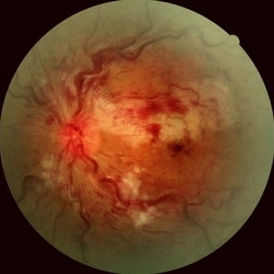
 Eals disease
Jan 26 2013 by Ratimir Lazic, MD, PhD
Color fundus photography of a 28-year-old male. Neovascularisations of the disc and retrohyaloid hemorrhages are seen together with intraretinal hemorrhages in macular area.
Photographer: Marko Lukic, MD
Imaging device: Zeis Visucam Lite 2
Condition/keywords: Eales disease, retinal vascular disorders, retrohyaloid hemorrhage
-
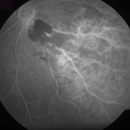
 Diabetic Retinopathy
Jun 29 2014 by Ratimir Lazic, MD, PhD
A FAG image of a 84-year-old female. Diabetic changes of the posterior pole and midperipheral retina can be seen. Mild dye leakage in macula with many hyperflorescent dots (microaneurisms) and hypoflorescent areas (intraretinal hemorrhages) can be seen.
Photographer: Marko Lukic, University Eye Clinic Svjetlost
Imaging device: Zeis Visucam Lite 2
Condition/keywords: diabetic retinopathy
-
 Chronic Retinal Vein Occlusion
Jul 8 2012 by Jeffrey S. Heier, MD
Chronic RVO with vascular changes, intraretinal hemorrhages
Imaging device: Zeiss
Condition/keywords: chronic retinal vein occlusion, intraretinal hemorrhage
-
---thumb.jpg/image-square;max$300,300.ImageHandler) Intraretinal Hemorrhage 1
Intraretinal Hemorrhage 1
Mar 20 2013 by Maurice F. Rabb
62-year- old white male with a small hemorrhage in the posterior segment in the right eye. In the left eye, the superior portion of the fundus was a uniform creamy white color with appearance of closure of the superior temporal artery. There were several areas of intraretinal hemorrhage and whitish lesions associated with the hemorrhage. There was vitreous haze, more marked inferiorly.
Condition/keywords: closure of superior temporal artery, intraretinal hemorrhage, vitreous haze, whitish lesions
-
---thumb.jpg/image-square;max$300,300.ImageHandler) intraretinal hemorrhage and retinal exudation
intraretinal hemorrhage and retinal exudation
Feb 15 2013 by From the Collections of Thomas M. Aaberg, MD and Thomas M. Aaberg Jr., MD
intraretinal hemorrhage and retinal exudation arising from microvascular damage associated with presumed herpesvirus infection.
Condition/keywords: macular edema, microangiopathy, retinal necrosis
-
---thumb.jpg/image-square;max$300,300.ImageHandler) Intraretinal Hemorrhage 2
Intraretinal Hemorrhage 2
Mar 20 2013 by Maurice F. Rabb
62-year- old white male with a small hemorrhage in the posterior segment in the right eye. In the left eye, the superior portion of the fundus was a uniform creamy white color with appearance of closure of the superior temporal artery. There were several areas of intraretinal hemorrhage and whitish lesions associated with the hemorrhage. There was vitreous haze, more marked inferiorly.
Condition/keywords: closure of superior temporal artery, intraretinal hemorrhage, vitreous haze, whitish lesions
-
---thumb.jpg/image-square;max$300,300.ImageHandler) peripheral retinal nonperfusion, capillary abnormalities, leaking retinal microaneurysms, and blocked fluorescence
peripheral retinal nonperfusion, capillary abnormalities, leaking retinal microaneurysms, and blocked fluorescence
Feb 15 2013 by From the Collections of Thomas M. Aaberg, MD and Thomas M. Aaberg Jr., MD
Mid-phase fluorescein angiograph showing peripheral retinal nonperfusion, capillary abnormalities, leaking retinal microaneurysms, and blocked fluorescence from intraretinal hemorrhage.
Condition/keywords: peripheral retinal nonperfusion, proliferative retinopathy
-
---thumb.jpg/image-square;max$300,300.ImageHandler) Terson's Syndrome
Terson's Syndrome
Oct 8 2013 by Maurice F. Rabb
39 year female with a long history of chronic back pain treated by a sequence of epidural injections. Following her last injection, she complained of a moderately severe protracted headache and had several attempts at placement of an epidural blood patch without success. Under general anesthesia, she underwent injection of a larger volume of saline in an attempt to stem a presumed CSF leak producing "spinal headache". In the left eye she demonstrated multiple superficial and deep intraretinal hemorrhages associated with mild disc swelling and a central scotoma. In the right eye she showed a posterior subhyaloid and sub-internal limiting membrane hemorrhage with buffy coat layering superiorly. The visual acuity measured hand motions OD, 20/200 OS. The patient underwent a surgical evacuation of the sub-ILM hemorrhage.
Condition/keywords: Terson's Syndrome
 Loading…