Search results (46 results)
-
 Open Funnel Retinal Detachment
Oct 13 2012 by Geoffrey G. Emerson, MD, PhD, FASRS
Open funnel retinal detachment
Condition/keywords: B scan ultrasound, open funnel RD
-
 Closed Funnel Retinal Detachment
Apr 9 2017 by Aliya Sultana
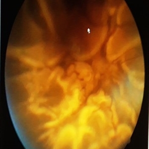
Fundus phtograph of an 51-year-old man with closed funnel rhegmatogenous retinal detachment presented to our department 6 weeks after cataract surgery. Posterior capsule rent noticed with vitreous in anterior chamber, condensed vitreous tag is incarcerated in side port wound.
Photographer: Dr Aliya Sultana , Assistant Professor,Sarojini Devi Eye Hospital, Hyderabad, Telangana. India.
-
 Funnel Retinal Detachment
Feb 2 2015 by Matt Poe, COA
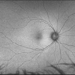
The patient presented with total vision loss for >2months. Patient had history of exudative ARMD with intravitreal injections. No surgical intervention was done due to the long standing detachment and patient health.
Photographer: Matt Poe, COA. Northwest Arkansas Retina Associates, Springdale, AR.
Condition/keywords: retinal defect
-
 Funnel Retinal Detachment With Proliferative Vitreoretinopathy
Oct 2 2013 by Jerald A. Bovino, MD
There is a total retinal detachment. The proliferative vitreoretinopathy has caused the retina to assume a funnel shape.
Condition/keywords: funnel, proliferative vitreoretinopathy (PVR), re-attached retinal detachment (RRD)
-
 Pediatric Tractional Retinal Detachment
Oct 23 2017 by Linda A Cernichiaro- Espinosa, MD
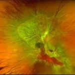
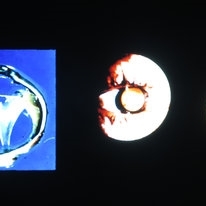
Color images and fluorescein angiography from two siblings. A and B from a 2 year old male. C and D a 5 year old male. Both fellow eyes have closed funnel total retinal detachments. They have characteristics from both familial exudative vitreoretinopathy (FEVR) and persistent fetal vasculature (PFV) within the NDP gene pathologies spectrum. This highlights the importance of genetic testing, prompt diagnosis and treatment.
Photographer: Abby Orcutt
Imaging device: RetCam III
Condition/keywords: exudative retinal detachment, familial exudative vitreoretinopathy (FEVR), neovascularization of the disc (NVD), pediatric retina, persistent fetal vasculature (PFV), tractional retinal detachment
-
 Total Retinal Detachment Due To Proliferative Vitreoretinopathy
Dec 10 2012 by Yale L. Fisher, MD
Funnel retinal detachment due to PVR - the patient is barely LP. Total detachments always connect and extend from the optic nerve.
Condition/keywords: proliferative vitreoretinopathy (PVR), video
-
 Closed-Funnel Retinal Detachment With Intraocular Melanoma
Oct 3 2013 by Jerald A. Bovino, MD
A mushroom shaped melanoma is present along with a closed funnel retinal detachment.
Condition/keywords: funnel, proliferative vitreoretinopathy (PVR)
-
 Closed Funnel Retinal Detachment
Oct 8 2019 by Olivia Rainey
Ultra-wide field pseudocolor image of a 57-year-old male with a closed funnel retinal detachment with anterior and posterior napkin rings affecting his left eye. Patient presented with klebsiella endophthalmitis in UK, and was in medically induced coma with tracheostomy. He awoke after sedation with loss of vision in both eyes, later developing a retinal detachment in both eyes. Prior inflammation attributable to prephthisical state and chronic funnel retinal detachment. The eye is inoperable and observation is recommended.
Photographer: Olivia Rainey and Amber Poss
Imaging device: Optos
Condition/keywords: blind eye, funnel, hypotony, klebsiella endopthalmitis, left eye, Optos
-
 Tractional vs Combined Tractional/Rhegmatogenous Retinal Detachment with Active Neovascularization OS
Jun 1 2018 by Hosam Attia, MD
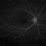
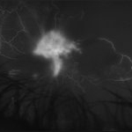
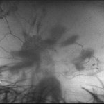
47-year-old African American, with history of diabetes mellitus of unknown duration and control, was referred for initial evaluation for conjunctival laceration in his left eye, following accidental finger nail injury, 6 days prior to presentation. - On exam, his vision was 20/50 OD and Bare HM/ LP OS. - Fundus color photos OD: No significant pathology, aside from attenuated vasculature OS: Chronic, Mac-Off, almost closed funnel tractional vs combined tractional/rhegmatogenous retinal detachment with large neovascularization (NVE) superiorly, detached ghost vessels, mild fresh vitreous hemorrhage, sub-retinal bands and inferior white vitreous debris from old hemorrhage (Not shown) - FA OD: No significant pathology aside from possible mild capillary non-perfusion in the extreme periphery, attenuated vasculature and possible tiny microaneurysms, nasally. OS: Extensive, wide spread capillary non- perfusion (correlate w/ detached Ghost vessels on color photos), and leakage from the NVE. - B/L Carotid Duplex was recommended due to the striking asymmetry in pathology with unknown medical history, diabetes duration and control etc (even in absence of any signs suggestive of possible ocular ischaemic syndrome OD)
Imaging device: Optos California
Condition/keywords: combined retinal detachment, tractional retinal detachment
-
 Chorioretinal Coloboma with Retinal Detachment
Dec 5 2020 by Niloofar Piri, MD
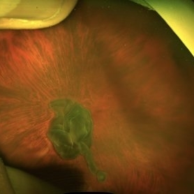
14-year-old female with 1q21.1 microdeletion syndrome and behavioral, intellectual, and systemic abnormalities, including congenital microcornea, iris coloboma, and chorioretinal and optic nerve coloboma presented with decreased vision. Right eye fundus taken with RetCam shows coloboma with retinal detachment. (Left eye showed white cataract with funnel RD on B-scan).
Photographer: Niloofar Piri MD, Douglas Snyder MD
Condition/keywords: chorioretinal coloboma, optic nerve coloboma
-
 Morning Glory Syndrome
Apr 14 2018 by Dhaivat Shah
7-year-old male patient presented to our OPD when the mother noticed that the child is not able to see clearly through the left eye. BCVA OD 6/6 OS 6/60. OU anterior segment normal. Fundus OD was normal, OS showed an enlarged, funnel-shaped excavation that incorporated the optic disc. The disc itself was enlarged, pink in color and had a surrounding area of peripapillary chorioretinal pigmentary changes which was sparing the fovea. MRI orbit/brain came out to be normal. OS diagnosed to be Morning glory syndrome. The child was prescribed full-time glasses to correct the anisometropia and occlusion/patching of the right(normal) eye 2 hours per day and guarded visual prognosis was explained.
Photographer: Miss Marina Parvin
Condition/keywords: Morning Glory Syndrome
-
 Tractional vs Combined Tractional/Rhegmatogenous Retinal Detachment with Active Neovascularization OS
Jun 1 2018 by Hosam Attia, MD
47-year-old African American, with history of diabetes mellitus of unknown duration and control, was referred for initial evaluation for conjunctival laceration in his left eye, following accidental finger nail injury, 6 days prior to presentation. - On exam, his vision was 20/50 OD and Bare HM/ LP OS. - Fundus color photos OD: No significant pathology, aside from attenuated vasculature OS: Chronic, Mac-Off, almost closed funnel tractional vs combined tractional/rhegmatogenous retinal detachment with large neovascularization (NVE) superiorly, detached ghost vessels, mild fresh vitreous hemorrhage, sub-retinal bands and inferior white vitreous debris from old hemorrhage (Not shown) - FA OD: No significant pathology aside from possible mild capillary non-perfusion in the extreme periphery, attenuated vasculature and possible tiny microaneurysms, nasally. OS: Extensive, wide spread capillary non- perfusion (correlate w/ detached Ghost vessels on color photos), and leakage from the NVE. - B/L Carotid Duplex was recommended due to the striking asymmetry in pathology with unknown medical history, diabetes duration and control, etc (even in absence of any signs suggestive of possible ocular ischemic syndrome OD)
Imaging device: Optos California
Condition/keywords: combined retinal detachment, neovascularization elsewhere (NVE), tractional retinal detachment
-
 Retinopathy of Prematurity
Feb 18 2015 by Andrea Arriola-Lopez, MD MSc
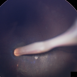
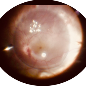
Closed funnel attached to posterior lens capsule.
Photographer: Andrea Elizabeth Arriola López, MSc. Asociación para Evitar la Ceguera, I.A.P. México D.F.
Imaging device: RetCam II
Condition/keywords: retinopathy of prematurity (ROP), stage 5
-
 Funnel Retinal Detachment
Jun 11 2023 by Ethan K Sobol, MD
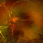
Intraoperative view of a funnel retinal detachment with proliferative vitreoretinoapthy in an eye with previous open globe injury. PVR membranes were peeled, and the retina was flattened and re-attached with an inferior relaxing retinotomy and silicone oil tamponade
Condition/keywords: intraoperative, open funnel RD, open globe injury, proliferative vitreoretinopathy (PVR)
-
 Retinal Detachment
Dec 18 2014 by H. Michael Lambert, MD
D3 PVR with funnel-shaped retinal detachment.
Condition/keywords: color fundus photograph
-
 Tractional vs Combined Tractional/Rhegmatogenous Retinal Detachment with Active Neovascularization OS
Jun 1 2018 by Hosam Attia, MD
47-year-old African American, with history of diabetes mellitus of unknown duration and control, was referred for initial evaluation for conjunctival laceration in his left eye, following accidental finger nail injury, 6 days prior to presentation. - On exam, his vision was 20/50 OD and Bare HM/ LP OS. - Fundus color photos OD: No significant pathology, aside from attenuated vasculature OS: Chronic, Mac-Off, almost closed funnel tractional vs combined tractional/rhegmatogenous retinal detachment with large neovascularization (NVE) superiorly, detached ghost vessels, mild fresh vitreous hemorrhage, sub-retinal bands and inferior white vitreous debris from old hemorrhage (Not shown) - FA OD: No significant pathology aside from possible mild capillary non-perfusion in the extreme periphery, attenuated vasculature and possible tiny microaneurysms, nasally. OS: Extensive, wide spread capillary non- perfusion (correlate w/ detached ghost vessels on color photos), and leakage from the NVE. - B/L Carotid Duplex was recommended due to the striking asymmetry in pathology with unknown medical history, diabetes duration and control, etc (even in absence of any signs suggestive of possible ocular ischemic syndrome OD)
Imaging device: Optos California
Condition/keywords: combined retinal detachment, tractional retinal detachment
-
 Tractional vs Combined Tractional/Rhegmatogenous Retinal Detachment with Active Neovascularization OS
Jun 1 2018 by Hosam Attia, MD
47-year-old African American, with history of diabetes mellitus of unknown duration and control, was referred for initial evaluation for conjunctival laceration in his left eye, following accidental finger nail injury, 6 days prior to presentation. - On exam, his vision was 20/50 OD and Bare HM/ LP OS. - Fundus color photos OD: No significant pathology, aside from attenuated vasculature OS: Chronic, Mac-Off, almost closed funnel Tractional vs Combined Tractional/Rhegmatogenous Retinal Detachment with large neovascularization (NVE) superiorly, detached ghost vessels, mild fresh vitreous hemorrhage, sub-retinal bands and inferior white vitreous debris from old hemorrhage (Not shown) - FA OD: No significant pathology aside from possible mild capillary non-perfusion in the extreme periphery, attenuated vasculature and possible tiny microaneurysms, nasally. OS: Extensive, wide spread capillary non- perfusion (correlate w/ detached Ghost vessels on color photos), and leakage from the NVE. - B/L Carotid Duplex was recommended due to the striking asymmetry in pathology with unknown medical history, diabetes duration and control, etc (even in absence of any signs suggestive of possible ocular ischaemic syndrome OD)
Imaging device: Optos California
Condition/keywords: combined retinal detachment, tractional retinal detachment
-
 Tractional vs Combined Tractional/Rhegmatogenous Retinal Detachment with Active Neovascularization OS
Jun 1 2018 by Hosam Attia, MD
47-year-old African American, with history of diabetes mellitus of unknown duration and control, was referred for initial evaluation for conjunctival laceration in his left eye, following accidental finger nail injury, 6 days prior to presentation. - On exam, his vision was 20/50 OD and Bare HM/ LP OS. - Fundus color photos OD: No significant pathology, aside from attenuated vasculature OS: Chronic, Mac-Off, almost closed funnel tractional vs combined tractional/rhegmatogenous retinal detachment with large neovascularization (NVE) superiorly, detached ghost vessels, mild fresh vitreous hemorrhage, sub-retinal bands and inferior white vitreous debris from old hemorrhage (not shown) - FA OD: No significant pathology aside from possible mild capillary non-perfusion in the extreme periphery, attenuated vasculature and possible tiny microaneurysms, nasally. OS: Extensive, wide spread capillary non- perfusion (correlate w/ detached Ghost vessels on color photos), and leakage from the NVE. - B/L Carotid Duplex was recommended due to the striking asymmetry in pathology with unknown medical history, diabetes duration and control, etc (even in absence of any signs suggestive of possible ocular ischemic syndrome OD)
Imaging device: Optos California
Condition/keywords: combined retinal detachment, tractional retinal detachment
-
 Tractional vs Combined Tractional/Rhegmatogenous Retinal Detachment with Active Neovascularization OS
Jun 1 2018 by Hosam Attia, MD
47-year-old African American, with history of diabetes mellitus of unknown duration and control, was referred for initial evaluation for conjunctival laceration in his left eye, following accidental finger nail injury, 6 days prior to presentation. - On exam, his vision was 20/50 OD and Bare HM/ LP OS. - Fundus color photos OD: No significant pathology, aside from attenuated vasculature OS: Chronic, Mac-Off, almost closed funnel Tractional vs Combined Tractional/Rhegmatogenous Retinal Detachment with large Neovascularization (NVE) superiorly, detached ghost vessels, mild fresh vitreous hemorrhage, sub-retinal bands and inferior white vitreous debris from old hemorrhage (Not shown) - FA OD: No significant pathology aside from possible mild capillary non-perfusion in the extreme periphery, attenuated vasculature and possible tiny microaneurysms, nasally. OS: Extensive, wide spread capillary non- perfusion (correlate w/ detached Ghost vessels on color photos), and leakage from the NVE. - B/L Carotid Duplex was recommended due to the striking asymmetry in pathology with unknown medical history, diabetes duration and control, etc (even in absence of any signs suggestive of possible ocular ischaemic syndrome OD)
Imaging device: Optos California
Condition/keywords: combined retinal detachment, tractional retinal detachment
-
 Diabetic TRD - Photos OS
Nov 17 2019 by John S. King, MD
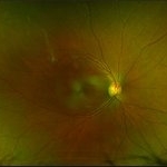
28-year-old white male with poorly controlled Type 1 DM, with a history of non-compliance with follow-ups, was referred with for DR with CME OS, and 3 weeks decrease vision OS. Va cc was 20/15 OD and HM OS. IOP 18/14. No NVI OU. Posteriorly, the right eye had macular exudates, no NVD, and a large area of NVE along the IT arcade. The left eye large NV plaque around disc, wrapping macula, with total RD with a posterior funnel appearance. The FA in the left eye showed severe peripheral and macular ischemia with diffuse leakage from a fibrovascualr plaque.
Photographer: Adriana Shelby
Imaging device: Optos CA
Condition/keywords: diabetic traction detachment, open funnel RD
-
 Slide 8-1
Mar 4 2019 by Lancaster Course in Ophthalmology
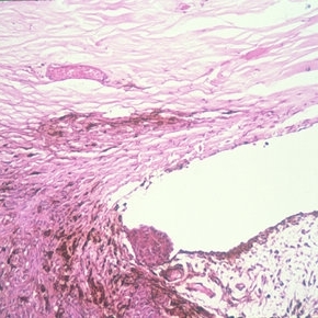
Left: Gross appearance of a funnel-shaped detachment of the vitreous. It remains attached at its base and at the optic nerve head. Center and right: Appearance of ring-shaped vitreous condensation, formerly attached at the optic nerve head. Such condensations can account for the visual symptom of floaters. (E.P. No. 21811)
Condition/keywords: detachment, operculum, vitreous
-
 Fundus Photo of Closed Funnel Retinal Detachment
Apr 10 2024 by Max D Schlesinger, MD
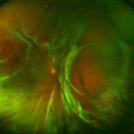
Wide-field funds photography of a closed funnel retinal detachment; patient had previously undergone 360 degree retinectomy in attempt to re-attach retina for a chronic retinal detachment, which was unsuccessful.
Condition/keywords: Closed funnel RD, detachment, Optos
-
 Closed-funnel-RD
Oct 27 2023 by Anand Temkar
Membranous echoes with high spikes with restricted after movements suggestive of retinal detachment ( closed funnel configuration )
Photographer: Dr.Anand Temkar- Retina Foundation, Ahmedabad
Condition/keywords: A-scan ultrasound, B scan ultrasound, Closed funnel RD
-
 PFC- Thor's Mjolnir in RD cases.
Jul 25 2020 by SANDEEP KUMAR
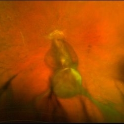
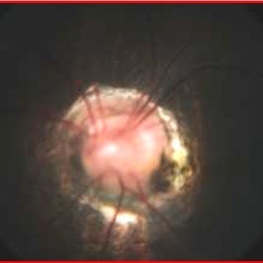
PFC in chronic retinal detachment has a vital role. The image is of a 12-year-old girl with open funnel RD. Characteristically PFCLs have high specific gravity ranging from 1.76 to 2.03, low surface tension, and viscosity. These physical properties make perfluorocarbon liquids an ideal for intraoperative tool in vitreoretinal surgery. With high specific gravity they flatten the detcahed retina and push the SRF anteriorly giving surgeon room for maneuvers.
Photographer: Dr Daraius Shroff . Shroff Eye Centre New Delhi
Condition/keywords: perfluorooctane
 Loading…