 Initializing download.
-
 By Dhaivat Shah
Sankara Nethralaya
Co-author(s): Dr Sudipta Das - Uploaded on Apr 14, 2018.
- Last modified by Caroline Bozell on Apr 17, 2018.
- Rating
- Appears in
- Imaging marvels
- Condition/keywords
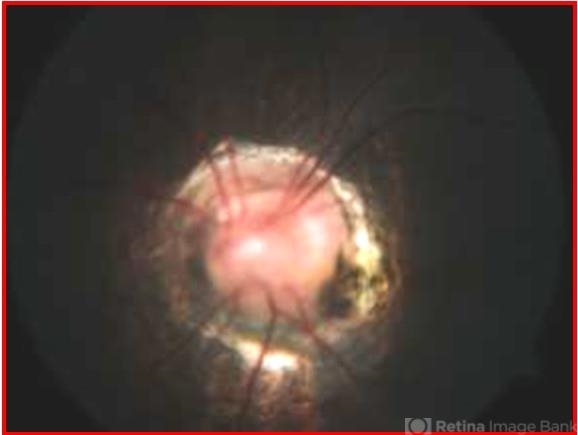
- Morning Glory Syndrome
- Photographer
- Miss Marina Parvin
- Description
- 7-year-old male patient presented to our OPD when the mother noticed that the child is not able to see clearly through the left eye. BCVA OD 6/6 OS 6/60. OU anterior segment normal. Fundus OD was normal, OS showed an enlarged, funnel-shaped excavation that incorporated the optic disc. The disc itself was enlarged, pink in color and had a surrounding area of peripapillary chorioretinal pigmentary changes which was sparing the fovea. MRI orbit/brain came out to be normal. OS diagnosed to be Morning glory syndrome. The child was prescribed full-time glasses to correct the anisometropia and occlusion/patching of the right(normal) eye 2 hours per day and guarded visual prognosis was explained.
---thumb.jpg/image-square;max$79,0.ImageHandler "Morning Glory of Disc")


