Search results (46 results)
-
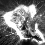
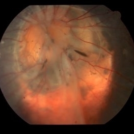
 Fundus Photo of Closed Funnel Retinal Detachment
Apr 10 2024 by Max D Schlesinger, MD
Wide-field funds photography of a closed funnel retinal detachment; patient had previously undergone 360 degree retinectomy in attempt to re-attach retina for a chronic retinal detachment, which was unsuccessful.
Condition/keywords: Closed funnel RD, detachment, Optos
-
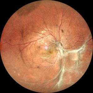
 Chorioretinal Coloboma with Retinal Detachment
Dec 5 2020 by Niloofar Piri, MD
14-year-old female with 1q21.1 microdeletion syndrome and behavioral, intellectual, and systemic abnormalities, including congenital microcornea, iris coloboma, and chorioretinal and optic nerve coloboma presented with decreased vision. Right eye fundus taken with RetCam shows coloboma with retinal detachment. (Left eye showed white cataract with funnel RD on B-scan).
Photographer: Niloofar Piri MD, Douglas Snyder MD
Condition/keywords: chorioretinal coloboma, optic nerve coloboma
-
 Chronic Open Funnel Retinal Detachment With Horse Shoe Tear
Feb 7 2024 by Harsh Vardhan Singh, MS
67 year old male with history of cataract surgery 1 year presented with old chronic retinal detachment with open funnel configuration with multiple breaks.
Photographer: Harsh Vardhan Singh
Imaging device: Clarus 700
Condition/keywords: chronic retinal detachment, Retinal Detachment, Retinal Detachment with multiple breaks
-
 Open Funnel Retinal Detachment
Oct 13 2012 by Geoffrey G. Emerson, MD, PhD, FASRS
Open funnel retinal detachment
Condition/keywords: B scan ultrasound, open funnel RD
-
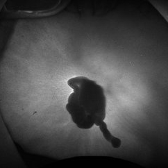
 Retinopathy of Prematurity
Feb 18 2015 by Andrea Arriola-Lopez, MD MSc
Closed funnel attached to posterior lens capsule.
Photographer: Andrea Elizabeth Arriola López, MSc. Asociación para Evitar la Ceguera, I.A.P. México D.F.
Imaging device: RetCam II
Condition/keywords: retinopathy of prematurity (ROP), stage 5
-
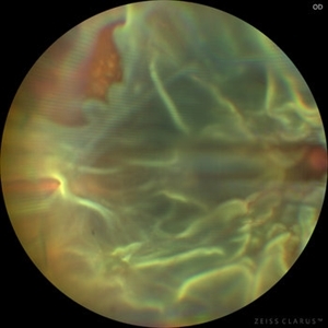
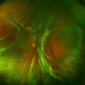
 360 retinotomy for combined closed funnel tractional and rhematogenous retinal detachment
Jan 1 2023 by Malek Yassine, MD
This is Fundus Autofluorecence, showing the residual hypoautofluorescent spots on the exposed choroid, relating to the previous panretinal photocoagulation, as well as the limits of the retinotomy with continuous laser which appeasr hypoautofluorecent with hyperautofluorecent margins.
Photographer: Malek Yassine, HMOED, Agadir, Morocco.
Imaging device: Zeiss Clarus
Condition/keywords: combined retinal detachment, rhegmatogenous retinal detachment, tractional retinal detachment
-
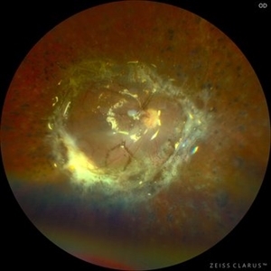 360 Retinotomy in a closed Funnel combined Tractional and rhegmatogenous retinal detachment
Jan 1 2023 by Malek Yassine, MD
This is the results at 6 months of a Bimanual 23 G-PPV with a very extensive and posterior 360 retinotomy for the management of a combined longstanding closed funnel RD, with submacular membranes, intraretinal PVR. Preop VA was a doubtful light perception. Borders of the retinotomy are stable at 6 months under 1300 Cs Silicon oil with some pigmented PVR developping the edges. Macula appears spared. Silicon oil emulsification droplets are well visualized beneath the superior temporal arcade.
Imaging device: Zeiss Clarus 700
Condition/keywords: combined retinal detachment, retinotomy, silicone oil
-
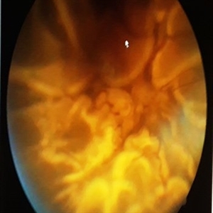
 Closed Funnel Retinal Detachment
Oct 8 2019 by Olivia Rainey
Ultra-wide field pseudocolor image of a 57-year-old male with a closed funnel retinal detachment with anterior and posterior napkin rings affecting his left eye. Patient presented with klebsiella endophthalmitis in UK, and was in medically induced coma with tracheostomy. He awoke after sedation with loss of vision in both eyes, later developing a retinal detachment in both eyes. Prior inflammation attributable to prephthisical state and chronic funnel retinal detachment. The eye is inoperable and observation is recommended.
Photographer: Olivia Rainey and Amber Poss
Imaging device: Optos
Condition/keywords: blind eye, funnel, hypotony, klebsiella endopthalmitis, left eye, Optos
-
 Closed Funnel Retinal Detachment
Apr 9 2017 by Aliya Sultana
Fundus phtograph of an 51-year-old man with closed funnel rhegmatogenous retinal detachment presented to our department 6 weeks after cataract surgery. Posterior capsule rent noticed with vitreous in anterior chamber, condensed vitreous tag is incarcerated in side port wound.
Photographer: Dr Aliya Sultana , Assistant Professor,Sarojini Devi Eye Hospital, Hyderabad, Telangana. India.
-
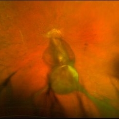
 Closed-Funnel Retinal Detachment With Intraocular Melanoma
Oct 3 2013 by Jerald A. Bovino, MD
A mushroom shaped melanoma is present along with a closed funnel retinal detachment.
Condition/keywords: funnel, proliferative vitreoretinopathy (PVR)
-
 Closed-funnel-RD
Oct 27 2023 by Anand Temkar
Membranous echoes with high spikes with restricted after movements suggestive of retinal detachment ( closed funnel configuration )
Photographer: Dr.Anand Temkar- Retina Foundation, Ahmedabad
Condition/keywords: A-scan ultrasound, B scan ultrasound, Closed funnel RD
-
 Diabetic TRD - Photos OS
Nov 17 2019 by John S. King, MD
28-year-old white male with poorly controlled Type 1 DM, with a history of non-compliance with follow-ups, was referred with for DR with CME OS, and 3 weeks decrease vision OS. Va cc was 20/15 OD and HM OS. IOP 18/14. No NVI OU. Posteriorly, the right eye had macular exudates, no NVD, and a large area of NVE along the IT arcade. The left eye large NV plaque around disc, wrapping macula, with total RD with a posterior funnel appearance. The FA in the left eye showed severe peripheral and macular ischemia with diffuse leakage from a fibrovascualr plaque.
Photographer: Adriana Shelby
Imaging device: Optos CA
Condition/keywords: diabetic traction detachment, open funnel RD
-
 Diabetic TRD OS - FA 1 Min
Nov 17 2019 by John S. King, MD
28-year-old white male with poorly controlled Type 1 DM, with a history of non-compliance with follow-ups, was referred with for DR with CME OS, and 3 weeks decrease vision OS. Va cc was 20/15 OD and HM OS. IOP 18/14. No NVI OU. Posteriorly, the right eye had macular exudates, no NVD, and a large area of NVE along the IT arcade. The left eye large NV plaque around disc, wrapping macula, with total RD with a posterior funnel appearance. The FA in the left eye showed severe peripheral and macular ischemia with diffuse leakage from a fibrovascualr plaque.
Photographer: Adriana Shelby
Imaging device: Optos CA
Condition/keywords: diabetic traction detachment, open funnel RD
-
 Extramacular TRD in Idiopathic Occlusive Vasculitis
Dec 5 2024 by Tejaswita Verma
Fundus photo showing extramacular TRD in a 16 year old boy with idiopathic occlusive vasculitis secondary to presumed IOTB. History of taking ATT for 6 months , Mantoux positive previously. Vision was 6/6P,other eye had funnel RD .
Photographer: DR. TEJASWITA VERMA
Imaging device: MIRANTE
Condition/keywords: tractional retinal detachment, vasculitis
-
 Fundus Autofluorescence of Closed Funnel Retinal Detachment
Apr 10 2024 by Max D Schlesinger, MD
Fundus autofluorescence of a closed funnel retinal detachment; patient had previously undergone 360 degree retinectomy in attempt to re-attach retina for a chronic retinal detachment, which was unsuccessful.
Condition/keywords: Autoflourescence, Closed funnel RD, detachment
-
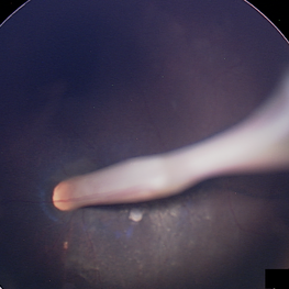
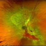
 Funnel Retinal Detachment
Jun 11 2023 by Ethan K Sobol, MD
Intraoperative view of a funnel retinal detachment with proliferative vitreoretinoapthy in an eye with previous open globe injury. PVR membranes were peeled, and the retina was flattened and re-attached with an inferior relaxing retinotomy and silicone oil tamponade
Condition/keywords: intraoperative, open funnel RD, open globe injury, proliferative vitreoretinopathy (PVR)
-
 Funnel Retinal Detachment
Feb 2 2015 by Matt Poe, COA
The patient presented with total vision loss for >2months. Patient had history of exudative ARMD with intravitreal injections. No surgical intervention was done due to the long standing detachment and patient health.
Photographer: Matt Poe, COA. Northwest Arkansas Retina Associates, Springdale, AR.
Condition/keywords: retinal defect
-
 Funnel Retinal Detachment With Proliferative Vitreoretinopathy
Oct 2 2013 by Jerald A. Bovino, MD
There is a total retinal detachment. The proliferative vitreoretinopathy has caused the retina to assume a funnel shape.
Condition/keywords: funnel, proliferative vitreoretinopathy (PVR), re-attached retinal detachment (RRD)
-
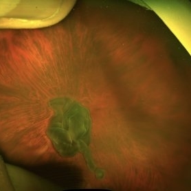
 Morning Glory
Jul 1 2022 by Geovanni Jassiel Rios, MD
Fundus Photograph 5 year old child with abnormal cup embryo-development. We can appreciate the radial vessel conformation and funnel shape nerve anomaly.
Photographer: Image Department Hospital de la Luz
Condition/keywords: Morning Glory Syndrome
-
 Morning Glory Disc
Sep 21 2023 by Ben Serar
Fundus photograph showing funnel shaped optic disc with radiating retinal vessels in a case of Morning glory syndrome.
Condition/keywords: Morning Glory Syndrome
-
 Morning Glory disc
Sep 14 2023 by Ben Serar
Fundus photograph showing Funnel shaped optic disc in a case of Morning Glory Syndrome.
Condition/keywords: Morning Glory Syndrome
-
 Morning Glory Disc
Sep 12 2023 by Ben Serar
Fundus photograph showing Funnel shaped optic disc in a case of Morning Glory Syndrome.
Condition/keywords: Morning Glory Syndrome
-
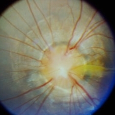
 Morning Glory Disc Anomaly
Feb 12 2024 by NIDHI PANWAR, MD FNB FICO
Fundus photograph of 43 year old male, hypertensive on medication, came for routine check up, and has been diagnosed to have poor vision left eye since childhood, denies any history of trauma. Vision left eye 6/18, Anterior segment normal, Fundus left eye shows excavated ,funnel-shaped optic nerve head, with central tuft of glial tissue obscuring the cup . The retinal vessels were seen emanating from the edge of disc in radial manner. In addition, the sectoral nasal retina shows localized area of hyperpigmented bony spicules like lesions. However, no history of nyctalopia or any other neurological disorder could be obtained.
Photographer: Nidhi Panwar, NMC Royal hospital, Sharjah , UAE
Imaging device: OPTOMAP
Condition/keywords: Morning Glory Anomaly, optic disc excavation
-
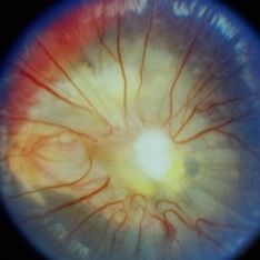
 Morning glory optic disc anomaly with retinal detachment
Sep 13 2022 by Min Kim, MD, PhD, MBA, FASRS
Fundus examination of this 5 year-old male shows large funneled optic nerve with conical excavation of the dysplastic optic disc. 360° macula-involving retinal detachment was observed. The best corrected visual acuity of the right eye was counting fingers 10cm.
Photographer: Min Kim, M.D.-Ph.D.-M.B.A. Gangnam Severance Hospital Yonsei University College of Medicine, Department of Ophthalmology
Imaging device: Optos Silverstone P200TxE
Condition/keywords: Morning Glory Anomaly, Morning Glory Syndrome
 Loading…