Search results (195 results)
-
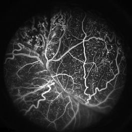
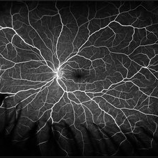
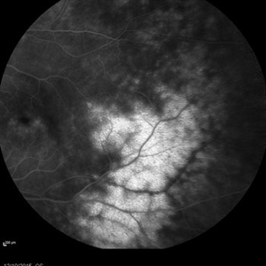
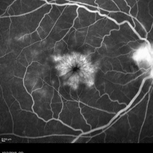
 Wyburn Mason Fluorescein Angiography
Jun 23 2018 by Caesar K. Luo, MD, FASRS
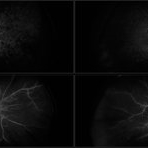
Fluorescein angiography of a 16-year-old female patient with unilateral ocular Wyburn-Mason syndrome without fluorescein leak.
Photographer: Joseph Trabucco, Progressive Vision Institute, Allentown, PA
Imaging device: Heidelberg Spectralis
Condition/keywords: fluorescein angiogram (FA), Wyburn-Mason
-
 Coats' Disease FA
Apr 27 2018 by Brenda Fallas
3-year-old boy with unilateral Coats' Disease FA photo.
Photographer: Brenda Fallas, Bascom Palmer Eye Institute, Miami, FL
Imaging device: Retcam III 130 degree lens
Condition/keywords: Coats' disease, FA early phase, fluorescein angiogram (FA), retinal telangiectasia
-
 Multiple Myeloma with Cytomegalovirus Retinitis
Apr 5 2018 by Kim Barrett
Ultra-wide field fluorescein angiogram of a 77-year-old male with multiple myeloma. Patient's angiogram presented significant peripheral retinal ischemia and cystoid macular edema. Patient tested positive for polymerase chain reaction, confirming cytomegalovirus retinitis. Patient is being treated with intravitreal ganciclovir and his current vision is 20/200.
Photographer: Kim Barrett, COA
Imaging device: Optos
Condition/keywords: cystoid macular edema (CME), fluorescein angiogram (FA), fluorescein leakage, intravitreal ganciclovir, myeloma, peripheral ischemia, positive polymerase chain reaction (PCR), ultra-wide field imaging
-
 Central Retinal Artery Occlusion
May 16 2017 by Olivia Rainey
Fluorescein angiogram of an 66-year-old female with a central retinal artery occlusion affecting her left eye.
Photographer: Olivia Rainey
Imaging device: Heidelberg Spectralis
Condition/keywords: 50 degrees, central retinal artery occlusion (CRAO), fluorescein angiogram (FA), left eye, mid phase, retinal ischemia
-
 Coats Disease
May 27 2016 by Olivia Rainey
Composite fluorescein angiogram of the left eye of a man with Coats Disease.
Photographer: Olivia Rainey
Imaging device: Heidelberg Spectralis
Condition/keywords: Coats' disease, composite, fluorescein angiogram (FA), fluorescein leakage, Heidelburg Spectralis
-
 Central Retinal Artery Occlusion
May 25 2017 by Olivia Rainey
UItra-widefield fluorescein angiography, taken at 6 minutes and 22 seconds, of an 73-year-old woman with a central retinal artery occlusion in her right eye.
Photographer: Olivia Rainey
Imaging device: Optos California
Condition/keywords: central retinal artery occlusion (CRAO), fluorescein angiogram (FA), ischemia, late phase, non-perfusion, Optos, ultra-wide field imaging
-
 Von Hippel-Lindau Syndrome with Retinal Hemangiomas
May 30 2017 by Olivia Rainey
Ultra-wide-field fluorescein angiogram of the left eye of an 29-year-old female with multiple retinal hemangiomas secondary to Von Hippel-Lindau Syndrome.
Photographer: Olivia Rainey
Imaging device: Optos California
Condition/keywords: fluorescein angiogram (FA), fluorescein leakage, left eye, Optos, retinal hemangioma, ultra-wide field imaging, Von Hippel-Lindau
-
 ROP FA OS
Apr 27 2018 by Brenda Fallas
4-month-old baby with regressed ROP post-Avastin.
Photographer: Brenda Fallas, Bascom Palmer Eye Institute, Miami, FL
Imaging device: RETCAM III 130 degree lens montage
Condition/keywords: FA late phase leakage, fluorescein angiogram (FA), retinopathy of prematurity (ROP)
-
 Central Retinal Artery Occlusion
May 25 2017 by Olivia Rainey
Ultra-wide field fluorescein angiography, taken at 42 seconds, of an 73-year-old female with a central retinal artery occlusion in her right eye.
Photographer: Olivia Rainey
Imaging device: Optos California
Condition/keywords: central retinal artery occlusion (CRAO), early phase, fluorescein angiogram (FA), ischemia, non-perfusion, Optos, ultra-wide field imaging
-
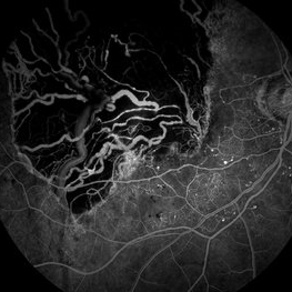
 Peripheral Retinal Ischemia
Apr 26 2018 by Olivia Rainey
Ultra-wide field fluorescein angiogram of a 55-year-old female with peripheral retinal ischemia affecting her left eye. CTA head and neck performed on 11/16/15 and showed calcified atherosclerotic plaque involving the intracranial internal carotid arteries with resulting luminal narrowing. Intracranial vertebral arteries have smooth luminal contours. CTA neck normal. Likely from internal carotid plaques. Sickle cell disease came back negative.
Photographer: Olivia Rainey
Imaging device: Optos California
Condition/keywords: fluorescein angiogram (FA), fluorescein leakage, left eye, Optos, retinal ischemia, ultra-wide field imaging
-
 ROP FA OD
Apr 27 2018 by Brenda Fallas
4-month-old baby with regressed ROP post-Avastin.
Photographer: Brenda Fallas, Bascom Palmer Eye Institute, Miami, FL
Imaging device: RETCAM III 130 degree lens mongtage
Condition/keywords: FA late phase leakage, fluorescein angiogram (FA), retina, retinopathy of prematurity (ROP)
-
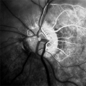
 Branch Retinal Artery Occlusion With Calcium Embolus at the Disc - Fundus Fluorescence Angiogram (FA)
Apr 7 2018 by Rameez N Hussain, MD
Acute branch retinal artery occlusion with a calcium embolus at the disc which is hyper-fluorescent in FA.
Photographer: DR RAMEEZ N HUSSAIN
Imaging device: ZEISS
Condition/keywords: branch retinal artery occlusion (BRAO), embolus, fluorescein angiogram (FA), retinal edema
-
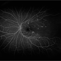
 Retinoschisis
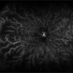
May 28 2016 by Olivia Rainey
Late image from a fluorescein angiogram of a patient's left eye with retinoschisis.
Photographer: Olivia Rainey
Imaging device: Heidelberg Spectralis
Condition/keywords: fluorescein angiogram (FA), retinoschisis, temporal retina
-
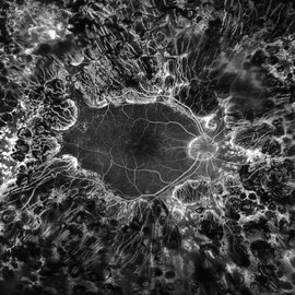
 Branch Retinal Artery Occlusion
Sep 11 2018 by Olivia Rainey
Ultra-wide field fluorescein angiogram of a 46-year-old male with a branch retinal artery occlusion affecting his left eye. The longstanding occlusion and has resulted in peripheral nonperfusion and neovascularization.
Photographer: Olivia Rainey
Imaging device: Optos
Condition/keywords: branch retinal artery occlusion (BRAO), fluorescein angiogram (FA), left eye, neovascularization (NV), non-perfusion, Optos
-
 Branch Retinal Artery Occlusion With Calcium Embolus at the Disc - Fundus Fluorescence Angiogram (FA)
Apr 7 2018 by Rameez N Hussain, MD
Acute branch retinal artery occlusion with a calcium embolus at the disc which is hyperfluorescent in FA.
Photographer: DR RAMEEZ N HUSSAIN
Imaging device: Zeiss
Condition/keywords: branch retinal artery occlusion (BRAO), embolus, fluorescein angiogram (FA), retinal edema
-
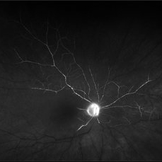
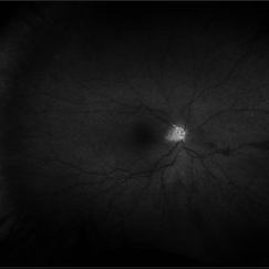
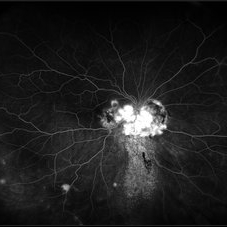
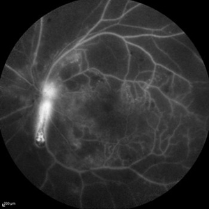 Optic Nerve Head Cannonball
Dec 15 2019 by Veer Singh, MS, FVRS, FMRF, FICO (Retina)
This is the fundus fluorescein angiography (FFA) of the left eye of a 62-year-old diabetic patient with proliferative diabetic retinopathy and neovascularization of disc who bled from the disc while he was undergoing an FFA procedure. The bleed from the disc gives the appearance of a cannonball fired from a cannon hence the caption "Optic Nerve Head Cannonball".
Photographer: Dr. Veer Singh
Imaging device: Heidelberg Spectralis HRA
Condition/keywords: fluorescein angiogram (FA), neovascularization of the disc (NVD), optic nerve head, proliferative diabetic retinopathy (PDR), vitreous hemorrhage
-
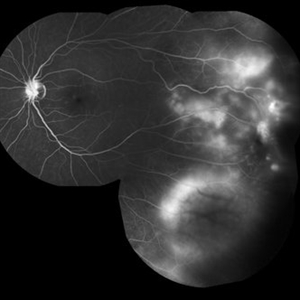
 Retinal Ischemia Secondary to Diabetic Retinopathy
Aug 29 2018 by Olivia Rainey
Fluorescein angiogram series of a 57-year-old male patient with proliferative diabetic retinopathy of the right eye. Patient has delayed AV transit with significant retinal ischemia and retinal capillary nonperfusion. The ischemia is extensive resulting in neovascularization of the iris and consequently neovascular glaucoma.
Photographer: Olivia Rainey
Imaging device: Optos
Condition/keywords: diabetes, disc hyperfluorescene, fluorescein angiogram (FA), non-perfusion, Optos, proliferative diabetic retinopathy (PDR), retinal ischemia, ultra-wide field imaging, vitreous hemorrhage
-
 Central Retinal Vein Occlusion
Jul 13 2018 by Olivia Rainey
Ultra-wide field fluorescein angiogram of a patient presenting with a central retinal vein occlusion their right eye.
Photographer: Olivia Rainey
Imaging device: Optos
Condition/keywords: central retinal vein occlusion (CRVO), fluorescein angiogram (FA), fluorescein leakage, hemorrhage, Optos, ultra-wide field imaging
-
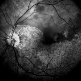
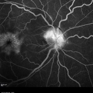
 Optic Disc Hemangioblastoma
May 30 2017 by Olivia Rainey
Ultra-wide-field fluorescein angiogram of the right eye of an 29-year-old female with an optic nerve hemangioblastoma secondary to Von Hippel-Lindau Syndrome.
Photographer: Olivia Rainey
Imaging device: Optos California
Condition/keywords: fluorescein angiogram (FA), fluorescein leakage, optic disc, Optos, retinal hemangioblastoma, ultra-wide field imaging, Von Hippel-Lindau
-
 Coats' disease early fluorescein angiogram of telangiectasia
Apr 3 2018 by Victor M Villegas, MD
6-year-old male with unilateral exudative retinopathy.
Photographer: Brenda Fallas
Imaging device: RetCam3
Condition/keywords: Coats' disease, FA early phase, fluorescein angiogram (FA), fluorescein leakage
-
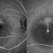 Central Serous Retinopathy
May 16 2017 by Olivia Rainey
Simultaneous fluorescein and indocyanine green angiography of an 37-year-old male with central serous retinopathy affecting his right eye. Patient's vision declined from 20/25 to 20/80 in the right eye. He elected for treatment with photodynamic therapy.
Photographer: Olivia Rainey
Imaging device: Heidelberg Spectralis
Condition/keywords: 30 degrees, central serous retinopathy (CSR), fluorescein angiogram (FA), fluorescein leakage, Heidelburg Spectralis, indocyanine green (ICG) angiography, late phase, mushroom cloud
-
 Cystoid Macular Edema Secondary to Panuveitis
Jan 15 2019 by Olivia Rainey
Fluorescein angiogram of a 55-year-old female with cystoid macular edema secondary to uveitis affecting her right eye. Patient was diagnosed with sarcoidosis.
Photographer: Olivia Rainey
Imaging device: Heidelberg Spectralis
Condition/keywords: 30 degrees, cystoid macular edema (CME), fluorescein angiogram (FA), fluorescein leakage, Heidelburg Spectralis, sarcoidosis, uveitis
-
 Choroidal Melanoma - Stable, Fluorescein Angiogram, Early Phase
Mar 13 2019 by James B. Soque, CRA, OCT-C, COA, FOPS
Early FA, right eye, with choroidal melanoma-stable, and a few tiny microaneurysms showing leakage in re-circulation phase.
Photographer: James Soque, CRA, OCT-C, FOPS
Imaging device: Topcon TRC-50DX with MERGE Eye Station software
Condition/keywords: FA early phase, fluorescein angiogram (FA), MERGE, microaneurysms
-
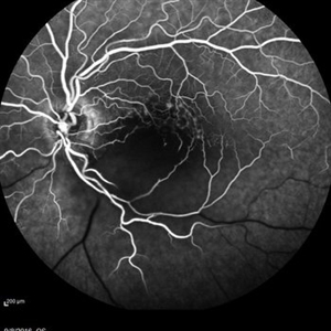
 Weiss Ring
Jan 15 2019 by Olivia Rainey
Fluorescein angiogram of a 55-year-old female with a Weiss ring affecting her right eye. Patient was diagnosed with sarcoidosis. She has cystoid macular edema secondary to panuveitis.
Photographer: Olivia Rainey
Imaging device: Heidelberg Spectralis
Condition/keywords: 30 degrees, cystoid macular edema (CME), fluorescein angiogram (FA), fluorescein leakage, Heidelburg Spectralis, optic nerve, sarcoidosis, uveitis, Weiss ring
-
 PDR; High Myopia; PRP
May 2 2019 by Carissa Hurdstrom
PDR; high myopia; PRP
Imaging device: Optos
Condition/keywords: fluorescein angiogram (FA), high myopia, pan-retinal photocoagulation (PRP), proliferative diabetic retinopathy (PDR)
 Loading…