Search results (32 results)
-
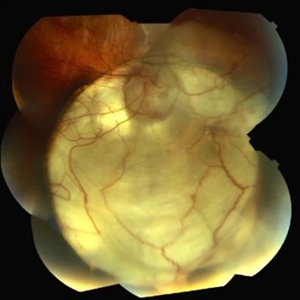
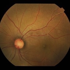
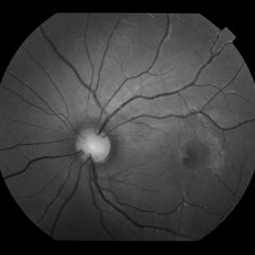
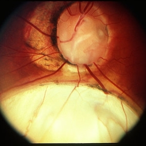 Coloboma
Mar 29 2013 by Henry J. Kaplan, MD
Optic disc and inferonasal choroidal coloboma in the same patient #2.
Condition/keywords: coloboma, coloboma of choroid, coloboma of optic disc
-
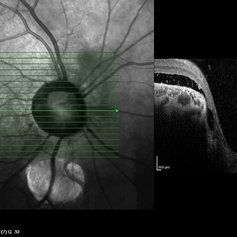 Coloboma of Disc & Choroid
Oct 6 2012 by Hamid Ahmadieh, MD
OCT image of a 25-year-old woman with serous retinal detachment secondary to coloboma of disc associated with coloboma of choroid.
Photographer: Hamid Ahmadieh, MD, Ophthalmic Research Center, Labbafinejad Medical Center, Shahid Beheshti University of Medical Sciences
Imaging device: Heidelberg Spectralis
Condition/keywords: coloboma of choroid, coloboma of optic disc, optical coherence tomography (OCT), serous retinal detachment
-
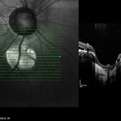 Coloboma of Disc & Choroid
Oct 6 2012 by Hamid Ahmadieh, MD
OCT image of a 25-year-old woman with a small coloboma of choroid associated with coloboma of disc.
Photographer: Hamid Ahmadieh, MD, Ophthalmic Research Center, Labbafinejad Medical Center, Shahid Beheshti University of Medical Sciences
Imaging device: Heidelberg Spectralis
Condition/keywords: coloboma of choroid, coloboma of optic disc, optical coherence tomography (OCT)
-
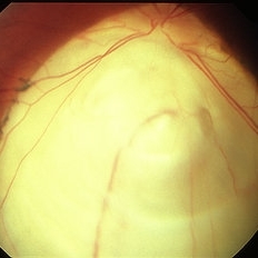
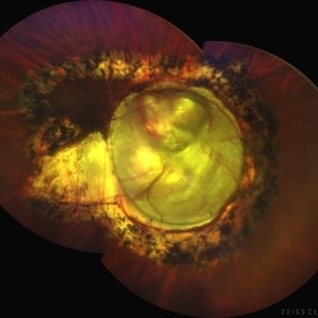
 Optic Disc Coloboma`
Mar 26 2018 by Purva Patwari
16-year-old female patient with vision of 6/60 presented with diminished vison. Other eye was normal.She had a normal birth history and developmental milestone. Look at the optic disc coloboma extending upto the macula. Intercalary membrane looks normal.
Photographer: Dr Purva Patwari, Patwari Retina Center, Ahmedabad, Gujarat , India
Imaging device: ZEISS VISU 500
Condition/keywords: coloboma, coloboma of optic disc, optic disc
-
 Coloboma
Mar 29 2013 by Henry J. Kaplan, MD
Coloboma involving optic nerve and inferior choroid.
Condition/keywords: coloboma of choroid, coloboma of optic disc
-
 Coloboma of Disc & Choroid
Oct 6 2012 by Hamid Ahmadieh, MD
OCT image of a 25-year-old woman with serous retinal detachment secondary to coloboma of disc associated with coloboma of choroid.
Photographer: Hamid Ahmadieh, MD, Ophthalmic Research Center, Labbafinejad Medical Center, Shahid Beheshti University of Medical Sciences
Imaging device: Heidelberg Spectralis
Condition/keywords: coloboma of choroid, coloboma of optic disc, optical coherence tomography (OCT), serous retinal detachment
-
 Retinal - Macular Coloboma
Mar 13 2014 by James B. Soque, CRA, OCT-C, COA, FOPS
Large retinal and optic nerve coloboma of a 31-year-old white male with 20/100 vision OS.
Photographer: James B Soque, CRA, COA
Imaging device: Topcon TRC 50DX, OIS 5 MP Camera,MERGE Software
Condition/keywords: coloboma of optic disc, color photo, macular coloboma
-
 Morning Glory Disc Anomaly
Aug 19 2017 by Mitzy E Torres Soriano, MD
A 10-year-old female patient with morning glory disc anomaly in her left eye.
Photographer: Mitzy E. Torres Soriano
Condition/keywords: coloboma of optic disc, coloboma of the optic nerve, Morning Glory Syndrome, optic disc, optic disc dysplasia
-
 Optic Nerve Coloboma
Nov 21 2014 by Thomas A. Ciulla, MD, MBA, FASRS
This 18-year-old woman has an optic nerve coloboma right eye with longstanding poor vision.
Photographer: Thomas Steele
Condition/keywords: coloboma of optic disc, coloboma of the optic nerve
-
 Coloboma
Sep 17 2015 by Jason S. Calhoun
Fundus photograph of young female with retinal coloboma.
Photographer: Jason Calhoun, Mayo Clinic, Department of Ophthalmology
Condition/keywords: coloboma of optic disc
-
 Optic Disc Coloboma
May 12 2017 by Nimrod Dar
9-year-old patient, noticed a gradual deterioration in her visual acuity at her LE (6/15). On her examination, an optic disc coloboma / pit can be seen. OCT scan revealed an intra retinal fluid and maculo schisis
Photographer: Nimrod Dar
Condition/keywords: coloboma, coloboma of optic disc, optic disc
-
 Optic Disc Coloboma
Jul 24 2019 by Haider Ali
16-year-old boy with horizontal nystagmus and decreased vision in both eyes.
Photographer: Dr Haider Ali Chaudhry, Madinah Teaching Hospital, Faisalabad
Condition/keywords: coloboma, coloboma of optic disc, coloboma of the optic nerve, excavation, Morning Glory Syndrome
-
 Cat Eye Syndrome
Feb 11 2020 by Sophia El Hamichi, MD
A 3-year-old female with cat eye syndrome including iris, chorioretinal and optic nerve colobomas. Note the CNV temporally to the optic nerve coloboma (blue arrows)
Photographer: Giselle De Oliveira, Bascom Palmer Eye Institute, Miami
Imaging device: RetCam
Condition/keywords: cat eye syndrome, chorioretinal coloboma, choroidal neovascularization (CNV), coloboma, coloboma of optic disc, optic nerve coloboma
-
 Optic Disc Coloboma
Jul 24 2019 by Haider Ali
16-year-old boy with horizontal nystagmus and decreased vision in both eyes.
Photographer: Dr Haider Ali Chaudhry, Madinah Teaching Hospital, Faisalabad
Condition/keywords: coloboma, coloboma of optic disc, coloboma of the optic nerve, excavation, Morning Glory Syndrome
-
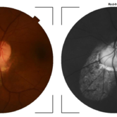
 Chorioretinal coloboma involving disc and macula
Mar 21 2022 by T. P . VIGNESH, MBBS,MS
Fundus photo of Right eye of a 55 year male patient revealing a fovea sparing well barraged chorioretinal coloboma involving the disc and the macula .
Photographer: Bharathi Singaravel
Imaging device: Zeiss Clarus
Condition/keywords: chorioretinal coloboma, coloboma of optic disc
-
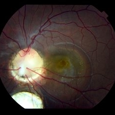 Colobomatous Optic Disc Maculopathy
Feb 13 2020 by Yoshihiro Yonekawa, MD, FASRS
Beautifully focused fundus photograph of a teenage girl with submacular fluid from a colobomatous optic disc.
Photographer: Netanya Lerner, COA, Wills Eye Hospital/Mid Atlantic Retina
Imaging device: Topcon
Condition/keywords: chorioretinal coloboma, coloboma of optic disc, congenital optic nerve pit, subretinal fluid
-
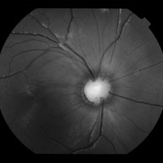
 Optic Disc Coloboma
Oct 17 2018 by Mehul A Shah
14-year-old girl at routine check-up and fundus picture found optic disc coloboma.
Photographer: MEHUL SHAH
Condition/keywords: coloboma of optic disc
-
 Coloboma involving the Optic nerve, Retina, and Choroid
Dec 6 2021 by Jesus Lozano, MD
78-year-old woman after prophylactic laser photocoagulation (PLP) for her RE Coloboma involving the optic nerve, retina, and choroid. At 6 month follow up, patient preserved her FC vision as it was before the procedure. Retina attached.
Photographer: Yair Bet Yosef, Hadassah Medical Center. Israel
Imaging device: Optos Silverstone fundus image
Condition/keywords: coloboma, coloboma of choroid, coloboma of macula, coloboma of optic disc, PLP, prophylactic photocoagulation
-
 Optic Disc Coloboma
Jul 24 2019 by Haider Ali
16-year-old boy with horizontal nystagmus and decreased vision in both eyes.
Photographer: Dr Haider Ali Chaudhry, Madinah Teaching Hospital, Faisalabad
Condition/keywords: coloboma, coloboma of optic disc, coloboma of the optic nerve, excavation, Morning Glory Syndrome
-
 Colobomatous Optic Disc Maculopathy
Feb 13 2020 by Yoshihiro Yonekawa, MD, FASRS
Fluorescein angiography, late frame, of a teenage girl with submacular fluid from a colobomatous optic disc. The camera is focused is on the elevated macula, and the disc is subtly defocused.
Photographer: Netanya Lerner, COA, Wills Eye Hospital/Mid Atlantic Retina
Imaging device: Topcon
Condition/keywords: chorioretinal coloboma, coloboma of optic disc, congenital optic nerve pit, subretinal fluid
-
 Colobomatous Optic Disc Maculopathy
Feb 13 2020 by Yoshihiro Yonekawa, MD, FASRS
EDI-OCT of a teenage girl with submacular fluid from a colobomatous optic disc. Note the subtle tracking of the subretinal fluid into the disc.
Photographer: Netanya Lerner, COA, Wills Eye Hospital/Mid Atlantic Retina
Imaging device: Topcon
Condition/keywords: chorioretinal coloboma, coloboma of optic disc, congenital optic nerve pit, subretinal fluid
-
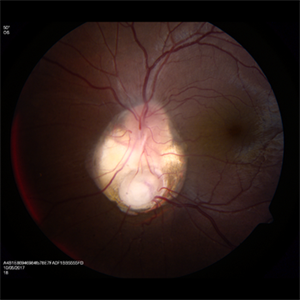
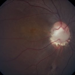
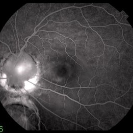
 Optic Disc Pit Associated with Optic Disc Coloboma and Retinochoroidal Coloboma
Jul 22 2020 by Deepak Bhojwani, MS
Fundus photograph of a 32-year-old male showing large optic disc pit in a colobomatous optic nerve head along with isolated inferior retino-choroidal coloboma. (A rare / coincidental occurrence of multiple congenital anomalies of optic disc and retina)
Photographer: DEEPAK BHOJWANI
Condition/keywords: coloboma of choroid, coloboma of optic disc, congenital optic nerve pit
-
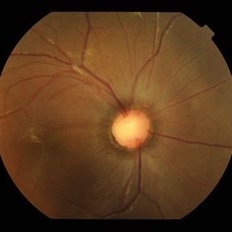
 Coloboma of Optic Disc
Apr 28 2019 by Bastián Schmidt Arias
Fundus photograph of an 63-year-old woman with retinal coloboma.
Photographer: Bastian Schmidt
Condition/keywords: coloboma of optic disc
-
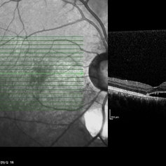
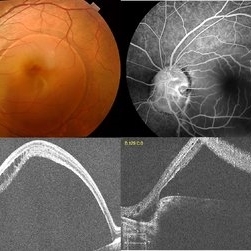 Optic Disc Pit with Coloboma (Hybrid Anomaly)
Jun 10 2021 by Janani Sreenivasan
Optic disc pit is a rare anomaly of the optic nerve head that can be associated with maculopathy leading to progressive visual deterioration. It belongs to the spectrum of congenital cavitary anomalies of the optic disc which encompasses extrapapillary cavitation, optic disc coloboma, and morning glory. Very rarely, optic disc pits are seen in combination with optic disc colobomas. Histopathologically, disc pit is defined as herniation of dysplastic retinal tissue into an excavation, rich in collagen, which can stretch into the subarachnoid space via a defect in the lamina cribrosa. Interestingly, this structural abnormality leading to a non-physiological communication between the intraocular and extraocular spaces is a common feature among all the congenital cavitary disc anomalies. Optic disc pit maculopathy is characterized by intraretinal and subretinal fluid at the area of macula. The origin of the retinal fluid remains unclear. Possible sources include the vitreous cavity, the subarachnoid space and the orbital space surrounding the dura. It has been estimated that approximately 25% to 75% of patients will develop serous detachment and/or retinoschisis of the central macula at some stage of their life. On fundus examination, ODPs typically appear as single grayish, round or oval depressions at the optic disc. Most commonly, they are detected at the inferotemporal segment of the disc, but may also be observed elsewhere, including the central area.The coexisting macular detachment can be related to lamellar or full-thickness macular holes, cystoid changes, retinal pigment epithelium atrophy and eventually to irreversible loss of vision,especially in longstanding cases. Herewith, we present a 32-years-old male patient presenting with an unusual combination of optic disc pit with maculopathy and optic disc coloboma (hybrid anomaly) in the same eye with corrected visual acuity of 3/60.
Photographer: Dr Janani Sreenivasan
Imaging device: Zeiss Cirrus HD-OCT
Condition/keywords: coloboma of optic disc, hybrid anomaly, macular detachment, optic disc, optic disc pit
 Loading…