Search results (232 results)
-
 Choroideremia
Sep 27 2012 by Michael P. Kelly, FOPS
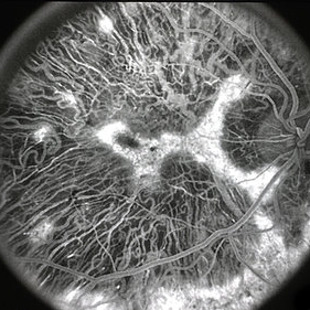
This is a 60 degree fluorescein angiogram image of choroideremia.
Photographer: Michael P. Kelly, FOPS Director, Duke Eye Labs, Duke University Hospital, Duke Eye Center, Durham, NC
Imaging device: Canon 60UV
Condition/keywords: choroideremia
-
 Choroideremia
Mar 29 2013 by Henry J. Kaplan, MD
Angiogram of the same patient in a later phase shows a small central island and multiple peripheral and a residual area inferiorly #2.
Condition/keywords: choroideremia
-
 Choroideremia
Jan 26 2013 by Ratimir Lazic, MD, PhD
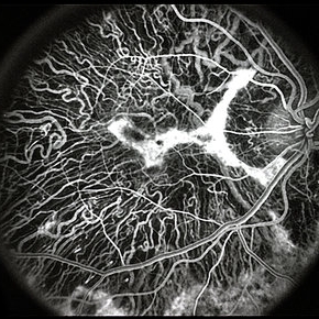
FAG image of a 66-year-old male. Diffuse chorioretinal atrophy is present. Large choroidal vessels can be seen due to atrophy of the RPE and choriocapilaris.
Photographer: Marko Lukic, MD
Imaging device: Zeis Visucam Lite 2
Condition/keywords: choroideremia, fundus photograph
-
 Choroideremia
Jan 26 2013 by Ratimir Lazic, MD, PhD
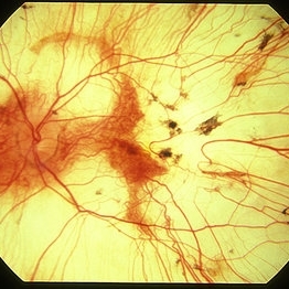
Color fundus photography of a 66-year-old male. Diffuse chorioretinal atrophy is seen. “Patches” or retinal tissue can be seen, some of it in macular area. Visual acuity on that eye is 0,15.
Photographer: Marko Lukic, MD
Imaging device: Zeis Visucam Lite 2
Condition/keywords: choroideremia, large choroidal vessels
-
 Choroideremia
Jan 26 2013 by Ratimir Lazic, MD, PhD
FAG image of a 66-year-old male. Diffuse chorioretinal atrophy is present. Large choroidal vessels can be seen. "Hyperflorescent" areas represent normal chorioretinal tissue.
Photographer: Marko Lukic, MD
Imaging device: Zeis Visucam Lite 2
Condition/keywords: choroideremia, fundus photograph
-
---thumb.jpg/image-square;max$300,300.ImageHandler) Choroideremia - Scleral Stain
Choroideremia - Scleral Stain
Feb 20 2013 by From the Collections of Thomas M. Aaberg, MD and Thomas M. Aaberg Jr., MD
External slit lamp photo of an eye with choroideremia exhibiting temporal scleral stain.
Condition/keywords: choroideremia, sclera, slit lamp photo
-
 Choroideremia
Mar 29 2013 by Henry J. Kaplan, MD
Angiogram of choroideremia shows hypofluorescence due to filling defect 9choriocapillary loss) except for a small central island #1.
Condition/keywords: choroideremia
-
 Choroideremia
Mar 29 2013 by Henry J. Kaplan, MD
Choroideremia, notice the residual small central island.
Condition/keywords: choroideremia
-
---thumb.jpg/image-square;max$300,300.ImageHandler) Choroideremia
Choroideremia
Feb 20 2013 by From the Collections of Thomas M. Aaberg, MD and Thomas M. Aaberg Jr., MD
Color fundus photo of a right eye with advanced choroideremia.
Condition/keywords: choroideremia, end stage, fundus photograph
-
---thumb.jpg/image-square;max$300,300.ImageHandler) Choroideremia
Choroideremia
Feb 20 2013 by From the Collections of Thomas M. Aaberg, MD and Thomas M. Aaberg Jr., MD
External slit lamp photo of an eye with choroideremia exhibiting temporal scleral stain looking to the superonasal direction so more of the temporal sclera is visible.
Condition/keywords: choroideremia, sclera, slit lamp photo
-
---thumb.jpg/image-square;max$300,300.ImageHandler) Choroideremia
Choroideremia
Feb 20 2013 by From the Collections of Thomas M. Aaberg, MD and Thomas M. Aaberg Jr., MD
Red free fundus photo of the same patient with choroideremia
Condition/keywords: choroideremia, red-free
-
---thumb.jpg/image-square;max$300,300.ImageHandler) Choroideremia
Choroideremia
Feb 20 2013 by From the Collections of Thomas M. Aaberg, MD and Thomas M. Aaberg Jr., MD
FA of a right eye with advanced choroideremia showing choroidal vessels.
Condition/keywords: choroideremia
-
 Choroideremia
Jun 27 2013 by Jason S. Calhoun
62-year-old female who has extensive retinal damage in both eyes. VA 20/50 right eye, HM, left eye. Spot in vision, which is PSC, both eyes.
Photographer: Jason S. Calhoun, Mayo Clinic Jacksonville, Florida
Imaging device: TOPCON TRC 50-EX
Condition/keywords: choroideremia
-
 Choroideremia With Periperal Pigment Changes, Drusenoid Flecks, Patchy Atrophy
Aug 1 2013 by From the Collections of Thomas M. Aaberg, MD and Thomas M. Aaberg Jr., MD
Choroideremia with periperal pigment changes, drusenoid flecks, patchy atrophy.
Condition/keywords: atrophy, choroideremia, drusenoid flecks
-
---thumb.jpg/image-square;max$300,300.ImageHandler) Choroideremia
Choroideremia
Feb 20 2013 by From the Collections of Thomas M. Aaberg, MD and Thomas M. Aaberg Jr., MD
Red free fundus photo of the same patient with choroideremia.
Condition/keywords: choroideremia, red-free
-
 Choroideremia
Dec 3 2014 by Andree Henaine-Berra, MD
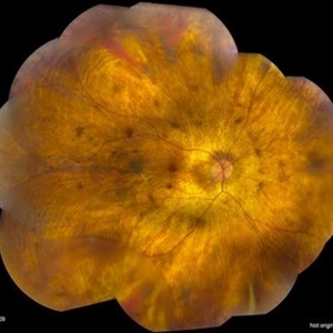
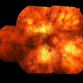
Fundus photograph of the left eye of a 33-year-old male with choroideremia, showing diffuse chorioretinal atrophy.
Photographer: Carlos Garcia, Hospital General "Dr. Manuel Gea Gonzalez", Mexico City
Condition/keywords: choroideremia
-
 Choriodemia
Jul 8 2013 by David W. Faber, MD
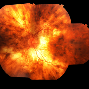
Fundus montage of a 50-year-old male.
Photographer: Donna Knight C.R.A., Rocky Mountain Retina Consultants, Sale Lake City, Utah
Condition/keywords: choroideremia
-
 Choroideremia With Periperal Pigment Changes, Drusenoid Flecks, Patchy Atrophy
Aug 1 2013 by From the Collections of Thomas M. Aaberg, MD and Thomas M. Aaberg Jr., MD
Choroideremia with periperal pigment changes, drusenoid flecks, patchy atrophy.
Condition/keywords: atrophy, choroideremia, drusenoid flecks
-
 Gyrate Atrophy
Oct 31 2018 by Dhaivat Shah
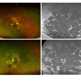
50-year-old male came in with complaint of daytime vision loss for a year and nighttime vision loss for more than 20 years, gradually increasing day by day. Fundus showed paving-stone like areas of atrophy of the RPE involving the macula which coalesces to form a characteristic scalloped border at the junction of normal and abnormal RPE. Gyrate atrophy is an autosomal recessive dystrophy caused by tenfold elevations of plasma ornithine, which is toxic to the RPE and choroid. Patients with gyrate atrophy have hyperpigmented fundi, with lobular loss of the RPE and choroid, normally sparing the fovea. The finding of generalized hyperpigmentation of the remaining RPE helps to clinically distinguish gyrate atrophy from choroideremia. Affected patients usually develop night blindness during the first decade of life and experience progressive loss of visual field and visual acuity later in the disease course. Early diagnosis is crucial because treatment in form of Arginine free diet and oral pyridoxine helps in slowing the progression of disease.
Imaging device: Optos
Condition/keywords: fundus autofluorescence (FAF), gyrate atrophy
-
 Choroideremia With Periperal Pigment Changes, Drusenoid Flecks, Patchy Atrophy
Aug 1 2013 by From the Collections of Thomas M. Aaberg, MD and Thomas M. Aaberg Jr., MD
Choroideremia with periperal pigment changes, drusenoid flecks, patchy atrophy.
Condition/keywords: atrophy, choroideremia, drusenoid flecks
-
 Choroidal atrophy, probable choroideremia
Apr 4 2013 by Jerald A. Bovino, MD
Choroidal atrophy, probable choroideremia
Condition/keywords: choroidal atrophy, choroideremia
-
 Chorioderemia
Jul 8 2013 by David W. Faber, MD
Fundus montage of a 50-year-old male.
Photographer: Donna Knight, C.R.A, Rocky Mountain Retina Consultants, Salt Lake City, Utah
Condition/keywords: choroideremia
-
 Choroideremia With Periperal Pigment Changes, Drusenoid Flecks, Patchy Atrophy
Aug 1 2013 by From the Collections of Thomas M. Aaberg, MD and Thomas M. Aaberg Jr., MD
Choroideremia with periperal pigment changes, drusenoid flecks, patchy atrophy.
Condition/keywords: atrophy, choroideremia, drusenoid flecks
 Loading…