 Initializing download.
-
 By Ratimir Lazic, MD, PhD
Eye Clinic Svjetlost
Co-author(s): Marko Lukic, MD - Uploaded on Jan 26, 2013.
- Last modified by Chayal Patel on Jan 29, 2013.
- Reviewed by Ratimir Lazic, MD, PhD
- Rating
- Appears in
- Choroideremia
- Condition/keywords
- choroideremia, fundus photograph
- Photographer
- Marko Lukic, MD
- Imaging device
-
Fundus camera
Zeis Visucam Lite 2 - Description
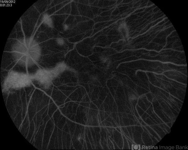
- FAG image of a 66-year-old male. Diffuse chorioretinal atrophy is present. Large choroidal vessels can be seen. "Hyperflorescent" areas represent normal chorioretinal tissue.
")
")
")


---thumb.JPG/image-square;max$79,0.ImageHandler "Welding arc maculopathy")


