File number: 879
Comments
-
 Michael P. Kelly, FOPS (September 30 2012)
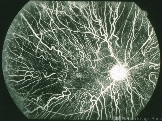
Thanks for the comments. Paula is right on as always. Additionally, this is a film angiogram that I scanned to electronic form for posting here and that may also have affected the final appearance. Thanks to you both.
-
 Paula Morris, CRA, FOPS (September 28 2012)
This is an early arterio-venous circulation phase image when the dye is highly concentrated in the vessels which would account for the bright exposure. Lovely image with excellent focus and great detail.
-
 Judy E. Kim, MD, FARVO, FASRS (September 27 2012)
Beautiful image of choroideremia, nicely demonstrating the choroidal vessels. A bit over exposed?
Sign in to comment.
 Initializing download.
-
 By Michael P. Kelly, FOPS
Duke University Hospital - Uploaded on Sep 27, 2012.
- Last modified by Michael P. Kelly, FOPS on Oct 5, 2012.
- Rating
- Appears in
- Miscellaneous
- Condition/keywords
- choroideremia
- Photographer
- Michael P. Kelly, FOPS Director, Duke Eye Labs, Duke University Hospital, Duke Eye Center, Durham, NC
- Imaging device
-
Fundus camera
Canon 60UV - Description
- This is a 60 degree fluorescein angiogram image of choroideremia.


---thumb.jpg/image-square;max$79,0.ImageHandler "Choroideremia")


---thumb.jpg/image-square;max$79,0.ImageHandler "Choroideremia - Scleral Stain")
---thumb.jpg/image-square;max$79,0.ImageHandler "Choroideremia")
---thumb.jpg/image-square;max$79,0.ImageHandler "Choroideremia")
---thumb.jpg/image-square;max$79,0.ImageHandler "Choroideremia")
---thumb.jpg/image-square;max$79,0.ImageHandler "Choroideremia")