Search results (150 results)
-
 Macular Choroidal Osteoma
Aug 17 2012 by Jonathan L. Prenner, MD
Macular choroidal osteoma in a 29-year-old woman
Condition/keywords: macular choroidal osteoma
-
 Choroidal Osteoma Plus CNV
Sep 2 2012 by Hamid Ahmadieh, MD
Color fundus photograph and OCT imaging of a 47-year-old man with a juxtafoveal CNV superimposed on a choroidal osteoma.
Photographer: Hamid Ahmadieh, Ophthalmic Research Center, Labbafinejad Medical Center
Imaging device: Topcon
Condition/keywords: choroidal neovascularization (CNV), choroidal osteoma, optical coherence tomography (OCT)
-
 Choroidal Osteoma with Choroidal Neovascularization
Aug 23 2012 by Gerardo Garcia-Aguirre, MD
Fundus photograph of the right eye of a 32 year-old male patient with choroidal osteoma and choroidal neovascularization. The left eye also has a choroidal osteoma.
Photographer: Ricardo Montoya, Asociación para Evitar la Ceguera en México
Condition/keywords: choroidal neovascularization (CNV), macular choroidal osteoma
-
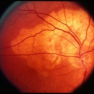 Choroidal Osteoma
Mar 29 2013 by Henry J. Kaplan, MD
Typical choroidal osteoma as yellow subretinal lesion around optic nerve with scalloped border and mild pigmentation on the surface.
Condition/keywords: choroidal osteoma
-
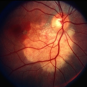 Choroidal Osteoma with CNV
Mar 29 2013 by Henry J. Kaplan, MD
Typical choroidal osteoma complicated by CNV as subretinal hemorrhage in the macular edge of the lesion.
Condition/keywords: choroidal neovascularization (CNV), choroidal osteoma
-
 Choroidal Osteoma and Secondary Choroidal Neovascular Membrane
Sep 21 2012 by Allen Chiang, MD, FASRS
Fundus photograph of a 44-year old woman with a choroidal osteoma complicated by secondary choroidal neovascular membrane, regressed after serial intravitreal bevacizumab injections. The tumor exhibits areas of decalcification.
Imaging device: Topcon
Condition/keywords: choroidal neovascularization (CNV), choroidal osteoma, macular choroidal osteoma
-
 Choroidal Osteoma 1
Oct 5 2012 by Ronald C. Gentile, MD
A cream colored choroidal osteoma involving the temporal macula in a young women
Photographer: The New York Eye & Ear Infirmary Department of Medical Imaging
Condition/keywords: choroidal tumor, macular choroidal osteoma
-
 Choroidal Osteoma + CNV
Mar 13 2013 by Hamid Ahmadieh, MD
Optical coherence tomography (OCT) of the right eye of a 13-year-old girl with decreased VA due to CNV secondary to choroidal osteoma.
Photographer: Naghmeh Nozhat, Negah Eye Center, Tehran
Imaging device: Topcon
Condition/keywords: choroidal neovascularization (CNV), choroidal osteoma, optical coherence tomography (OCT)
-
 Choroidal Osteoma 5
Oct 5 2012 by Ronald C. Gentile, MD
B scan ultrasonography with representative A scan of the macular choroidal osteoma. The B scan reveals a characteristic highly reflective plaque consistent with its bone-like calcium composition that persists with low gain. The A scan reveals a large spike.
Photographer: The New York Eye & Ear Infirmary Department of Medical Imaging
Condition/keywords: B scan ultrasound, choroidal tumor, macular choroidal osteoma
-
---thumb.jpg/image-square;max$300,300.ImageHandler) Osteoma B Scan
Osteoma B Scan
Apr 18 2014 by Susanna S. Park, MD, PhD
72-year-old woman noted with a mid-peripheral amelanotic choroidal lesion with minimal elevation which shows marked shadowing on ultrasonography consistent with a choroidal osteoma. Optic nerve is shown in the bottom of the image.
Photographer: Ellen Redenbo, University of California Davis Eye Center
Condition/keywords: B scan ultrasound
-
 Choroidal Osteoma
Dec 28 2015 by P. Mahesh Shanmugam, MBBS, DO, FRCSEd, PhD, FAICO
Choroidal osteoma - yellow subretinal minimally elevated lesion with scalloped margins and pigmentation on surface. Peripheral part of the lesion is orange in color, the older central part yellow in color.
Condition/keywords: choroidal osteoma
-
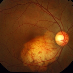
 Choroidal Osteoma
Oct 4 2012 by Anat Loewenstein, MD
Orange, pale yellow, plate-like choroidal lesion with a bone density.
Photographer: Galit Yair-Pur
Condition/keywords: choroidal osteoma
-
 Choroidal Osteoma
Aug 23 2012 by Gerardo Garcia-Aguirre, MD
Fundus photograph of the left eye of a 32 year-old male patient showing a choroidal osteoma. The right eye also has a choroidal osteoma with choroidal neovascularization.
Photographer: Ricardo Montoya, Asociación para Evitar la Ceguera en México
Condition/keywords: macular choroidal osteoma
-
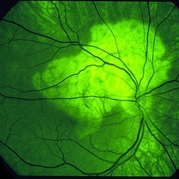 Choroidal Osteoma
Mar 29 2013 by Henry J. Kaplan, MD
Autofluorescence in choroidal osteoma.
Condition/keywords: autofluorescence imaging, choroidal osteoma
-
 Choroidal Osteoma + CNV
Mar 13 2013 by Hamid Ahmadieh, MD
Color fundus photograph the right eye of a 13-year-old girl with decreased VA due to CNV secondary to choroidal osteoma.
Photographer: Naghmeh Nozhat, Negah Eye Center, Tehran
Imaging device: Topcon
Condition/keywords: choroidal neovascularization (CNV), choroidal osteoma
-
 Choroidal Osteoma 2
Oct 5 2012 by Ronald C. Gentile, MD
Magnified view of the macular choroidal osteoma with visible internal vascularity.
Photographer: The New York Eye & Ear Infirmary Department of Medical Imaging
Condition/keywords: choroidal tumor, macular choroidal osteoma
-
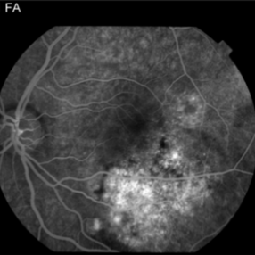
 Choroidal Osteoma 4
Oct 5 2012 by Ronald C. Gentile, MD
Fluorescein angiography of the macular choroidal osteoma in the late phase of the angiogram with late staining of the tumor highlighting its negative staining internal vascularity.
Photographer: The New York Eye & Ear Infirmary Department of Medical Imaging
Condition/keywords: macular choroidal osteoma
-
 Choroidal Osteoma Or Hemangioma
-
 Choroidal osteoma case 1 no 1
Jan 11 2013 by Alex P. Hunyor, MD
Bilateral choroidal osteoma in a young female. Image 1 of 4 showing growth of the osteoma over a 2-year period in the left eye.
Condition/keywords: choroidal osteoma
-
 Choroidal Osteoma 3
Oct 5 2012 by Ronald C. Gentile, MD
Fluorescein angiography of the macular choroidal osteoma in the early arterial-venous phase highlighting its internal vascularity.
Photographer: The New York Eye & Ear Infirmary Department of Medical Imaging
Condition/keywords: macular choroidal osteoma
-
---thumb.jpg/image-square;max$300,300.ImageHandler) Choroidal osteoma case 1 no 3
Choroidal osteoma case 1 no 3
Jan 11 2013 by Alex P. Hunyor, MD
Bilateral choroidal osteoma in a young female. Image 3 of 4 showing growth of the osteoma over a 2-year period in the left eye.
Condition/keywords: choroidal osteoma
-
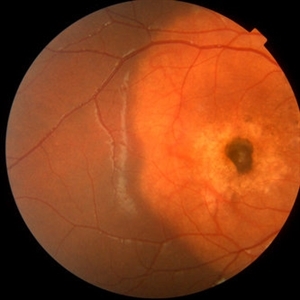
 Choroidal Osteoma
Nov 21 2014 by Thomas A. Ciulla, MD, MBA, FASRS
This 13-year-old girl presented with mild painless progressive blurring of central vision left eye over the past several months. Visual acuity was 20/25. In the affected left eye, retinal examination revealed a relatively flat, lightly pigmented lesion, with well-defined and scalloped edges. Clumps of associated pigment were noted. This OCT image shows subretiinal fluid just inferior to the fovea. Choroidal osteoma can be associated with the development of subretinal neovascularization (particularly at the edges of the osteoma).
Photographer: Thomas Steele
Condition/keywords: choroidal neovascular membrane (CNVM), choroidal neovascularization (CNV), choroidal osteoma, macular choroidal osteoma
-
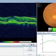
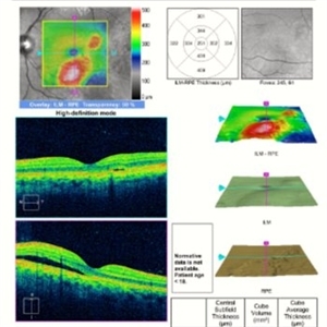 Choroidal Osteoma
Nov 21 2014 by Thomas A. Ciulla, MD, MBA, FASRS
This OCT image shows subretiinal fluid just inferior to the fovea. Choroidal osteoma can be associated with the development of subretinal neovascularization (particularly at the edges of the osteoma).
Condition/keywords: choroidal neovascular membrane (CNVM), choroidal neovascularization (CNV), choroidal osteoma, macular choroidal osteoma
-
---thumb.jpg/image-square;max$300,300.ImageHandler) Choroidal osteoma case 1 no 2
Choroidal osteoma case 1 no 2
Jan 11 2013 by Alex P. Hunyor, MD
Bilateral choroidal osteoma in a young female. Image 2 of 4 showing growth of the osteoma over a 2-year period in the left eye.
Condition/keywords: choroidal osteoma
-
---thumb.jpg/image-square;max$300,300.ImageHandler) Choroidal Osteoma
Choroidal Osteoma
Apr 29 2013 by Subijay Sinha
Ultrasonography shows a curvilinear plaque of high amplitude with shadowing in the juxtapapillary area.
Condition/keywords: calcification, shadow, ultrasound
 Loading…