Search results (162 results)
-
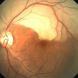
 Branch Retinal Artery Occlusion
Sep 21 2012 by Allen Chiang, MD, FASRS
Fundus photograph of a 27-year old male with a branch retinal artery occlusion. Systemic medical evaluation identified anti-phospholipid antibody syndrome.
Imaging device: Topcon
Condition/keywords: antiphospholipid antibody syndrome, branch retinal artery occlusion (BRAO)
-
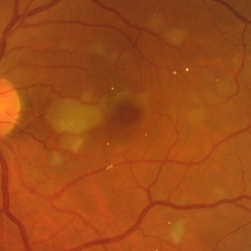
 BRAO
BRAO
Jun 29 2014 by John S. King, MD
BRAO.
Photographer: Wayne A Ladlee Jr
Condition/keywords: branch retinal artery occlusion (BRAO), embolic
-
 Hollenhorst Plaque
Sep 18 2016 by John T. Thompson, MD
Color photo of Hollenhorst plaque at branch of inferotemporal artery.
Imaging device: Zeiss FF4
Condition/keywords: branch retinal artery occlusion (BRAO), hollenhorst plaque
-
 Hollenhorst Plaque in Eye with BRAO
Oct 1 2012 by Jeffrey G. Gross, MD, FASRS
Hollenhorst plaque in eye with BRAO.
Condition/keywords: branch retinal artery occlusion (BRAO), hollenhorst plaque
-
 BRAO d/t cat scratch disease - LE fundus photograph
Jan 2 2013 by Roy Schwartz, MD
A 38 year-old-male complained of a grey spot in visual field in his left eye. On clinical exam BRAO in LE, as seen in picture. Serology for bartonella was positive.
Photographer: Galit Yair-Pur
Condition/keywords: branch retinal artery occlusion (BRAO), cat scratch retinitis
-
 Branch Retinal Artery Occlusion With Concurrent Central Retinal Vein Occlusion
Oct 5 2016 by Larry M Puthenparambil, MD
Branch retinal artery occlusion with concurrent central retinal vein occlusion.
Photographer: Stacey Groom
Imaging device: Topcon
Condition/keywords: branch retinal artery occlusion (BRAO), central retinal vein occlusion (CRVO)
-
 BRAO d/t cat scratch disease - RE fundus photograph
Jan 2 2013 by Roy Schwartz, MD
A 38-year-old male complained of a grey spot in visual field in his left eye. On clinical exam BRAO in LE, and a CWS in RE, as seen in this photograph. Serology for bartonella was positive.
Photographer: Galit Yair-Pur
Condition/keywords: branch retinal artery occlusion (BRAO), cat scratch retinitis
-
 BRAO with Cilioretinal Sparing
Oct 1 2012 by Jeffrey G. Gross, MD, FASRS
BRAO with cilioretinal sparing.
Condition/keywords: branch retinal artery occlusion (BRAO), cilioretinal sparing
-
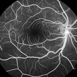
 Branch Retinal Artery Occlusion - FA
Sep 21 2012 by Allen Chiang, MD, FASRS
Fluorescein angiogram of a 27-year old male with a branch retinal artery occlusion demonstrates interruption of arterial flow and retrograde venous filling. Systemic medical evaluation identified anti-phospholipid antibody syndrome.
Imaging device: Topcon
Condition/keywords: antiphospholipid antibody syndrome, branch retinal artery occlusion (BRAO)
-
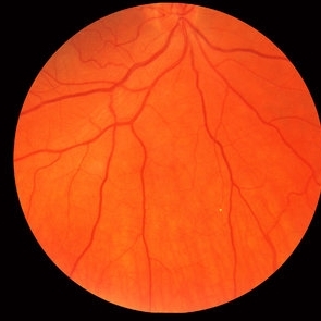
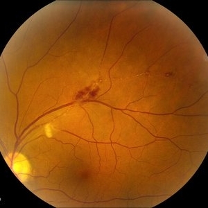
 Branch Retinal Artery Occlusion
Mar 29 2013 by Henry J. Kaplan, MD
Inferotemporal BRAO.
Condition/keywords: branch retinal artery occlusion (BRAO)
-
 BRAO d/t cat scratch disease - FA 00:18 min.
Jan 2 2013 by Roy Schwartz, MD
A 38-year-old male complained of a grey spot in visual field in his left eye. On clinical exam BRAO in LE, confirmed by FA, as seen in picture. Image shows delayed filling of artery. Serology for bartonella was positive.
Photographer: Galit Yair-Pur
Condition/keywords: branch retinal artery occlusion (BRAO), cat scratch retinitis
-
 BRAO d/t cat scratch disease - FA 5:23 min.
Jan 2 2013 by Roy Schwartz, MD
A 38-year-old male complained of a grey spot in visual field in his left eye. On clinical exam BRAO in LE, confirmed by FA, as seen in picture. The image was taken after late filling of artery. CWS blocks proximal part of artery. Serology for bartonella was positive.
Photographer: Galit Yair-Pur
Condition/keywords: branch retinal artery occlusion (BRAO), cat scratch retinitis
-
 Branch Retinal Artery Occlusion FFA
May 4 2014 by Neha Goel, MS DNB FRCS (Glasg)
FFA of the patient confirming inferotemporal BRAO and also demonstrating the embolus in the retinal artery at the disc.
Photographer: Neha Goel
Imaging device: Zeiss Visucam
Condition/keywords: arterial embolus, branch retinal artery occlusion (BRAO)
-
 Branch Retinal Artery Occlusion
Oct 2 2013 by Jerald A. Bovino, MD
There is a hollenhorst plaque causing a branch retinal artery occlusion. The patient has scars from prior panretinal laser photocoagulation.
Condition/keywords: branch retinal artery occlusion (BRAO), hollenhorst plaque, pan-retinal photocoagulation (PRP)
-
 Branch Retinal Artery Occlusion With Calcium Embolus at the Disc - Fundus Photo
Apr 7 2018 by Rameez N Hussain, MD
Acute branch retinal artery occlusion with a calcium embolus at the disc with retinal whitening in the area of retinal edema.
Photographer: DR RAMEEZ N HUSSAIN
Imaging device: zeiss
Condition/keywords: branch retinal artery occlusion (BRAO), embolus, fundus photograph, retinal edema
-
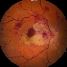
 Multiple Branch Retinal Artery Occlusion ( BRAO)
May 31 2014 by Hamid Ahmadieh, MD
Color fundus photograph of the left eye of a 65-year-old man with sudden drop of vision due to multiple BRAO following carotid artery stenting leading to multiple emboli.
Photographer: Solmaz Shahmohammad, Negah Eye Center, Tehran
Condition/keywords: branch retinal artery occlusion (BRAO), color fundus photograph, retinal cloudy swelling
-
---thumb.jpg/image-square;max$300,300.ImageHandler) Branch Retinal Artery Occlusion
Branch Retinal Artery Occlusion
Jun 6 2013 by Sharon Fekrat, MD FACS FASRS
Fundus photograph of a branch retinal artery occlusion, left eye.
Photographer: Duke Eye Imaging, Duke University Eye Center, Durham, NC
Condition/keywords: branch retinal artery occlusion (BRAO)
-
 BRAO Rianto AF
Apr 12 2014 by Sjakon G Tahija, MD
Auto fluorescence fundus image of a 70-year-old man with a superior temporal branch retinal artery occlusion. The emboli can be very clearly seen as the white dot of AF blockage.
Photographer: Avris Siahaan
Imaging device: Heidelberg Spectralis
Condition/keywords: branch retinal artery occlusion (BRAO)
-
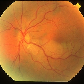
 Arterial Occlusion
Jul 14 2013 by Jason S. Calhoun
Patient in with vision loss in the lower quadrant of his visual field. Fundus photo shows arterial occlusion superior to the optic nerve
Photographer: Jason S. Calhoun, Department of Ophthalmology, Mayo Clinic Jacksonville, Florida
Imaging device: TOPCON TRC 50-EX
Condition/keywords: branch retinal artery occlusion (BRAO)
-
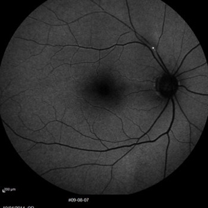
 BRAO
BRAO
Jun 29 2014 by John S. King, MD
Deceased arterial flow in laminar phase of FA.
Photographer: Wayne A Ladlee Jr
Condition/keywords: branch retinal artery occlusion (BRAO), embolic
-
 Brach Retinal Artery Occlusion
Oct 2 2013 by Jerald A. Bovino, MD
There is a hollenhorst plaque causing a branch retinal artery occlusion. The patient has scars from prior panretinal laser photocoagulation.
Condition/keywords: branch retinal artery occlusion (BRAO), hollenhorst plaque, pan-retinal photocoagulation (PRP)
-
 AIDS - BAOI
Apr 8 2013 by Howard Schatz, MD
III AIDS - BAOI (BAO-AIDS)
Condition/keywords: AIDS, branch retinal artery occlusion (BRAO)
-
 Branch Artery Occlusion
Jul 14 2013 by Jason S. Calhoun
Superior branch artery occlusion in the left eye
Photographer: Jason S. Calhoun, Department of Ophthalmology, Mayo Clinic Jacksonville, Florida
Imaging device: TOPCON TRC 50-EX
Condition/keywords: branch retinal artery occlusion (BRAO)
-
 Multiple Acute Embolic Branch Retinal Artery Occlusions
May 27 2015 by Darin R. Goldman, MD
60-year-old phakic male with multiple retinal emboli found to have a patent foramen ovale, which was repaired surgically with no further retinal occlusive episodes.
Condition/keywords: branch retinal artery occlusion (BRAO), cherry red spot, embolus, retinal microembolism
-
 Combined Artery and Vein Occlusion
Jun 27 2013 by Jason S. Calhoun
Patient comes in with decreased vision in both eyes. VA 20/200-OD, 20/60-OS. Fundus exam shows great amount of macular edema due to artery and vein occlusions. There is some neovascularization on the optic nerve in the right eye. Patient was treated with Eylea injection in the left eye today and will return for Eylea injection in the right eye.
Photographer: Jason S. Calhoun, Mayo Clinic Jacksonville, Florida
Imaging device: TOPCON TRC 50-EX
Condition/keywords: branch retinal artery occlusion (BRAO), branch retinal vein occlusion (BRVO)
 Loading…