Search results (169 results)
-
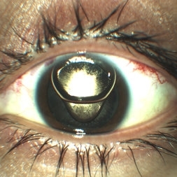
 Ozurdex implant
Aug 23 2012 by Daniel A. Adelberg, MD, FASRS
Anterior Segment photograph of a 50 year old with Uveitis and Cystoid Macular Edema status post Intravitreal injection of an Ozurdex dexamethasone implant
Photographer: Robert Ramsey, Southwestern Eye Center, Mesa Arizona
Condition/keywords: Ozurdex implant
-
000---thumb.jpg/image-square;max$300,300.ImageHandler) Anterior Segment Photo of Emulsified Silicone Oil
Anterior Segment Photo of Emulsified Silicone Oil
Dec 25 2013 by Dong Yoon Kim, MD
47-year-old woman underwent vitrectomy and silicone oil tampoande for tractional retinal detachment due to proliferative diabetic retinopathy. 8 months after silicone oil tamponade, silicone oil was emulsified. And emulsified silicone oil was observed at anterior chamber.
Condition/keywords: silicone oil, tractional retinal detachment
-
 Iris Nevus
Apr 11 2016 by Kathy Karsten, COT
Anterior segment photo of an iris nevus with a peaked pupil observed over time for growth.
Photographer: KATHY KARSTEN
Imaging device: TOPCON 50-DX
Condition/keywords: iris, nevus
-
 Glaukomflecken
Oct 23 2017 by Claire Kiernan, MD
Slit lamp photograph of a 59-year-old man with recent-onset severe eye pain noted to have glaukomflecken consistent with recent episode of angle closure glaucoma.
Photographer: Steve Moser, University of Tennessee Hamilton Eye Institute; Joe Mastellone, University of Tennessee Hamilton Eye Institute
Condition/keywords: angle-closure glaucoma interval, glaucoma anterior segment anomalies
-
---thumb.jpg/image-square;max$300,300.ImageHandler) Birdshot Choroidopathy
Birdshot Choroidopathy
Oct 9 2013 by Maurice F. Rabb
Forty two year old white female first noted flashing lights in her left eye at the age of 30. Although she had many previous eye examinations for low grade myopia, she had never had a dilated fundus examination. The evaluation twelve years ago disclosed 20/20 acuity in each eye with a myopic correction, an afferent pupillary defect on the left, no evidence of anterior segment inflammation in either eye, a full field on the right and markedly constricted field on the left, fundus pigmentary abnormalities extending beyond the equator in each eye, and narrow vessels with pigment migration into the retina in the left eye only.
Condition/keywords: birdshot choroidopathy
-
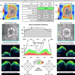 OCT in Patient With IIH Showing Thickened RNFL
Jan 16 2019 by John S. King, MD
18-year-old African American female with increased BMI with a history of headaches, nausea, transient diplopia and vision loss that she notices when getting up from her bed (and goes away after standing upright) for the last two weeks. Went to PCP and was treated for the flu, and after no improvement and visual symptoms known, was sent to ED. MRI did not show any masses and showed empty sella turcia. Vision 20/30 OD and 20/20 OS; no RAPD; IOP 15OU; no anterior segment or vitreous inflammation; discs are elevated with obscuration of the disc margins and some of the smaller vessels; there are no SVPs; there are mild Patton's lines temporally (see Initial Photos). The optic disc cube shows 360 degrees of RNFL thickening (see OCT). Was referred to near-ophthalmologist, Dr. Doyle. She obtained additional work-up, and LP opening pressure was high, and MRV showed bilateral transverse sinus stenosis. Patient showed steady improvement with medical therapy, that included weight loss and oral diamox. On her last visit with Dr. Doyle, vision has remained stable at 20/20-20/25 without an enlarged blindspot; there are SVPs and optic disc edema has resolved (see Post Treatment Photos); she is currently on 1000 mg of diamox and has lost 15 pounds, and no stinting procedure needed.
Imaging device: Cirrus
Condition/keywords: benign idiopatic intracranial hypertension, optic disc edema, papilledema
-
 pupillary block; periph uveitis
Feb 14 2013 by From the Collections of Thomas M. Aaberg, MD and Thomas M. Aaberg Jr., MD
diffuse and slit-beam anterior segment photographs showing pupillary-block angle closure associated with uveitis. pupillary block; angle closure; uveitis; posterior synechiae
Condition/keywords: angle closure, posterior synechiae, pupillary block, uveitis
-
 X-linked ocular albinism slide 1
Oct 22 2012 by Ronald C. Gentile, MD
Anterior segment photo revealed lightly pigmented iris and yellow-white lashes. His complexion was much lighter then both his parents and unaffected siblings.
Photographer: The New York Eye & Ear Infirmary Department of Medical Imaging
Condition/keywords: Nettleship-Falls ocular albinism, ocular albinism
-
 Metallic intraocular foreign body - colour composite image
Dec 25 2012 by Alex P. Hunyor, MD
4-up colour images: 1. anterior segment photo, 2. normal appearing posterior pole image, 3. temporal view showing streak of vitreous haemorrhage, and 4. superotemporal view of impact site where metallic IOFB disrupted superotemporal branch retinal vein leading to vitreous haemorrhage. The IOFB is located in the inferior vitreous cavity, obscured by haemorrhage (see associated CT scan).
Condition/keywords: intraocular foreign body
-
 Mild Patton's Lines in IIH - Initial Photos
Jan 16 2019 by John S. King, MD
18-year-old African American female with increased BMI with a history of headaches, nausea, transient diplopia and vision loss that she notices when getting up from her bed (and goes away after standing upright) for the last two weeks. Went to PCP and was treated for the flu, and after no improvement and visual symptoms known, was sent to ED. MRI did not show any masses and showed empty sella turcia. Vision 20/30 OD and 20/20 OS; no RAPD; IOP 15OU; no anterior segment or vitreous inflammation; discs are elevated with obscuration of the disc margins and some of the smaller vessels; there are no SVPs; there are mild Patton's lines temporally (see Initial Photos). The optic disc cube shows 360 degrees of RNFL thickening (see OCT). Was referred to near-ophthalmologist, Dr. Doyle. She obtained additional work-up, and LP opening pressure was high, and MRV showed bilateral transverse sinus stenosis. Patient showed steady improvement with medical therapy, that included weight loss and oral diamox. On her last visit with Dr. Doyle, vision has remained stable at 20/20-20/25 without an enlarged blindspot; there are SVPs and optic disc edema has resolved (see Post Treatment Photos); she is currently on 1000 mg of diamox and has lost 15 pounds, and no stinting procedure needed.
Photographer: Gretchen Harper
Imaging device: Topcon 50
Condition/keywords: idiopathic intracranial hypertension, optic disc edema, papilledema, Patton's Lines
-
---thumb.jpg/image-square;max$300,300.ImageHandler) Floaters
Floaters
Oct 9 2013 by Maurice F. Rabb
KR is a 25 year old white female who presented with a one month history of floaters OD. Past ocular and systemic history were unremarkable. On clinical examination, the visual acuity was 20/20 OU, and the anterior segments were normal. There was a very mild degree of vitreous cell OD, though no cystoid macular edema nor vasculitis. A lobulated white mass was noted overlying the vitreous base inferotemporally OD (thickness 3.3mm). There was no calcification, though prominent cysts were noted on the surface of the lesion. A fluorescein angiogram, echogram, and CT scan were obtained, along with a thorough systemic evaluation.
Condition/keywords: floaters
-
 Chronic Central Serous Chorioretinopathy (CSCR)
Nov 15 2014 by Rita Couceiro, MD, MS
53-year-old black male, with no relevant prior medical history, complained of bilateral blurry vision for the previous 16 years. On examination, visual acuity was 20/50 on the right eye (OD) and 20/100 on the left eye (OS). Anterior segment evaluation was unremarkable. Fundoscopy revealed pigmentary changes near the macular area in both eyes, with a mottling configuration, suggesting chronic CSCR. Fluorescein angiography showed an ink-blot pattern, with leakage superior to the fovea in OD and nasal to the fovea in OS.
Photographer: Telma Gala - Hospital de Santa Maria, Lisbon, Portugal
Condition/keywords: chronic central serous chorioretinopathy (CSCR)
-
---thumb.jpg/image-square;max$300,300.ImageHandler) Floaters
Floaters
Oct 9 2013 by Maurice F. Rabb
KR is a 25 year old white female who presented with a one month history of floaters OD. Past ocular and systemic history were unremarkable. On clinical examination, the visual acuity was 20/20 OU, and the anterior segments were normal. There was a very mild degree of vitreous cell OD, though no cystoid macular edema nor vasculitis. A lobulated white mass was noted overlying the vitreous base inferotemporally OD (thickness 3.3mm). There was no calcification, though prominent cysts were noted on the surface of the lesion. A fluorescein angiogram, echogram, and CT scan were obtained, along with a thorough systemic evaluation.
Condition/keywords: floaters
-
---thumb.jpg/image-square;max$300,300.ImageHandler) Birdshot Choroidopathy
Birdshot Choroidopathy
Oct 9 2013 by Maurice F. Rabb
Forty two year old white female first noted flashing lights in her left eye at the age of 30. Although she had many previous eye examinations for low grade myopia, she had never had a dilated fundus examination. The evaluation twelve years ago disclosed 20/20 acuity in each eye with a myopic correction, an afferent pupillary defect on the left, no evidence of anterior segment inflammation in either eye, a full field on the right and markedly constricted field on the left, fundus pigmentary abnormalities extending beyond the equator in each eye, and narrow vessels with pigment migration into the retina in the left eye only.
Condition/keywords: birdshot choroidopathy
-
 Serpigenous Choroidopathy in a 68-Year-Old Male
Feb 15 2013 by Roy Schwartz, MD
A 68-year-old healthy male presented with a few years of decreased vision bilaterally. Visual acuity in OD was 1/36 and in OS 20/40. Anterior segments were normal except for bilateral mild nuclear sclerosis and pseudoexfoliation in OS. In the fundus of OD a large atrophy with pigmentary scars were seen in the macula and nasally to the optic disc while OS presented with the same clinical picture but an island of normal appearing retina was seen in the fovea. On fluorscein angiography no leakage was shown. A diagnosis of Serpigenous choroidopathy was made.
Photographer: Galit Yair-Pur
Condition/keywords: macula serpiginous choroidopathy, serpiginous choroiditis
-
---thumb.jpg/image-square;max$300,300.ImageHandler) CMV Retinitis
CMV Retinitis
Oct 7 2013 by Maurice F. Rabb
Thirty one year old man with AIDS referred for an evaluation of treatment of CMV retinitis. In, addition, he had a history of cryptococcal meningitis being treated with Amphotericin. On examination, his visual acuity was 20/20 in both eyes. The anterior segments and vitreous wer quiet. There is a superior nasal CMV retinitis lesion in the periphery of the left eye. Both eyes had multiple deep chorioretinal lesions as noted on the enclosed photographs.
Condition/keywords: CMV retinitis
-
 Color Fundus Photograph of Macular Infarction Secondary to Subonjunctival Gentamicin Injection
May 16 2014 by Arwa Azmeh, MD, PhD
A 20-year-old male suffered from diplopia since age one. He was diagnosed to have acquired fourth nerve palsy in his left eye. VA at time of diagnosis was 20/20 in OU and Fundus exam was WNL in OU. His history revealed no other complaints. 3 days ago he underwent left superior oblique tucking for relief of his diplopia.The surgery was uneventful and at the end of surgery subconjunctival gentamicin was injected. Immediately following surgery his VA in OS decreased from 20/20 to complete loss of central vision and sensation of HM from the periphery. He was referred to us 3 days after surgery. At time of referral fundus exam of his left eye revealed macular infarction with cherry red spot appearance with few retinal hemorrhages, mild optic disc edema and CWS surrounding optic disc. Peripheral retina had normal color and appearance. The vitreous was clear. Anterior segment was quiet. IOP was WNL. Macular OCT was consistent with macular infarction. FA revealed delay in central retinal artery filling as fluorescein started to appear in the arteries at the level of the optic disc at 28 sec, and in the retinal veins at 38 sec. Macular area remained to be non-perfused throughout the whole FA. In late phases staining of blood vessels walls was noticed. The "wipe out" of large vessels and capillaries persisted in the central area. OCT through foveal area showed diffuse thickening of the retina with severe elevation in the fovea, reduced backscattering from the outer layers of the retina and enhanced reflectivity from the inner retina, due to ischemia. Complete blood count and cardiovascular study were WNL. The final diagnosis was macular infarction secondary to subconjunctival gentamicin injection.
Imaging device: OCT
Condition/keywords: macular infarction, subconjunctival gentamicin
-
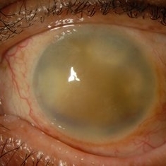 Corneal Decompensation Following Endophthalmitis
Jul 12 2014 by Philip J. Polkinghorne, MD
This patient presented following a tap and inject for endophthalmitis. Unfortunately the cornea decompensated as fibrin became organized in the anterior segment.
Condition/keywords: corneal decompensation, endophthalmitis
-
---thumb.jpg/image-square;max$300,300.ImageHandler) CMV Retinitis
CMV Retinitis
Oct 7 2013 by Maurice F. Rabb
Thirty one year old man with AIDS referred for an evaluation of treatment of CMV retinitis. In, addition, he had a history of cryptococcal meningitis being treated with Amphotericin. On examination, his visual acuity was 20/20 in both eyes. The anterior segments and vitreous wer quiet. There is a superior nasal CMV retinitis lesion in the periphery of the left eye. Both eyes had multiple deep chorioretinal lesions as noted on the enclosed photographs.
Condition/keywords: CMV retinitis
-
 Anterior Chamber Gas and PFC Migration
Jun 21 2018 by Maria Stephanie R. Jardeleza, MD
Anterior segment photographs of 30-year-old male who underwent superior rhegmatogenous retinal detachment repair with intraocular gas tamponade. Perfluorocarbon was used to flatten the macula to prevent a macular fold and was removed during PFC/air exchange. Post operative week two visit shows gas migration into the anterior chamber with retained PFC layered in a tear drop shape posterior to the gas bubble and anterior to the lens. Patient had been maintaining face down positioning.
Photographer: Andy Zepeda, COA, Retina Clinic, San Antonio Eye Center, San Antonio, TX
Condition/keywords: retained perfluorocarbon, retina surgery complications, vitreous substitutes
-
 Color Photo of Optic Disc Capillary Hemangioblastoma
Mar 18 2014 by Arwa Azmeh, MD, PhD
Color fundus photograph of an 48-year-old male who complained of decreased visual acuity in his right eye over the last few months. Systemically the patient was healthy. His VA was OD Cf 3m, OS 20/20. Anterior segments were WNL in OU. IOP was WNL in OU. Fundus exam OD revealed unpigmented mass over the optic disc with retinal venous tortuosity at its edges with a ring of thick HYE surrounding it and shallow RD in this area extending to the foveal area. Several few small retinal hemorrhages were seen in the far retinal periphery which were explained to be caused by venous stasis due the optic disc tumor.
Condition/keywords: color photo, optic disc, retinal hemangioblastoma
-
 Giant Retinal Tear
Apr 1 2016 by Nichole Lewis
Giant retinal tear montaged on Anterior Segment due to the Detachment being very bullous.
Photographer: Nichole Lewis - Pennsylvania Retina Specialists, Camp Hill, PA
Condition/keywords: giant retinal tear, retinal tear
-
 Serpigenous Choroidopathy in a 68-Year-Old Male
Feb 15 2013 by Roy Schwartz, MD
A 68-year-old healthy male presented with a few years of decreased vision bilaterally. Visual acuity in OD was 1/36 and in OS 20/40. Anterior segments were normal except for bilateral mild nuclear sclerosis and pseudoexfoliation in OS. In the fundus of OD a large atrophy with pigmentary scars were seen in the macula and nasally to the optic disc while OS presented with the same clinical picture but an island of normal appearing retina was seen in the fovea. On fluorscein angiography no leakage was shown. A diagnosis of Serpigenous choroidopathy was made.
Photographer: Galit Yair-Pur
Condition/keywords: macula serpiginous choroidopathy, serpiginous choroiditis
-
 Retained perfluorocarbon in the anterior segment
Dec 19 2012 by Eric A. Postel, MD
color photograph of the anterior segment of an aphakic eye with retained perfluorocarbon below the intraocular gas bubble
Condition/keywords: retained perfluorocarbon
-
 IOFB-Endophthalmitis Slide 2
Oct 22 2012 by Ronald C. Gentile, MD
Anterior segment examination revealed a self sealing peripheral corneal wound at the 7:30 position with iris defect. There was a small hypopyon layering in the inferior angle. CT scan revealed a small intra-ocular foreign body.
Photographer: The New York Eye & Ear Infirmary Department of Medical Imaging
Condition/keywords: intraocular foreign body
 Loading…