 Initializing download.
-
 By Alex P. Hunyor, MD
Retina Associates - Uploaded on Dec 25, 2012.
- Last modified by Chayal Patel on Dec 26, 2012.
- Reviewed by Chayal Patel
- Rating
- Appears in
- Intraocular foreign body
- Condition/keywords
- intraocular foreign body
- Imaging device
- Fundus camera
- Description
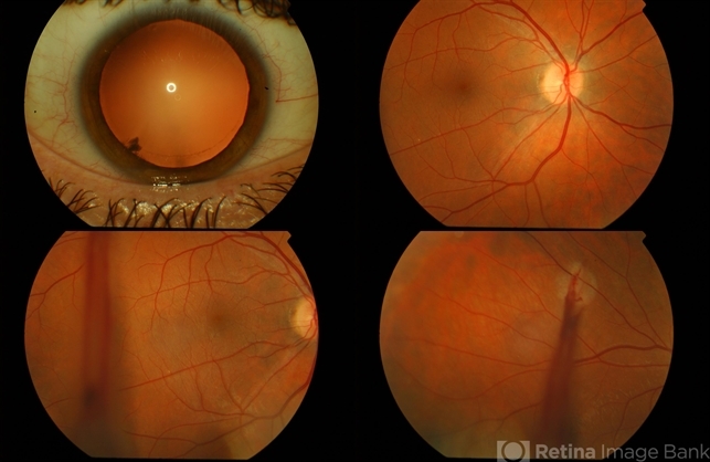
- 4-up colour images: 1. anterior segment photo, 2. normal appearing posterior pole image, 3. temporal view showing streak of vitreous haemorrhage, and 4. superotemporal view of impact site where metallic IOFB disrupted superotemporal branch retinal vein leading to vitreous haemorrhage. The IOFB is located in the inferior vitreous cavity, obscured by haemorrhage (see associated CT scan).


