Search results (438 results)
-
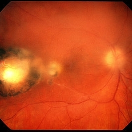
 Stage 5 Retinopathy of Prematurity (ROP)
Oct 9 2012 by Audina M. Berrocal, MD FASRS
Advanced APROP with Stage 5 and vascularly active.
Photographer: Ditte Hess CRA, BPEI
Imaging device: RETCAM
Condition/keywords: retinopathy of prematurity (ROP), stage 5
-
 Toxoplasma Retinochoroiditis
Feb 25 2013 by Henry J. Kaplan, MD
Toxoplasmosis, right eye: reactivation of congenital toxoplasmosis as an active retinitis lesion with overlying vitritis adjacent to an old scar.
Condition/keywords: toxoplasmosis chorioretinitis, toxoplasmosis reactivation
-
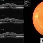 Chronic Active Central Serous Chorioretinopathy (CSCR)
Sep 11 2012 by Hamid Ahmadieh, MD
Color fundus photograph and OCT image of a 30-year-old man with chronic active CSCR.
Photographer: Hamid Ahmadieh, MD, Ophthalmic Research Center, Labbafinejad Medical Center, Shahid Beheshti University of Medical Sciences
Imaging device: Topcon
Condition/keywords: central serous chorioretinopathy (CSCR), optical coherence tomography (OCT)
-
 Retinal Angiomatous Proliferation in Age-Related Macular Degeneration with Subretinal Neovascularization
Sep 24 2012 by James B. Soque, CRA, OCT-C, COA, FOPS
75-year-old white male with classic SRN with RAP. Lesion OD is active, and patient is receiving anti-VEGF treatment. Mid phase FA, 50 Deg, Mag 2x.
Photographer: James Soque, CRA, COA, Island Retina, Shirley, NY, USA
Imaging device: Topcon TRC 50 DX, OIS 5.0 MP Color, FA Camera, OIS Software
Condition/keywords: age-related macular degeneration (AMD), fundus autofluorescence (FAF), leakage, retinal angiomatous proliferation (RAP), subretinal neovascularization (SRNV)
-
 Cytomegalovirus Retinitis, Active, with Papillary Involvement
Sep 27 2012 by Jeffrey G. Gross, MD, FASRS
CMV retinitis active with papilary involvement, inferotemporal arcade.
Condition/keywords: inferotemporal arcade
-
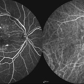 Multifocal CSCR 2
Sep 2 2012 by Hamid Ahmadieh, MD
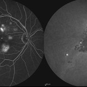
Early-phase FA and ICG angiograms of a 36-year-old man with an active multifocal CSCR.
Photographer: Hamid Ahmadieh, Ophthalmic Research Center, Labbafinejad Medical Center
Imaging device: Heidelberg Spectralis
Condition/keywords: central serous chorioretinopathy (CSCR), indocyanine green (ICG) angiography
-
 PDR with Active NVD
Oct 8 2012 by Jeffrey G. Gross, MD, FASRS
PDR with active NVD and preretinal hemorrhage, mild VH and partial PRP.
Condition/keywords: neovascularization of the disc (NVD), preretinal hemorrhage, scatter laser photocoagulation, vitreous hemorrhage
-
---thumb.jpg/image-square;max$300,300.ImageHandler) Multifocal Choroiditis
Multifocal Choroiditis
Feb 26 2013 by Henry J. Kaplan, MD
Multifocal choroiditis, MFC, inactive scars in the periphery.
Condition/keywords: multifocal choroiditis
-
 Cytomegalovirus Retinitis with Papillary Involvement
Oct 9 2012 by Jeffrey G. Gross, MD, FASRS
CMV retinitis with papillary involvement, active.
Condition/keywords: active, papillary involvement
-
 Cytomegalovirus Papillitis
Oct 10 2012 by Jeffrey G. Gross, MD, FASRS
CMV, papillitis, active, CF.
Condition/keywords: active, chorioretinal fold, papillitis
-
 Toxoplasma chorioretinitis 1
Jan 11 2013 by Alex P. Hunyor, MD
Toxoplasmosis 1 - chorioretinal scar from previous toxoplasma chorioretinitis. See image 2 - recurrent todo adjacent to this scar
Condition/keywords: inactive toxoplasmosis, ocular toxoplasmosis, toxoplasmosis, toxoplasmosis retinitis
-
---thumb.jpg/image-square;max$300,300.ImageHandler) Multifocal Choroiditis and Panuveitis Syndrome
Multifocal Choroiditis and Panuveitis Syndrome
Feb 26 2013 by Henry J. Kaplan, MD
Multifocal choroiditis and panuveitis: left eye. Acute stage: haziness of the media due to vitritis and multiple active yellow and also inactive choroidal lesions are present.
Condition/keywords: multifocal choroiditis
-
 Active CNVM
Jul 11 2016 by Manish Nagpal, MD, FRCS (UK), FASRS
Colour photo showing an active CNVM.
Photographer: pooja barot
Condition/keywords: choroidal neovascular membrane (CNVM), optical coherence tomography (OCT)
-
 Multifocal CSCR 1
Sep 2 2012 by Hamid Ahmadieh, MD
Fundus autofluorescence of a 36-year-old man with an active multifocal CSCR.
Photographer: Hamid Ahmadieh, Ophthalmic Research Center, Labbafinejad Medical Center
Imaging device: Heidelberg Spectralis
Condition/keywords: central serous chorioretinopathy (CSCR)
-
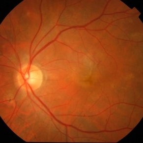
 Proliferative Diabetic Retinopathy - Neovascularization on the Disc
Aug 23 2012 by Gerardo Garcia-Aguirre, MD
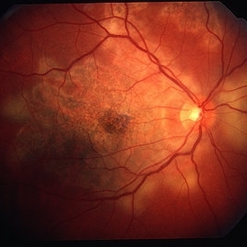
Fundus photograph of the left eye of a 51-year-old female with proliferative diabetic retinopathy that has been treated with panretinal photocoagulation, but is still active. Note the neovascularization on the disc.
Photographer: Noemí Hernández, Asociación para Evitar la Ceguera en México
Condition/keywords: neovascularization of the disc (NVD)
-
 CSCR Mushroom Cloud
Feb 25 2015 by James J. Bedrick, MD
Late transit FA of a large active subfoveal CSCR leak. Focus is on peri-foveal vessels to give sense of height of large serous RD of macula. This patient presented with a BCVA of 20/200 and fluorescein and historic evidence of prior episodes of leakage. After discussion of known treatment options including observation, he was initially treated with rifampin and had partial resolution to 20/70 BCVA but this was short-lived with reaccumulation of the large serous detachment within 3 months. He then received sub-threshold micro-pulse laser photocoagulation with an 810 nm diode laser which resulted 1 month later in complete drying of the serous detachment and BCVA of 20/25.
Photographer: Diana Bodnar, COT
Imaging device: Topcon 50X with OIS capture station
Condition/keywords: CSCR subfoveal leak
-
 Multifocal CSCR
Sep 2 2012 by Hamid Ahmadieh, MD
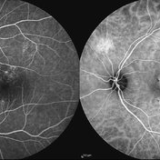
Late-phase FA and ICG angiograms of a 36-year-old man with an active multifocal CSCR.
Photographer: Hamid Ahmadieh, Ophthalmic Research Center, Labbafinejad Medical Center
Imaging device: Heidelberg Spectralis
Condition/keywords: central serous chorioretinopathy (CSCR), indocyanine green (ICG) angiography
-
 PDR with Active NVD
Oct 8 2012 by Jeffrey G. Gross, MD, FASRS
PDR with active NVD and preretinal hemorrhage.
Condition/keywords: neovascularization of the disc (NVD), preretinal hemorrhage
-
 Active Neovascular AMD With Disciform Scar
Apr 30 2015 by Mitzy E Torres Soriano, MD
Active neovascular AMD with disciform scar.
Photographer: Mitzy E. Torres Soriano, MD; Centro medico Cagua-Estado Aragua. Venezuela
Imaging device: TOPCON
Condition/keywords: disciform scar, disciform with hemorrhage, neovascular age-related macular degeneration (AMD), wet age-related macular degeneration (wet AMD)
-
 Cytomegalovirus Retinitis with Papillary Involvement
Oct 9 2012 by Jeffrey G. Gross, MD, FASRS
CMV retinitis with papillary involvement, active.
Condition/keywords: active, papillary involvement
-
 Chronic Active Central Serous Chorioretinopathy (CSCR)
Sep 11 2012 by Hamid Ahmadieh, MD
Late phase FA & ICG angiography images of a 30-year-old man with chronic active CSCR.
Photographer: Hamid Ahmadieh, MD, Ophthalmic Research Center, Labbafinejad Medical Center, Shahid Beheshti University of Medical Sciences
Imaging device: Heidelberg Spectralis
Condition/keywords: central serous chorioretinopathy (CSCR), indocyanine green (ICG) angiography
-
 Serpiginous Choroiditis
Feb 25 2013 by Henry J. Kaplan, MD
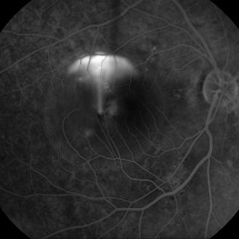
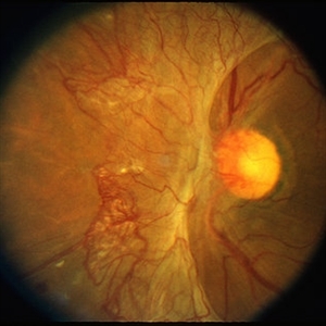
Serpiginous choroiditis, left eye #2. Active edematous lesion visible moving toward the fovea.
Condition/keywords: serpiginous choroiditis
-
 Advanced Active PDR
Mar 29 2013 by Henry J. Kaplan, MD
Extensive NVD-FPD and NVE-FPE in a diabetic patient.
Condition/keywords: foveal photoreceptor defect, FPE, neovascularization (NV), neovascularization of the disc (NVD)
-
 Serpiginous Choroiditis
Feb 25 2013 by Henry J. Kaplan, MD
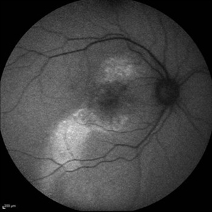
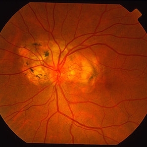
Serpiginous choroiditis, right eye. Both active and inactive lesions clearly visible; active lesions are the yellowish subretinal area most prominant nasal to optic nerve head and also around the inferior arcade and temporal to the macular lesion.
Condition/keywords: serpiginous choroiditis
-
 Chronic Central Serous Chorioretinopathy
Sep 26 2012 by Hamid Ahmadieh, MD
Autofluorescence imaging of the right eye of a 50-year-old man with active chronic CSCR and BCVA of 20/100.
Photographer: Hamid Ahmadieh, MD, Ophthalmic Research Center, Labbafinejad Medical Center, Shahid Beheshti University of Medical Sciences
Imaging device: Heidelberg Spectralis
Condition/keywords: autofluorescence imaging, chronic central serous chorioretinopathy (CSCR)
 Loading…