Search results (438 results)
-
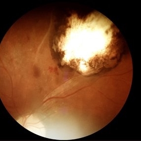
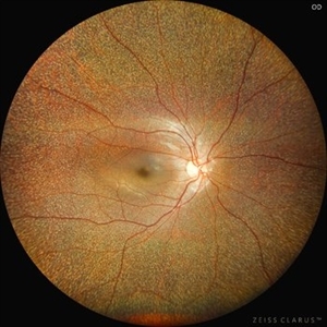
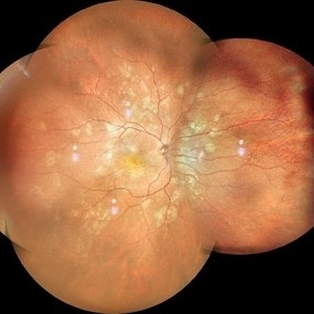
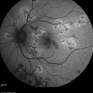 Active multifocal choroiditis
May 26 2025 by Moazzam Parvez
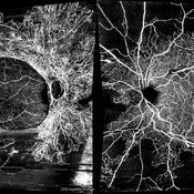
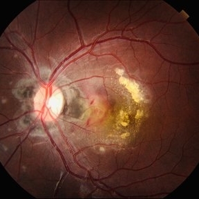
Auto fluorescence photograph of an 43 year old man with active choroiditic lesion present in the left eye with recurrence
Photographer: Dr Moazzam Parvez , Netralayam , Kolkata
Imaging device: Heidelberg Spectralis
Condition/keywords: active choroididtis, choroiditi
-
 Neovascular AMD with Active CNV
May 22 2025 by Kimberly Wakester
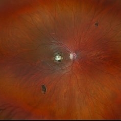
Optomap RGB of an 82-year-old man with Neovascular AMD with Active CNV and Dry AMD in the right eye. There is advanced atrophic changes without subfoveal involvement located temporally to the fovea. Patient is to continue follow up care with dilated exam, repeat OCT, and treatment of intravitreal injection of Vabysmo every 5 weeks at this time.
Photographer: Kimberly Wakester, COA, OCT-C
Imaging device: Optos California
Condition/keywords: advanced geographic atrophy, dry age-related macular degeneration (dry AMD), neovascular age-related macular degeneration (AMD)
-
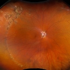
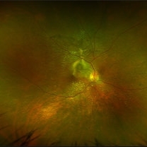 VKH Pseudotumor – Chronic Subretinal Fibrosis
May 11 2025 by Felipe Murati
Ultra-widefield fundus image from a 36-year-old woman with chronic VKH syndrome showing a pseudotumor-like subretinal fibrotic lesion in the right eye. The lesion developed after multiple relapses and remained stable over a 1-year follow-up with immunosuppressive treatment including prednisone, mycophenolate mofetil, and adalimumab. No active choroiditis or exudative detachment was observed. Multimodal imaging was essential for disease monitoring.
Photographer: Felipe A. Murati, MD, University of Arizona
Imaging device: Optos California ultra-widefield retinal imaging system, single-capture, color fundus modality.
Condition/keywords: adalimumab, chronic inflammation, granulomatous uveitis, OCT, Optos ultra-widefield imaging, pseudotumor, subretinal fibrosis, VKH, Vogt-Koyanagi-Harada
-
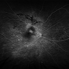 VKH Pseudotumor – Fluorescein Angiography
May 11 2025 by Felipe Murati
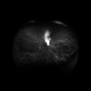
Fluorescein angiography image from a 36-year-old woman with chronic Vogt-Koyanagi-Harada (VKH) syndrome showing a pseudotumor-like lesion with late-phase staining and no active leakage. The image highlights subretinal fibrosis in the right eye, stable under long-term immunosuppressive therapy with mycophenolate mofetil and adalimumab. No signs of active choroiditis are present, confirming a quiescent phase.
Photographer: Felipe A. Murati, MD, University of Arizona
Imaging device: Optos California, fluorescein angiography modality
Condition/keywords: choroiditis, Fluorescein angiography, granulomatous uveitis, Optos FA, pseudotumor, subretinal fibrosis, VKH, Vogt-Koyanagi-Harada
-
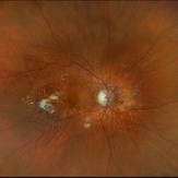
 Retinocoroiditis Inactiva Por Toxoplasmosis
Apr 28 2025 by Paulina Araujo
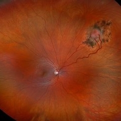
Fundus photography demonstrates a 2-disc-diameter chorioretinal scar in the superior temporal arcade, consistent with inactive toxoplasmic retinochoroiditis. The lesion exhibits pigmented borders and central atrophy, with adjacent splinter hemorrhages and vascular sheathing. No vitreous inflammation or active satellite lesions are present.
Photographer: Paulina D.Araujo Martínez, Asociación para Evitar la Ceguera en México I.A.P., Hospital Dr Luis Sánchez Bulnes.
Condition/keywords: toxoplasmosis chorioretinitis
-
 Toxocara Granuloma
Apr 18 2025 by Chellarani Kumarasamy, MD
10 year old girl referred for starbismus and red free image showed well demarcted inactive lesion.
Condition/keywords: toxocara granuloma
-
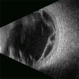 Proliferative Vitreoretinopathy
Apr 17 2025 by Gustavo Uriel Fonseca Aguirre
This B-mode transverse ultrasound scan depicts a post-vitrectomy eye with recurrent retinal detachment in a patient with diabetic retinopathy history. The image reveals fresh vitreous cavity hemorrhage and subretinal bleeding, along with subretinal proliferative bands (PVR strands). These findings indicate complicated tractional re-detachment with active hemorrhagic components.
Photographer: Gustavo U. Fonseca Aguirre, Hospital Conde de Valenciana, Ciudad de México
Condition/keywords: proliferative vitreoretinopathy (PVR)
-
 Actively Bleeding NVE
Apr 1 2025 by Jordyn Beckman
47 year old woman presented with actively bleeding NVE temporally on exam with complaints of foggy vision and floaters.
Photographer: Jordyn Beckman, Retina Consultants of Carolina, P.A.
Imaging device: Optos California
Condition/keywords: active bleeding, Elevated retinal neovascularization, vitreous hemorrhage
-
 Shunt Vessels
Apr 1 2025 by Korey Starkey
62-year-old patient presented with stable CRVO in the left eye. FA performed that day shows delayed AV transit is present, this has compensated with shunt vessels at the disc. However there is no evidence of active leakage. OS vision 20/25.
Photographer: Korey Starkey
Imaging device: Topcon
Condition/keywords: central retinal vein occlusion (CRVO), fundus photograph, optic nerve, shunts vessels, Topcon
-
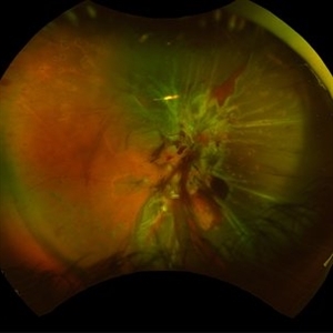 Combined Traction and Rhegmatogenous Retinal Detachment From Proliferative Diabetic Retinopathy
Mar 27 2025 by Nikhil K Bommakanti, MD
A middle-aged patient presented with a combined traction and rhegmatogenous retinal detachment.
Condition/keywords: Active PDR Tractional retinal Detachment, PDR, Retinal Detachment, rrd, TRD
-
 Bilateral Proliferative Diabetic Retinopathy OU
Feb 21 2025 by Drew Mitchell
OCT-Angiography 8x8 Montage OU. PDR with active NVE OD. 37 year old male with no visual complaints. Vision is 20/20 in both eyes.
Photographer: Drew Mitchell OCT-C
Imaging device: Zeiss Cirrus 5000
Condition/keywords: CIRRUS 5000 ANGIOPLEX, Diabetes, NVE, OCT Angiography, proliferative diabetic retinopathy (PDR)
-
 Diabetic Tractional Retinal Detachment Involving the Macula OD
Feb 21 2025 by Kaitlyn Anderson
57-year-old female. Diabetic Tractional Retinal Detachment involving the Macula OD. Active Proliferative Diabetic Retinopathy
Photographer: Kaitlyn Anderson TN Retina Nashville TN
Imaging device: Optos Fluorescein Angiogram
Condition/keywords: Active PDR Tractional retinal Detachment
-
 Inactive Chorioretinal Scars
Dec 11 2024 by Virginia Gebhart
30 year old female with chorioretinal and macula scars. Appears post-infectious, most likely toxoplasmic. No active inflammatory changes or choroidal neovascularization. Will continue to monitor. Central vision limited by macula scar, BCVA 20/100
Photographer: Virginia Gebhart, Retina Consultants of Carolina
Imaging device: Optos California
Condition/keywords: chorioretinal scar, inactive toxoplasmosis
-
 Reactive Retinal Astrocytic Tumor
Dec 6 2024 by Virginia Gebhart
27 year old female self-referred for continued follow-up care of hemangioma of retina. Previous genetic testing negative for Von Hippel-Lindau. Pt recently diagnosed with Ehlers-Danlos Arthroclasia, most likely reactive retinal astrocytic tumor. Tumor is stable and surrounded by good laser barricade, will continue to observe.
Photographer: Virginia Gebhart, Retina Consultants of Carolina
Imaging device: Optos California
Condition/keywords: feeder vessel, hemangioma, RRAT
-
 Benign Familial Fleck Retina
Dec 2 2024 by KANWALJEET HARJOT MADAN, M.S. (Ophthalmology); FAICO (Vitreous - Retina)
This is fundus picture of a 21 year old female patient who had come for refractive surgery consultation. Her best corrected vision in both eyes was 20/20. She had myopic astigmatism in both eyes. Fundus exam revealed presence of multiple yellowish white flecks spread throughout retina sparing macular area in both eyes. Her color vision was normal. Electroretinogram and electrooculogram were normal. She gave no history of night blindness. A diagnosis of Benign Familial Fleck Retina was made. She was also advised ocular exam of her parents and elder brother which was normal.
Photographer: Dr. Kanwaljeet Harjot Madan, M.S. (Ophthalmologist) Fellow in Vitrous & Retina. Thind Eye Hospital, Jalandhar City. Punjab. India
Imaging device: Zeiss Clarus
Condition/keywords: Benign familial fleck retina, Night Blindness
-
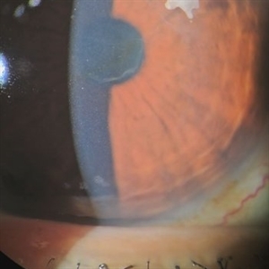
 New Iris Melanoma
Oct 10 2024 by Virginia Gebhart
56 year old male with new amelanotic melanoma emanating from the ciliary body through the posterior iris epithelium. CT scan showed no evidence of metastatic disease. Pt scheduled for radioactive plaque and tumor biopsy
Photographer: Virginia Gebhart, Retina Consultants of Carolina
Imaging device: Samsung Galaxy
Condition/keywords: amelanotic melanoma, iris melanoma
-
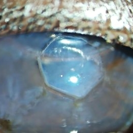 Radial Keratotomy
Sep 28 2024 by DR Rohit Gupta
A slit lamp photograph of 46 year-old female patient operated for high hypermetropia 10 years back . On slit lamp examination hexagonal pattern of radial incisions can be seen.
Photographer: Dr Rohit gupta
Condition/keywords: hypermetropia, hyperopia, Radial keratotomy, refractive surgery
-
 POHS/Schlaegel Lines
Sep 19 2024 by Virginia Gebhart
46 year old female with h/o Histoplasmosis. Multiple punched out chorioretinal scars with Schlaegel lines. No evidence of CNV or active inflammation. VA 20/20
Photographer: Virginia Gebhart, Retina Consultants of Carolina
Imaging device: Optos California
Condition/keywords: chorioretinal scar, histoplasmosis, presumed ocular histoplasmosis syndrome (POHS), Schlaegel
-
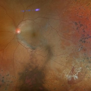
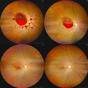 Valsalva Retinopathy
Sep 10 2024 by KANWALJEET HARJOT MADAN, M.S. (Ophthalmology); FAICO (Vitreous - Retina)
These are fundus pictures of a young 32 years male who presented with sudden decrease in vision in right eye after lifting heavy weights. His vision in right eye was hand movements close to face. He had no systemic history or any history of trauma. He was diagnosed to have Valsalva Retinopathy. His hematological investigations were normal. He refused any active intervention. He was kept under observation and his vision improved to 20/20 within 4 weeks.
Photographer: Dr. Kanwaljeet Harjot Madan, M.S. (Ophthalmologist) Fellow in Vitrous & Retina. Thind Eye Hospital, Jalandhar City. Punjab. India
Imaging device: Zeiss Clarus
Condition/keywords: valsalva retinopathy
-
 Multifocal Choroiditis
Jul 13 2024 by Tejaswita Verma
RE fundus montage of a 34 y/o male showing old and active hypopigmented lesions with macular involvement .He presented with DOV since a month,treated with oral steroids for 15 days elsewhere,with BCVA of CF2mt and positive Mantoux test.
Photographer: DR. TEJASWITA VERMA
Imaging device: MIRANTE
Condition/keywords: multifocal choroiditis
-
 Active Proliferative Diabetic Retinopathy
Jul 12 2024 by Korey Starkey
Fluorescein angiogram performed on 35 year old female with active proliferative diabetic retinopathy. Patient has peripapillary vascular loop and history of PRP treatment in both eyes. Patients left eye vision measured at Dcc20/200-1 at this visit.
Photographer: Korey Starkey
Imaging device: Optos
Condition/keywords: FLUORESCEIN ANGIOGRAPHY, hyperfluorescence, laser scarring, Optos, proliferative diabetic retinopathy (PDR), sea fan, ultra-wide field imaging, vascular loop
-
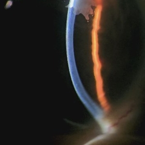
 Ink Blot Epithelial Ingrowth Post-LASIK Refractive Surgery
Jun 29 2024 by Luai Abu-Ismail, MD
Anterior segment photo of a 45-year-old female patient presented 12-year post-LASIK surgery.
Photographer: Dr. Luai Abu-Ismail, Ophthalmology Department, Islamic Hospital.
Imaging device: Slit lamp biomicroscope photo taken by Smart phone camera.
Condition/keywords: cornea, corneal scars and opacities, flap, LASIK
-
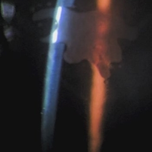 Ink Blot Epithelial Ingrowth Post-LASIK Refractive Surgery
Jun 29 2024 by Luai Abu-Ismail, MD
Anterior segment photo of a 45-year-old female patient presented 12-year post-LASIK surgery.
Photographer: Dr. Luai Abu-Ismail, Ophthalmology Department, Islamic Hospital.
Imaging device: Slit lamp biomicroscope photo taken by Smart phone camera.
Condition/keywords: complication, cornea, corneal scars and opacities, epithelial ingrowth, LASIK, LASIK FLAP, refractive surgery
-
 Serpiginous Choroidopathy
Apr 21 2024 by César Adrián Gómez Valdivia, MD
Gray-yellowish subretinal infiltrates that usually spread centrifugally from the peripapillary region in a serpiginous (snake-like) manner. Active lesions show a leading edge and resolve with subsequent RPE and choriocapillary atrophy.
Photographer: @eyemissu2
Imaging device: TOPCON TRC-50DX
Condition/keywords: macula serpiginous choroidopathy
 Loading…