Search results (521 results)
-
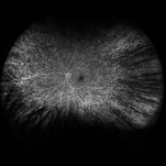
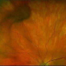
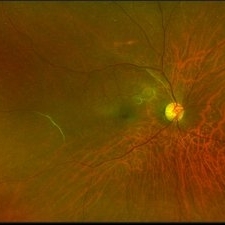
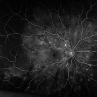 "Starry Sky" Fundus in Vogt-Koyanaki-Harada Syndrome
Jan 10 2018 by Peter H. Tang, MD, PhD
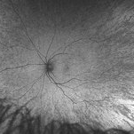
Fluorescein angiography imaging of a 27-year-old male with acute inflammation as part of Vogt-Koyanagi-Harada Syndrome.
Imaging device: Optos California
Condition/keywords: chorioretinal inflammations, retina, uveitis, Vogt-Koyanagi-Harada
-
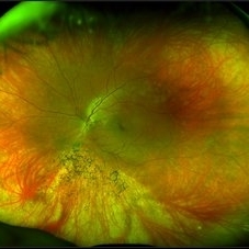
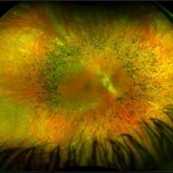 Retinitis Pigmentosa
May 26 2017 by Olivia Rainey
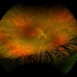
Ultra-wide-field pseudocolor image of the right eye of an 39-year-old female with Retinitis Pigmentosa. She had slightly atypical appearance due to asymmetry: sectoral atrophy in left eye, compared to 360 degree bone spicule formation in right eye. Ddx: Pigmentary degeneration vs infection vs X-linked RP carrier due to asymmetry. Recommended genetic testing through My Retina Tracker, as well as visual field and ERG testing. Patient's vision was sc20/100 PH 20/70 in the right eye and sc20/80 PH 20/40 in the left.
Photographer: Olivia Rainey
Imaging device: Optos California
Condition/keywords: bone spicule, fundus photograph, Optos, peripheral bone spicules, pseudocolor, retinitis pigmentosa, ultra-wide field imaging
-
 Optos Picture With Speculum: Dislocated Natural Lens
Oct 9 2018 by John S. King, MD
55-year-old white female with history of pathologic myopia+, lattice (laser), SB OU (1990s), and dislocated natural lenses OU that had been watched for years. In the fellow eye she developed phacolytic glaucoma and a PPV, PPL was performed. Plan for both eyes are monitoring. I wanted to get a good picture of her lens today with the optos machine, as the other pics had artifact from the lower lid. It worked out well to use a speculum in the left eye. Vision cc is 20/400 J1+ OD and 20/40 J2 OS; aphakic OU; vitreous clear OD; dislocated lens OS (see pic); retinas attached.
Photographer: Maisee Yang
Imaging device: Optos California
Condition/keywords: dislocated crystalline lens, pathologic myopia, scleral buckle, staphyloma
-
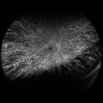
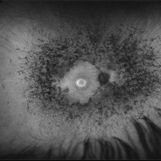
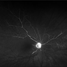
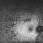 Retinitis Pigmentosa - Autofluorescence OD
Jun 18 2018 by Hosam Attia, MD
Ultra-wide fundus auto-fluorescence photograph of a 38-year-old African, American female with degenerative myopia, unilateral RP variant, depicting extensive mid-peripheral bone spicules hypo-autofluorescence, extending further into the periphery w/ relative sparing of the macula OD VF 30-V showed severe peripheral constriction OD, enlarged BS OS and OCT showed severe ellipsoid zone degeneration with saucerization and cystoid macular degeneration with no obvious late macular leakage on FA (Both, not shown)
Imaging device: Optos California
Condition/keywords: bone spicule, peripheral bone spicules, retinitis pigmentosa
-
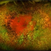
 "Mud-Splatter" of Posterior Pole and Peripheral Radial Streaks in a Carrier of Ocular Albinism
Jan 22 2019 by John S. King, MD
14-year-old healthy white female with family history of ocular albinism was seen by Dr. Hruby for a second opinion. Father and some of his brothers were positive for a history of ocular albinism. Va cc 20/30 J1+ OU; no nystagmus; no TIDs; no foveal hypoplasia. A "mud-spatter" appearance to the posterior pole was present, along with peripheral alternating streaks (photo). Dr. Hruby agreed that this was most likely a carrier of Ocular Albinism Type-1 (XR; GPR143 mutation), and possible genetic testing/counselling was discussed.
Photographer: Gretchen Harper
Imaging device: Optos California
Condition/keywords: Nettleship-Falls ocular albinism, ocular albinism
-
 "Mud-Splatter" of Posterior Pole and Peripheral Radial Streaks in a Carrier of Ocular Albinism
Jan 22 2019 by John S. King, MD
14-year-old healthy white female with family history of ocular albinism was seen by Dr. Hruby for a second opinion. Father and some of his brothers were positive for a history of ocular albinism. Va cc 20/30 J1+ OU; no nystagmus; no TIDs; no foveal hypoplasia. A "mud-spatter" appearance to the posterior pole was present, along with peripheral alternating streaks. Hypoautofluorescent areas correspond to hyperpigmented areas of retinal pigment epithelium, and vice versa (see photo). Dr. Hruby agreed that this was most likely a carrier of Ocular Albinism Type-1 (XR; GPR143 mutation), and possible genetic testing/counselling was discussed.
Photographer: Gretchen Harper
Imaging device: Optos California
Condition/keywords: Nettleship-Falls ocular albinism, ocular albinism
-
 "Mud-Splatter" of Posterior Pole and Peripheral Radial Streaks in a Carrier of Ocular Albinism
Jan 22 2019 by John S. King, MD
14-year-old healthy white female with family history of ocular albinism was seen by Dr. Hruby for a second opinion. Father and some of his brothers were positive for a history of ocular albinism. Va cc 20/30 J1+ OU; no nystagmus; no TIDs; no foveal hypoplasia. A "mud-spatter" appearance to the posterior pole was present, along with peripheral alternating streaks (photo). Dr. Hruby agreed that this was most likely a carrier of Ocular Albinism Type-1 (XR; GPR143 mutation), and possible genetic testing/counselling was discussed.
Photographer: Gretchen Harper
Imaging device: Optos California
Condition/keywords: Nettleship-Falls ocular albinism, ocular albinism
-
 Macula-Sparing GRT RRD
Jul 6 2017 by Andrew A. Moshfeghi, MD, MBA, FASRS
Wide-field fundus photograph of a 43-year-old myopic man with a history of lattice retinal degeneration status posterior barrier laser performed elsewhere who presented with a giant-retinal tear associated retinal detachment of the right eye.
Photographer: Jay Jiang, University of Southern California Roski Eye Institute
Imaging device: Optos California
Condition/keywords: acute retinal detachment, giant retinal tear, lattice degeneration
-
 "Mud-Splatter" of Posterior Pole and Peripheral Radial Streaks in a Carrier of Ocular Albinism
Jan 22 2019 by John S. King, MD
14-year-old healthy white female with family history of ocular albinism was seen by Dr. Hruby for a second opinion. Father and some of his brothers were positive for a history of ocular albinism. Va cc 20/30 J1+ OU; no nystagmus; no TIDs; no foveal hypoplasia. A "mud-spatter" appearance to the posterior pole was present, along with peripheral alternating streaks (photo). Dr. Hruby agreed that this was most likely a carrier of Ocular Albinism Type-1 (XR; GPR143 mutation), and possible genetic testing/counselling was discussed.
Photographer: Gretchen Harper
Imaging device: Optos California
Condition/keywords: Nettleship-Falls ocular albinism, ocular albinism
-
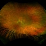
 Acute Retinal Necrosis secondary to Herpes Zoster Ophthalmicus
Jan 9 2018 by Olivia Rainey
Ultra-wide field Optos pseudocolor montage of an 40-year-old female presenting with acute retinal necrosis secondary to herpes zoster ophthalmicus affecting her right eye.
Photographer: Olivia Rainey
Imaging device: Optos California
Condition/keywords: acute retinal necrosis, color fundus photograph, Herpes zoster, montage, Optos, ultra-wide field imaging
-
 "Mud-Splatter" of Posterior Pole and Peripheral Radial Streaks in a Carrier of Ocular Albinism
Jan 22 2019 by John S. King, MD
14-year-old healthy white female with family history of ocular albinism was seen by Dr. Hruby for a second opinion. Father and some of his brothers were positive for a history of ocular albinism. Va cc 20/30 J1+ OU; no nystagmus; no TIDs; no foveal hypoplasia. A "mud-spatter" appearance to the posterior pole was present, along with peripheral alternating streaks that are very prominent on this late phase FA OS. Dr. Hruby agreed that this was most likely a carrier of Ocular Albinism Type-1 (XR; GPR143 mutation), and possible genetic testing/counselling was discussed.
Photographer: Gretchen Harper
Imaging device: Optos California
Condition/keywords: Nettleship-Falls ocular albinism, ocular albinism
-
 "Mud-Splatter" of Posterior Pole and Peripheral Radial Streaks in a Carrier of Ocular Albinism
Jan 22 2019 by John S. King, MD
14-year-old healthy white female with family history of ocular albinism was seen by Dr. Hruby for a second opinion. Father and some of his brothers were positive for a history of ocular albinism. Va cc 20/30 J1+ OU; no nystagmus; no TIDs; no foveal hypoplasia. A "mud-spatter" appearance to the posterior pole was present, along with peripheral alternating streaks, which are very prominent in this late phase FA of the right eye. Dr. Hruby agreed that this was most likely a carrier of Ocular Albinism Type-1 (XR; GPR143 mutation), and possible genetic testing/counselling was discussed.
Photographer: Gretchen Harper
Imaging device: Optos California
Condition/keywords: Nettleship-Falls ocular albinism, ocular albinism
-
 "Mud-Splatter" of Posterior Pole and Peripheral Radial Streaks in a Carrier of Ocular Albinism
Jan 22 2019 by John S. King, MD
14-year-old healthy white female with family history of ocular albinism was seen by Dr. Hruby for a second opinion. Father and some of his brothers were positive for a history of ocular albinism. Va cc 20/30 J1+ OU; no nystagmus; no TIDs; no foveal hypoplasia. A "mud-spatter" appearance to the posterior pole was present, along with peripheral alternating streaks (photo). Dr. Hruby agreed that this was most likely a carrier of Ocular Albinism Type-1 (XR; GPR143 mutation), and possible genetic testing/counselling was discussed.
Photographer: Gretchen Harper
Imaging device: Optos California
Condition/keywords: Nettleship-Falls ocular albinism, ocular albinism
-
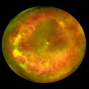 Varicella Zoster Virus Retinitis
Jan 17 2019 by Netan Choudhry, MD, FRCS(C) FASRS
Wide-field montage image of a patient with unilateral varicella-zoster virus retinitis following ingestion of animal feces in Colombia.
Photographer: Carmelina Timboli, Vitreous Retina Macula Specialists of Toronto
Imaging device: Optos California (Optos PLC, Edinburgh, UK)
Condition/keywords: retinitis, varicella zoster virus (VZV)
-
 Proliferative Diabetic Retinopathy with Pre-retinal Hemorrhage
Jan 16 2018 by Olivia Rainey
Ultra-wide field pseudo-color image of an 57-year-old male with a large pre-retinal hemorrhage secondary to proliferative diabetic retinopathy affecting his left eye.
Photographer: Olivia Rainey
Imaging device: Optos California
Condition/keywords: color fundus photograph, diabetic mellitus, hemorrhage, left eye, neovascularization (NV), Optos, proliferative diabetic retinopathy (PDR), pseudocolor, ultra-wide field imaging
-
 Varix of Vortex Vein - Optos Photo - Holding Superonasal Gaze
Jan 29 2019 by John S. King, MD
69-year-old white female found to have a "supero-nasal darkened elevation" in the left eye, during cataract evaluation. Patient was referred to Dr. Hruby, who confirmed that the elevation was a varix of the vortex vein; photos were obtained and thought to be good to put on Retina Image Bank. On the optos camera, the area of elevation was not seen in primary gaze. This is a picture of patient holding a gaze up and nasal. Varix seen supero-nasal.
Photographer: Karin Aletter
Imaging device: Optos California
Condition/keywords: vortex vein, vortex vein varix
-
 Retinitis Pigmentosa
May 26 2017 by Olivia Rainey
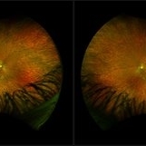
Ultra-wide-field pseudocolor image of the left eye of an 39-year-old female with Retinitis Pigmentosa. She had slightly atypical appearance due to asymmetry: sectoral atrophy in left eye, compared to 360 degree bone spicule formation in right eye. Ddx: Pigmentary degeneration vs infection vs X-linked RP carrier due to asymmetry. Recommended genetic testing through My Retina Tracker, as well as visual field and ERG testing. Patient's vision was sc20/100 PH 20/70 in the right eye and sc20/80 PH 20/40 in the left eye.
Photographer: Olivia Rainey
Imaging device: Optos California
Condition/keywords: bone spicule, fundus photograph, left eye, Optos, peripheral bone spicules, pseudocolor, retinitis pigmentosa, ultra-wide field imaging
-
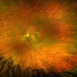
 Retinitis Pigmentosa with Asteroid Hyalosis
Jan 16 2018 by Olivia Rainey
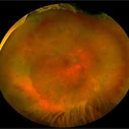
Ultra-wide field pseudocolor fundus photograph of an 75-year-old female with Retinitis Pigmentosa with Asteroid Hyalosis affecting her right eye.
Photographer: Olivia Rainey
Imaging device: Optos California
Condition/keywords: asteroid hyalosis, color fundus photograph, Optos, pseudocolor, retinitis pigmentosa, ultra-wide field imaging
-
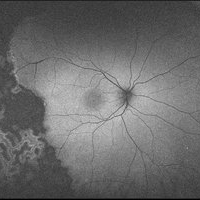
 Retinitis Pigmentosa
May 26 2017 by Olivia Rainey
Ultra-wide-field pseudocolor image of the left eye of an 39-year-old female with Retinitis Pigmentosa. She had slightly atypical appearance due to asymmetry: sectoral atrophy in left eye, compared to 360 degree bone spicule formation in right eye. Ddx: Pigmentary degeneration vs infection vs X-linked RP carrier due to asymmetry. Recommended genetic testing through My Retina Tracker, as well as visual field and ERG testing. Patient's vision was sc20/100 PH 20/70 in the right eye and sc20/80 PH 20/40 in the left eye.
Photographer: Olivia Rainey
Imaging device: Optos California
Condition/keywords: autofluorescence imaging, bone spicule, hyperautofluorescent ring, hypoautofluorescence, Optos, peripheral bone spicules, retinitis pigmentosa, ultra-wide field imaging
-
 Vascular Sheathing
Dec 19 2019 by Lauren Schuler
Ultra-wide field pseudocolor fundus photograph of a 68-year-old female with vascular sheathing affecting her right eye. This was noted on initial exam on 1/15/16, at patient's first appointment. This remains unchanged and patient is asymptomatic at this time.
Photographer: Lauren Schuler
Imaging device: Optos California
Condition/keywords: fundus photograph, ghost vessels, pseudocolor, vascular sheathing of retina
-
 Retina Dialysis Associated Retinal Detachment
Aug 22 2018 by Luis J Haddock, MD
Optos color fundus photography showing nasal macula-on retinal detachment associated to a superonasal chronic retinal dialysis. This is a 19-year-old male who presented to glaucoma for elevated IOP. On dilated fundus exam retinal detachment was noted. Extended DFE showed nasal macula-on retinal detachment associated to a nasal retinal dialysis with peripheral vitreous contraction. The patient reported remote history of BB gun injury to his left eye at 5-years-old.
Imaging device: Optos California
Condition/keywords: blunt trauma, retinal dialysis
-
 Central Retinal Artery Occlusion
May 25 2017 by Olivia Rainey
UItra-widefield fluorescein angiography, taken at 6 minutes and 22 seconds, of an 73-year-old woman with a central retinal artery occlusion in her right eye.
Photographer: Olivia Rainey
Imaging device: Optos California
Condition/keywords: central retinal artery occlusion (CRAO), fluorescein angiogram (FA), ischemia, late phase, non-perfusion, Optos, ultra-wide field imaging
-
 Pseudophakic RRD, S/P Buckle/Vit. w/ Residual Gas Fish Eggs OD
May 23 2018 by Hosam Attia, MD
71-year-old male, s/p combined buckle vitrectomy for recurrent, macula-off, rhegmatogenous retinal detachment, with residual gas fish eggs OD.
Imaging device: Optos California Ultra-Wide Field Fundus Camera
Condition/keywords: encircling scleral buckle, gas bubble, intraocular gas, intravitreal gas bubble
-
 Myopic Degeneration
Jul 3 2018 by Armando L. Oliver, MD
Myopic Degeneration
Photographer: Moises Castro
Imaging device: Optos California
Condition/keywords: pathologic myopia, posterior staphyloma
-
 Familial Exudative Vitreoretinopathy
May 16 2017 by Olivia Rainey
Ultra-wide-field autofluorescence image of an 10-year-old male with familial exudative vitreoretinopathy s/p laser.
Photographer: Olivia Rainey
Imaging device: Optos California
Condition/keywords: autofluorescence imaging, familial exudative vitreoretinopathy (FEVR), laser scarring, Optos, ultra-wide field imaging
 Loading…