Search results (436 results)
-
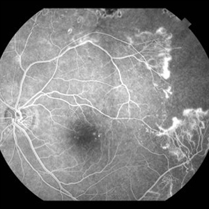 Sickle Cell Retinopathy with Sea Fans (angiography)
Aug 24 2012 by Geoffrey G. Emerson, MD, PhD, FASRS
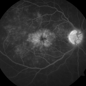
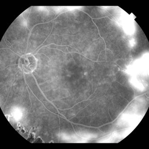
Fluorescein angiography (early/mid phase) of a 40-year-old man with African heritage and sickle SC disease. Sea fans are present temporal to the macula (leaking fluorescein).
Photographer: Geoffrey G. Emerson, MD, PhD, Retina Center, Minneapolis
Condition/keywords: sea fan, sickle cell retinopathy
-
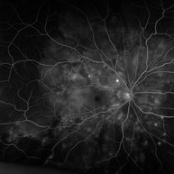 "Starry Sky" Fundus in Vogt-Koyanaki-Harada Syndrome
Jan 10 2018 by Peter H. Tang, MD, PhD
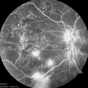
Fluorescein angiography imaging of a 27-year-old male with acute inflammation as part of Vogt-Koyanagi-Harada Syndrome.
Imaging device: Optos California
Condition/keywords: chorioretinal inflammations, retina, uveitis, Vogt-Koyanagi-Harada
-
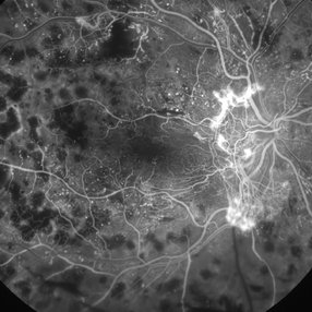 Fluorescein Angiogram of the Case with Proliferative Diabetic Retinopathy
Mar 21 2013 by Yusuke Oshima, MD, PhD
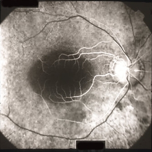
Fluorescein angiography demonstrates a prominent neovascular network at the disc with an enlarged avascular zone at the macula.
Photographer: Yusuke Takada, Osaka University Graduate School of Medicine
-
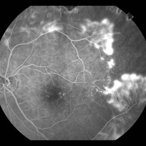 Sickle Cell Retinopathy with Sea Fans (angiogram)
Aug 24 2012 by Geoffrey G. Emerson, MD, PhD, FASRS
Fluorescein angiography (mid phase) of a 40-year-old man with African heritage and sickle SC disease. Sea fans are present temporal to the macula.
Photographer: Geoffrey Emerson, MD, PhD, Retina Center, Minneapolis
Condition/keywords: sea fan, sickle cell retinopathy
-
 CME-FFA
Apr 28 2015 by Neha Goel, MS DNB FRCS (Glasg)
Fundus fluorescein angiography of the right eye showing flower-petal appearance of the leakage.
Photographer: Neha Goel
Imaging device: Zeiss visucam
Condition/keywords: cystoid macular edema (CME)
-
 Sea Fan Neovascularisation
Apr 27 2015 by Neha Goel, MS DNB FRCS (Glasg)
Fluorescein angiography of the left eye of a 40-year-old male.
Photographer: Neha Goel
Imaging device: Zeiss visucam
Condition/keywords: Eales disease, neovascularization elsewhere (NVE), vasculitis
-
 Polypoidal Choroidal Vasculopathy-FA
Aug 27 2012 by Young Hee Yoon, MD, PhD
Fluorescein Angiography(FA) image of a 56-year-old woman. Her best-corrected visual acuity was 20/30.
Photographer: Soo Hyun Cho, Asan Medical Center
Imaging device: Heidelberg
Condition/keywords: polypoidal choroidal vasculopathy (PCV)
-
 Ocualr Ischemic Syndrome
Sep 22 2012 by Hamid Ahmadieh, MD
Wide field fluorescein angiography of a 66-year-old man with ocular ischemic syndrome due to severe stenosis of the right internal carotid artery.
Photographer: Hamid Ahmadieh, MD, Ophthalmic Research Center, Labbafinejad Medical Center, Shahid Beheshti University of Medical Sciences
Imaging device: HRA
Condition/keywords: carotid artery occlusion, ocular ischemic syndrome
-
 FFA - PDR
Mar 30 2018 by Lanin Chen
Fundus fluorescein angiography photo of the left eye of a 62-year-old woman with history of Type 2 diabetes mellitus since 20 years showing proliferative diabetic retinopathy.
Photographer: Lanin Chen
Condition/keywords: fundus autofluorescence (FAF), proliferative diabetic retinopathy (PDR)
-
 Retinitis pigmentosa AD Slide 4
Oct 22 2012 by Ronald C. Gentile, MD
Early fluorescein angiography revealed early hyper-fluorescence surrounding the fovea consistent with retinal pigment epithelial (RPE) depigmentation. This accentuated the contrast between the normal blockage (hypo-fluorescence) of the macular luteal pigment from the surrounding RPE window defect (hyper-fluorescence).
Photographer: The New York Eye & Ear Infirmary Department of Medical Imaging
Condition/keywords: retinitis pigmentosa
-
 Retinitis Pigmentosa With Hemangioma CF
Dec 15 2016 by Manish Nagpal, MD, FRCS (UK), FASRS
Fluorescein angiography OS of a patient having retinitis pigmentosa with a hemangioma inferiorly.
Condition/keywords: hemangioma, retinitis pigmentosa
-
 Ischemic Branch Retinal Vein Occlusion With Compensatory Collateral Vessels
Jul 8 2015 by Kathy Karsten, COT
Heidelberg fluorescein angiography picture of ischemic branch retinal vein occlusion with compensatory collateral vessels in 30-year-old woman.
Photographer: Kathy Karsten, COT
Imaging device: Heidelberg capturing system
Condition/keywords: ischemia
-
 Preretinal Hemorrhage due to Proliferative Diabetic Retinopathy
Oct 17 2012 by Sharon Fekrat, MD FACS FASRS
Fluorescein angiography of right eye with preretinal hemorrhage from neovascularization elsewhere associated with proliferative diabetic retinopathy. Note associated fibrosis.
Photographer: John Reaves, Ophthalmic Photographer, Durham VA Medical Center, Durham, NC
Condition/keywords: preretinal hemorrhage
-
 Sickle Cell Retinopathy with Sea Fans (angiography)
Aug 24 2012 by Geoffrey G. Emerson, MD, PhD, FASRS
Fluorescein angiography (early phase) of a 40-year-old man with African heritage and sickle SC disease. Sea fans are present temporal to the macula.
Photographer: Geoffrey Emerson, MD, PhD, Retina Center, Minneapolis
Condition/keywords: sea fan, sickle cell retinopathy
-
 Sickle Cell Retinopathy with Sea Fans (angiogram)
Aug 24 2012 by Geoffrey G. Emerson, MD, PhD, FASRS
Fluorescein angiography (late phase) of a 40-year-old man with African heritage and sickle SC disease. Sea fans are present around the macula (profusely leaking fluorescein dye).
Photographer: Geoffrey Emerson, MD, PhD, Retina Center, Minneapolis
Condition/keywords: sea fan
-
 Retinitis Pigmentosa
Oct 17 2014 by Avris Romario Diparaja Siahaan
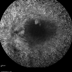
A fundus fluorescein angiography of a 25-year-old woman with retinitis pigmentosa in both of her eyes.
Photographer: Renjer Daniel Roring, Klinik Mata Nusantara
Imaging device: Heidelberg Spectralis
Condition/keywords: retinitis pigmentosa, ultra-wide field imaging
-
---thumb.jpg/image-square;max$300,300.ImageHandler) Preretinal Hemorrhage in Proliferative Diabetic Retinopathy
Preretinal Hemorrhage in Proliferative Diabetic Retinopathy
Oct 17 2012 by Sharon Fekrat, MD FACS FASRS
Fluorescein angiography of right eye with preretinal hemorrhage from neovascularization elsewhere associated with proliferative diabetic retinopathy.
Photographer: John Reaves, Ophthalmic Photographer, Durham VA Medical Center, Durham, NC
Imaging device: Fluorescein angiography
Condition/keywords: preretinal hemorrhage, subhyaloid hemorrhage
-
 Coats Disease
Apr 16 2015 by Rita Couceiro, MD, MS
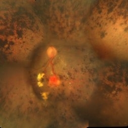
Fundus photograph and fluorescein angiography pictures of a 13-year-old girl with Coats Disease, showing abnormal telangiectatic vessels and intense exsudation in the inferior retinal periphery of the left eye.
Condition/keywords: Coats' disease, retinal telangiectasia
-
 Retinal Angiomatous Proliferation
Oct 11 2012 by Gabriela Lopezcarasa Hernandez, MD
70-year-old male with diagnostic of RAP by OCT and ICG fluorescein angiography
Photographer: Azucena Rios, Macula Retina Consultores Mexico
Imaging device: Heidelberg Spectralis
Condition/keywords: retinal angiomatous proliferation (RAP)
-
 Retinoblastoma
Sep 13 2013 by Maria Ana Martinez-Castellanos, MD
Fundus photograph, fluorescein angiography and OCT of the macula and of the tumor of a 2-years-old boy with retinoblastoma.
Photographer: Maria A. Martinez-Castellanos. Asociacion para Evitar la Ceguera en Mexico
Imaging device: RetCAm II
Condition/keywords: leakage, optical coherence tomography (OCT), pediatric tumor, retinoblastoma
-
 Juvenile X-linked Retinoschisis
Jan 11 2014 by Caesar K. Luo, MD, FASRS
RetCam fluorescein angiography of child with JXLRS.
Photographer: Caesar Luo, Progressive Vision Institute, PA
Condition/keywords: juvenile retinoschisis
-
 Fibrovascular PED
May 2 2013 by Henry J. Kaplan, MD
Fundus photograph and fluorescein angiography of a fibrovascular PED with a typical notch on F/A.
Condition/keywords: exudative age-related macular degeneration, fibrovascular pigment epithelial detachment (PED), vascularized pigment epithelial detachment (PED)
-
---thumb.jpg/image-square;max$300,300.ImageHandler) Polypoidal Choroidal Vasculopathy: Case 1 - Image 1 of 7
Polypoidal Choroidal Vasculopathy: Case 1 - Image 1 of 7
Oct 4 2012 by Gregg T. Kokame, MD, MMM, FASRS
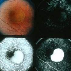
Fluorescein Angiography image of a 57-year-old woman with treatment-naive polypoidal choroidal vasculopathy. Series of images provides an comparative view of the same condition while utilizing a variet of different imaging procedures.
Photographer: Andrew Yuen, Retina Consultants of Hawaii
Imaging device: Heidelberg Spectralis
Condition/keywords: fluorescein leakage, polypoidal choroidal vasculopathy (PCV)
-
---thumb.jpg/image-square;max$300,300.ImageHandler) Ocular Histoplasmosis Syndrome (OHS)
Ocular Histoplasmosis Syndrome (OHS)
Oct 8 2013 by Maurice F. Rabb
Thirty six year old white male stated that approximately 5 years earlier he had a blurry spot in his left eye that went away spontaneously after 3 months. Three years later the spot returned. He was seen by a local ophthalmologist who noted two "histo spots" in the left eye. Over the next 6 months his vision deteriorated from 20/30 to 20/200 in the left eye. A week prior to being seen at the UIHC he noted bulginess in his right eye. Visual acuity without correction was 20/15 OD, 20/200 OS. Color fundus photography and fluorescein angiography were performed and the patient was treated with argon laser photocoagulation. Vision decreased to 20/30 following laser surgery, but within two weeks returned to 20/15 and remained that way over the next two years. OVer the following 15 years the patient did well although he developed a recurrence in the untreated left eye and periodically he experienced vague changes in his central field.
Condition/keywords: ocular histoplasmosis syndrome (OHS)
-
 Retinitis pigmentosa AD Slide 5
Oct 22 2012 by Ronald C. Gentile, MD
Late fluorescein angiography revealed some fading of the early hyper-fluorescence surrounding the fovea consistent with retinal pigment epithelial depigmentation without any late leakage or CME.
Photographer: The New York Eye & Ear Infirmary Department of Medical Imaging
Condition/keywords: retinitis pigmentosa
 Loading…