Search results (1033 results)
-
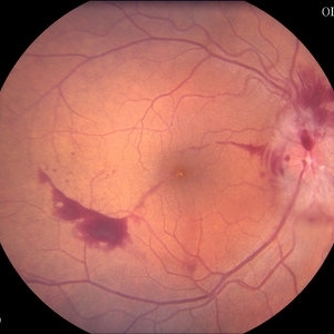
 PDR with Traction RD of Macular
Oct 8 2012 by Jeffrey G. Gross, MD, FASRS
PDR with traction RD of macular, post-op, 20/40.
Condition/keywords: 20/40, macular, post-op, tractional retinal detachment
-
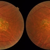 Central Areolar Choroidal Dystrophy
Jul 7 2015 by Hamid Ahmadieh, MD
Color fundus photograph of both eyes of a 58-year-old man with progressive loss of vision. VA OD is 20/60 and VA OS is 20/400.
Photographer: Soulmaz Shahmohammad, Negah Eye Center, Tehran, Iran
Imaging device: Topcon
Condition/keywords: central areolar choroidal dystrophy (CACD), color fundus photograph
-
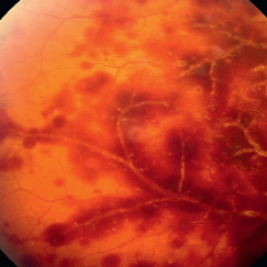 Acute Retinal Periphlebitis and Panuveitis OD
Jul 16 2014 by Deepayan Kar
Hemorrhagic retinopathy + frosted retinal angitis OD. Exudative sheathing of the major retinal blood vessels Px 25-year-old woman (VA-OD 20/40/ OS 20/25): recovering from URTI. AC ++ Vit Cells +.
Photographer: Deepayan Kar
Condition/keywords: exudative sheathing, frosted branch angiitis, hemorrhage, retinopathy
-
 Macular Pseudohole
Jul 7 2015 by Hamid Ahmadieh, MD
Color fundus photograph and optical coherence tomography of the left eye of a 72-year-old woman with blurred vision due to epiretinal membrane. VA OS is 20/40 . Macular pseudohole is visible.
Photographer: Shabnam Poureh, Negah Eye Center, Tehran, Iran
Imaging device: Topcn
Condition/keywords: color fundus photograph, macular pseudohole, optical coherence tomography (OCT)
-
 Gonioscopy; Scattered Peripheral Anterior Synechiae
Jul 8 2013 by Jason S. Calhoun
Patient came in for evaluation for glaucoma. Patient also has a history of uveitis. Last flare up was back in 1990. Patient's VA was 20/30, Right eye and 20/40-1, Left eye. Slit Lamp Gonioscopy reveals iris bow with scattered PAS around the angles of the anterior chamber. You can also see pigmentation in the trabecular meshwork. Patient will follow up in 3 months.
Photographer: Jason S. Calhoun, Department of Ophthalmology, Mayo Clinic Jacksonville, Florida
Condition/keywords: gonioscopy, goniosynechiae
-
 Retinitis Pigmentosa
May 26 2017 by Olivia Rainey
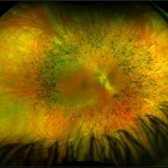
Ultra-wide-field pseudocolor image of the right eye of an 39-year-old female with Retinitis Pigmentosa. She had slightly atypical appearance due to asymmetry: sectoral atrophy in left eye, compared to 360 degree bone spicule formation in right eye. Ddx: Pigmentary degeneration vs infection vs X-linked RP carrier due to asymmetry. Recommended genetic testing through My Retina Tracker, as well as visual field and ERG testing. Patient's vision was sc20/100 PH 20/70 in the right eye and sc20/80 PH 20/40 in the left.
Photographer: Olivia Rainey
Imaging device: Optos California
Condition/keywords: bone spicule, fundus photograph, Optos, peripheral bone spicules, pseudocolor, retinitis pigmentosa, ultra-wide field imaging
-
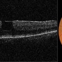
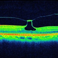
 Severe vitreomacular traction
Dec 23 2012 by Alex P. Hunyor, MD
OCT scan of the right eye of an 82-year-old male with 20/40 vision despite severe vitreomacular traction (VMT).
Condition/keywords: vitreomacular traction (VMT)
-
 Optos Picture With Speculum: Dislocated Natural Lens
Oct 9 2018 by John S. King, MD
55-year-old white female with history of pathologic myopia+, lattice (laser), SB OU (1990s), and dislocated natural lenses OU that had been watched for years. In the fellow eye she developed phacolytic glaucoma and a PPV, PPL was performed. Plan for both eyes are monitoring. I wanted to get a good picture of her lens today with the optos machine, as the other pics had artifact from the lower lid. It worked out well to use a speculum in the left eye. Vision cc is 20/400 J1+ OD and 20/40 J2 OS; aphakic OU; vitreous clear OD; dislocated lens OS (see pic); retinas attached.
Photographer: Maisee Yang
Imaging device: Optos California
Condition/keywords: dislocated crystalline lens, pathologic myopia, scleral buckle, staphyloma
-
 Bilateral Macular Star
Mar 27 2014 by Jason S. Calhoun
Young female patient in with blurred vision in both eyes. VA is 20/40 in both eyes. Fundus photos show visible macular star centrally in both eyes. This is a result of Bilateral Neuroretinitis due to cat scratch.
Photographer: Jason S. Calhoun, Mayo Clinic Jacksonville, Department of Ophthalmology
Imaging device: TOPCON TRC 50-EX
Condition/keywords: macular star, neuroretinitis
-
 Macular Hole
Sep 20 2012 by Jeffrey G. Gross, MD, FASRS
Macular hole, Stage 3, pre-op 20/400
Condition/keywords: macular hole, pre-op
-
---thumb.jpg/image-square;max$300,300.ImageHandler) "Spots" In The Central Visual Zone
"Spots" In The Central Visual Zone
Oct 14 2013 by Maurice F. Rabb
A 26 year old healthy female who had been aware of decreased vision in OS for 5 days before the initial examination. When questioned specifically about OD, she did admit to being aware of some "spots" in the central visual zone. Her past ocular history is negative for eye disease and the family history is negative for retinal and macular disease. The patient is in excellent general health. She had a recent upper respiratory infection and is presently disabled because of a herniated disc. Uncorrected vision OD is 20/20 and OS is 20/400, improving to 20/100- with pinhole. The findings of significance are noted in the posterior poles.
Condition/keywords: spots in the central visual zone
-
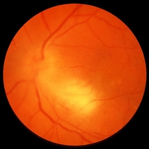
 Sarcoid Granuloma of Optic Nerve
Oct 9 2012 by Jeffrey G. Gross, MD, FASRS
Sarcoid granuloma of optic nerve, 20/400.
Condition/keywords: 20/400, autoimmunity, sarcoid granuloma, sarcoidosis
-
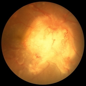
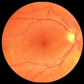
 Venous Stasis Retinopathy
Feb 20 2015 by H. Michael Lambert, MD
Scattered hemorrhages associated with venous stasis retinopathy in eye with ipsilateral total internal carotid artery occlusion. Vision is 20/40.
Condition/keywords: arterial occlusion, venous stasis retinopathy
-
---thumb.jpg/image-square;max$300,300.ImageHandler) "Spots" In The Central Visual Zone
"Spots" In The Central Visual Zone
Oct 14 2013 by Maurice F. Rabb
A 26 year old healthy female who had been aware of decreased vision in OS for 5 days before the initial examination. When questioned specifically about OD, she did admit to being aware of some "spots" in the central visual zone. Her past ocular history is negative for eye disease and the family history is negative for retinal and macular disease. The patient is in excellent general health. She had a recent upper respiratory infection and is presently disabled because of a herniated disc. Uncorrected vision OD is 20/20 and OS is 20/400, improving to 20/100- with pinhole. The findings of significance are noted in the posterior poles.
Condition/keywords: spots in the central visual zone
-
 Severe vitreomacular traction
Dec 23 2012 by Alex P. Hunyor, MD
Colour fundus image of the right eye of an 82-year-old male with 20/40 vision despite severe vitreomacular traction (VMT).
Condition/keywords: vitreomacular traction (VMT)
-
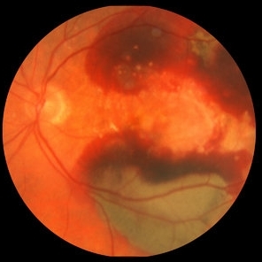 ARMD with Subretinal Hemorrhages and Macular Scarring
Oct 16 2012 by Jeffrey G. Gross, MD, FASRS
ARMD with subretinal hemorrhages and macular scarring, 20/400.
Condition/keywords: 20/400, macular scar, subretinal hemorrhage
-
 Macular Pucker Post-Op
Oct 8 2012 by Jeffrey G. Gross, MD, FASRS
Macular pucker, post-op, 20/40.
Condition/keywords: 20/40, macular pucker, post-op
-
 VMT, Vitreo Macular Traction Syndrome, SD OCT
Apr 18 2013 by James B. Soque, CRA, OCT-C, COA, FOPS
79-year-old white female, VA sc 20/40, VMT OS, diagnosed on exam and SD OCT. Accompanying FC, RF, and FA photos also reveal a BRVO OS.
Photographer: James B. Soque, CRA, COA, Island Retina, Shirley, NY
Imaging device: HD 5 Line Scan, Zeiss Cirrus SD OCT
Condition/keywords: branch retinal vein occlusion (BRVO), vitreomacular traction (VMT)
-
 Macular Degeneration with Large Submacular Hemorrhage
Oct 10 2012 by Joseph M. Civantos, MD
Acute drop in vision to 20/400 with large hemorrhage.
Condition/keywords: submacular hemorrhage
-
 Toxoplasmosis Chorioretinitis
Oct 10 2012 by Jeffrey G. Gross, MD, FASRS
Toxoplasmosis chorioretinitis, 20/400, + APD.
Condition/keywords: 20/400, afferent pupillary defect (APD), toxoplasmosis chorioretinitis
-
 Macular Hole Post-Op
Sep 27 2012 by Jeffrey G. Gross, MD, FASRS
Macular hole post-op, hole closed 20/40.
Condition/keywords: 20/40, hole closed, macular hole, post-op
-
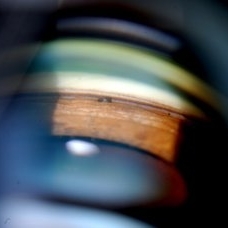
---thumb.JPG/image-square;max$300,300.ImageHandler) Rubeosis Iridis
Rubeosis Iridis
Jul 8 2013 by Jason S. Calhoun
Patient presents with rubeosis iridis in the right eye due to neovascular glaucoma. VA is 20/40 in the right eye. Will follow up in 3 months.
Photographer: Jason S. Calhoun, Department of Ophthalmology, Mayo Clinic Jacksonville, Florida
-
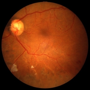
 Diabetic Papillitis
Jun 28 2013 by Jason S. Calhoun
Patient was about to undergo surgery for CNS aneurysm. Patient woke up with little spots which never cleared up. Patients VA was 20/30-OD and 20/40-OS. Both eyes appeared to have disc edema with hemorrhages in the right eye. Ordered a CT of the brain to make sure the aneurysm didn't ruptured.
Photographer: Jason S. Calhoun, Mayo Clinic Jacksonville, Florida
Imaging device: TOPCON TRC 50-EX
Condition/keywords: diabetic mellitus
 Loading…