Search results (1033 results)
-
 Sialidosis
Jul 10 2025 by Jessilla Phou
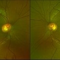
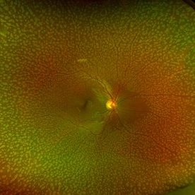
These are fundus photographs capturing an 18 year old male with Type 1 Sialidosis, a rare inherited lysosomal storage disorder caused by a deficiency in the neuraminidase 1 (Neu1) enzyme. Currently, there are fewer than 1,000 people in the USA who have this disorder. It is characterized by a cherry red spot in the macula which occurs when lipids accumulate in the retinal ganglion cells. This causes the macula to appear red as seen in these fundus images. The patient presented at our office with ataxia, depth perception issues, and slow reaction time. His visual acuity was 20/40, suggestive of early stage Sialidosis.
Photographer: Jessilla Phou
Imaging device: Optos California
Condition/keywords: cherry red spot, fundus photograph, Sialidosis
-
 LCA Type 2
Apr 10 2025 by Joshua Friedman
LCA Type 2 (RPE65) showing characteristic hypoautofluorescence and retinal thinning. 8F with best corrected visual acuity of 20/400 (OD) and 20/150 (OS). Small white intraretinal spots and RPE mottling are visible on color fundus photography. Blue light autofluorescence reveals near-complete loss of signal, while OCT demonstrates widespread outer retinal thinning.
Photographer: Stephen Tsang, MD, PhD
Condition/keywords: Leber Congenital Amaurosis
-
 Toxic Maculopathy (Elmiron)
Apr 9 2025 by Virginia Gebhart
79 year old male with toxic maculopathy from long term use of Elmiron (15+ yrs.) On exam there is stippled RPE changes, pigment clumping, and subretinal deposits. BCVA 20/100 | 20/40.
Photographer: Virginia Gebhart, Retina Consultants of Carolina
Imaging device: Optos California
Condition/keywords: autofluorescence imaging, cystoid macular degeneration, Elmiron Toxicity, Toxic Maculopathy
-
 Emulsified Silicone Oil
Apr 3 2025 by Andrew A. Moshfeghi, MD, MBA, FASRS
This is an 87 year- old male with 3.5 year history of retained silicone oil following treatment of late-onset recurrent retinal detachment 18 years following prior primary scleral buckle repair. Robust emulsified silicone oil aggregates are appreciated. Visual acuity is 20/400.
Photographer: Tammy Schoenholz, University of Southern California.
Imaging device: Zeiss Clarus
Condition/keywords: emulsified silicone oil
-
 Solar Retinopathy
Apr 1 2025 by Isaac Agranoff
OCT scan of 18-year-old male presenting with 20/40 BCVA OU and bilateral focal outer retinal subfoveal defects. Patient reported long-term history of frequent sungazing, has stopped within past 6-9 months.
Photographer: Isaac Agranoff
Imaging device: Heidelberg Spectralis
Condition/keywords: solar retinopathy
-
 Iris Discoloration
Apr 1 2025 by Korey Starkey
4 year-old patient sent for genetic testing to rule out possibility of Waardenburg syndrome with Hirschsprung disease. Left eye iris has no discoloration present, vision in both eyes is 20/40.
Photographer: Korey Starkey
Imaging device: Topcon
Condition/keywords: external, external photography, iris, topcon
-
 Ozurdex in AC
Apr 1 2025 by Korey Starkey
90-year-old patient with an Ozurdex implant that migrated into the AC and with the cornea decompensating. Patient recommended for urgent surgery to remove implant. Vision OD at this visit was CF @ 2ft, most recent visit vision is 20/400, PH 20/25.
Photographer: Korey Starkey
Imaging device: Topcon
Condition/keywords: anterior chamber, corneal decompensation, external, external photography, Ozurdex implant, Topcon
-
 Elmiron Toxicity
Mar 25 2025 by Toolie Winters
Fundus autofluorescence image of a 69-year-old woman with toxic maculopathy OU due to Elmiron usage. Patient stopped using Elmiron in the late 2010s after having been on it for 17 years. The patient has areas of outer retinal and RPE atrophy temporal to fovea that have expanded compared to photos from two years ago. At the time of this appointment, her VA OD was sc20/40-1+2 PH20/30 and VA OS was scCF @ 1 foot.
Photographer: Toolie Winters
Imaging device: Heidelberg Spectralis
Condition/keywords: Elmiron Toxicity, FAF, fundus autofluorescence (FAF), Heidelburg Spectralis, Pentosan Toxicity, Toxic Maculopathy
-
 Proliferative Sickle Cell Retinopathy
Jan 27 2025 by Virginia Gebhart
61 year-old with proliferative sickle cell retinopathy s/p cryotherapy to peripheral fibrotic NV. Eye is stable with resolving exudates and maturing cryo scar. BCVA 20/40
Photographer: Virginia Gebhart, Retina Consultants of Carolina
Imaging device: Optos California
Condition/keywords: cryotherapy, fibrotic neovascularization, sickle cell retinopathy
-
 Choroidal Melanoma 3 Ways
Jan 16 2025 by Virginia Gebhart
RGB/FA/ICG of 76 year old female with a new choroidal melanoma. Pt scheduled for plaque radiation. BCVA 20/400
Photographer: Virginia Gebhart, Retina Consultants of Carolina
Imaging device: Optos California
Condition/keywords: fluorescein angiogram (FA), indocyanine green (ICG) angiography, OPTOS CALIFORNIA RGB
-
 Combined Traction Rhegmatogenous Detachment
Oct 17 2024 by Hemanth Murthy, MBBS, MD, FASRS
A 68 year old male presented with a shadow in the left eye since 3 days. He was a known diabetic and hypertensive for 20 years. Vision was 20/40 in right eye and 20/60 in left eye. Fundus examination showed Proliferative diabetic retinopathy in right eye and Proliferative diabetic retinopathy with combined traction rhegmatogenous detachment in left eye.
Photographer: Mr Veda Vyas
Condition/keywords: combined retinal detachment, proliferative diabetic retinopathy (PDR)
-
 Retinitis Pigmentosa
Oct 16 2024 by Virginia Gebhart
74 year old female with bone spicule pigmentation associated with Retinitis Pigmentosa. Pt diagnosed at age 53, relatively asymptomatic prior to diagnosis. Pt reports gradual vision loss over 10+ years. BCVA 20/40
Photographer: Virginia Gebhart, Retina Consultants of Carolina
Imaging device: Optos California
Condition/keywords: bone spicule, retinitis pigmentosa, retinitis pigmentosa (RP) dystrophy
-
 Macula On Retinal Detachment
Jul 5 2024 by Zach Seim
This is an Optos fundus photo of a 67 year old female with a Macula On Retinal Detachment. Patient presented with VA DCC 20/40-1.
Photographer: Zach Seim
Imaging device: Optos California
Condition/keywords: macula on, Optos, OPTOS CALIFORNIA, right eye
-
 Geographic Atrophy
Apr 22 2024 by Angela Rico
59 year-old female with MM1 Mitochondrial Genetic Defect. V/A- OD: 20/25, OS:20/40
Photographer: Angela Rico M.D.
Condition/keywords: Dystrophy of the Retinal Pigment Epithelium
-
 Subretinal Gas After Pneumatic Retinopexy
Mar 6 2024 by James P Dossett, MD
Pseudocolor fundus photograph of a 68-year-old man who presented with a macula-on rhegmatogenous retinal detachment with a single horseshoe tear at 12 o'clock. Pneumatic retinopexy was performed with cryopexy to the retinal break. He returned to clinic three days later and the entire SF6 gas bubble was noted to have migrated to the subretinal space through the retinal break. Pars plana vitrectomy was performed that day with retinal reattachment and improvement in vision to 20/40 now 6 months postoperatively.
Imaging device: Optos
Condition/keywords: pneumatic retinopexy, subretinal gas bubble
-
 Dry AMD
Jan 25 2024 by Virginia Gebhart
79 year old female with intermediate dry AMD. Small area of geographic atrophy superior, large drusen and stippled RPE changes. BCVA 20/40
Photographer: Virginia Gebhart
Imaging device: Topcon
Condition/keywords: age-related macular degeneration (AMD), dry age-related macular degeneration (dry AMD), geographic atrophy
-
 Scalloped Choroidal Atrophy
Jan 8 2024 by Zach Seim
An ultra-widefield fluorescein angiogram of a 90 year old female with Scalloped Choroidal Atrophy affecting both eyes. Patient's vision at the time of the image was Dcc 20/40 OD. Genetic test pending.
Photographer: Zach Seim
Imaging device: OPTOS California
Condition/keywords: atrophy, choroidal atrophy, fluorescein angiogram (FA), Fluorescein angiography, optic nerve, OPTOS CALIFORNIA, retina, right eye, ultra-wide field imaging
-
 Layer Cake; Sub-retinal, Pre-retinal, Vitreous Hemorrhages
Dec 5 2023 by Virginia Gebhart
73 year old female with sub-retinal, pre-retinal, and vitreous hemorrhages all in OD. Will consider sx if blood does not clear on its own. Vision 20/40
Photographer: Virginia Gebhart
Imaging device: Topcon
Condition/keywords: pre-retinal hemorrhage, retinal macroaneurysm, subretinal hemorrhage, subretinal blood, vitreous hemorrhage
-
 Severe NPDR
Oct 24 2023 by Virginia Gebhart
Fluorescein angiogram of left eye in 60-year-old male with severe non-proliferative diabetic retinopathy with extensive macular edema. Most recent A1c is 11. Vision 20/400. Injection of Eylea given
Photographer: Virginia Gebhart
Imaging device: Topcon
Condition/keywords: diabetic macular edema, Diabetic Retinopathy, fluorescein angiogram (FA), Fluorescein angiography
-
 Congenital Retinal Macrovessel
Oct 13 2023 by Jacob D. Grodsky, MD
41 y/o male who presented with acute onset of blurred vision OD. Visual acuity was 20/200 OD; 20/25 OS. Examination was consistent with congenital retinal macrovessel through the macula with intraretinal hemorrhage as seen in the fundus photo. Intravitreal bevacizumab was injected, and visual acuity improved to 20/40 at 4-week follow-up. MRA head and neck was ordered to rule out other vascular anomalies.
Condition/keywords: congenital retinal macrovessel, RETINAL MACROVESSEL
-
 Chorioretinitis with Overlying Vitreous Stranding/Vitritis
Mar 23 2023 by Isaac Agranoff
Fundus photograph of a 37-year-old woman presenting with chorioretinitis with overlying vitreous stranding/vitritis that has remained unchanged for multiple years. Patient presented with irritation and blurred vision and her vision was 20/40 OD. The OCT revealed evidence of low-grade inflammation and the recommend treatment was anti-inflammatory eye drops at this time and to obtain second opinion with another physician in the office.
Photographer: Isaac Agranoff, Technician
Imaging device: Optos California
Condition/keywords: chorioretinal scar, chorioretinitis, inflammation, Optos, ultra-wide field imaging, vitritis
-
 Benign familial Fleck Retina-left eye
Feb 2 2023 by Hemanth Murthy, MBBS, MD, FASRS
12 year boy first born of consanguineous marriage, came for routine eye check up with BCVA 20/40 OU. He has no night blindness. His OCT showed thickening of the RPE with dome like elevations involving the ellipsoid layer. Dark adapted ERG showed normal 'b' wavesPhotopic ERG showed reduced 'a' and b waves.
Photographer: Veda Vyas
Imaging device: Optos Daytona
Condition/keywords: Benign familial Fleck Retina
-
 Benign Familial Fleck Retina
Feb 2 2023 by Hemanth Murthy, MBBS, MD, FASRS
12 year boy first born of consanguineous marriage, came for routine eye check up with BCVA 20/40 OU. He has no night blindness. His OCT showed thickening of the RPE with dome like elevations involving the ellipsoid layer. Dark adapted ERG showed normal 'b' wavesPhotopic ERG showed reduced 'a' and b waves.
Photographer: Veda Vyas
Imaging device: Optos Daytona
Condition/keywords: Benign familial fleck retina
-
 Subretinal Hemorrhage with Chorioretinal Macular Scars
Sep 28 2022 by Denica Rodriguez
Ultra-widefield pseudocolor fundus photograph of a 59 year old female with Subretinal Hemorrhage with Chorioretinal Macular Scars affecting her left eye. The physician presumes the etiology is CNV from adjacent scarring/choroidal rupture. Patient has history of ocular trauma with cricket ball at age 10-12 years old. She suspects that she might have suffered choroidal rupture, which has resulted in secondary CNV and hemorrhage that we are seeing today. She recommends treatment with Eylea sample injection in a series of 4 at a 4-5 week interval. The patient's vision at the time of her appointment was Dcc20/40-2 PHNI.
Photographer: Denica Rodriguez, COA
Imaging device: Optos California
Condition/keywords: antiVEGF therapy, chorioretinal scar, choroidal neovascular membrane (CNVM), fundus photography, left eye, macular scar, Optos, peripheral drusen, pseudocolor, secondary CNV, subretinal hemorrhage, ULTRA WIDE FIELD, ultra-wide field imaging
-
 Methotrexate Bubble following Intravitreal Injection for PVR
Sep 21 2022 by Zach Seim
Ultra-widefield fundus photograph of an 81 year old female with a Methotrexate bubble following an Intravitreal Injection for Proliferative Vitreoretinopathy. Patient has been presenting to the office for two week interval Methotrexate injections in her left eye. The image was taken prior to her eighth injection which revealed a residual Methotrexate bubble in her inferior retinal image. This patient was seeing "lots" of floaters, as well as having visual acuity of cc20/400 cc20/200 PH.
Photographer: Zach Seim
Imaging device: OPTOS California
Condition/keywords: bubble, fundus photograph, fundus photography, intravitreal injection, left eye, methotrexate, nasal retina, Optos, proliferative vitreoretinopathy (PVR), pseudocolor, ultra-wide field imaging
 Loading…