Search results (81 results)
-
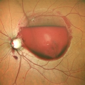
 Valsalva Retinopathy
Jan 26 2017 by JEFFERSON R SOUSA, Tecg.º (Biomedical Systems Technology)
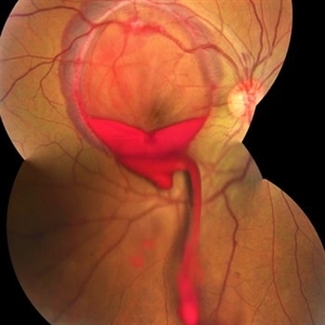
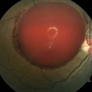
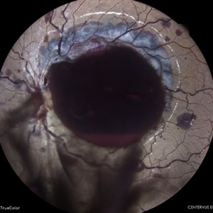
Male patient, 23-years-old, with low visual acuity in the right eye. In the ocular examination of the retinography, intense subhyaloidal hemorrhage. 2 minutes after laser application.
Photographer: JEFFERSON R SOUSA - Suel Abujamra Institute - São Paulo - Brazil
Imaging device: Topcon TRC-50 DX, Imaginet, 35 degree field. Flash 36 / Mosaic with four images.
Condition/keywords: subhyaloid hemorrhage, valsalva retinopathy
-
 Valsalva Retinopathy
Dec 20 2021 by Unnati Vishwanath Shukla, M. S. ,DNB, FVRS FNERF, MNAMS,PhD Scholar(Retina)
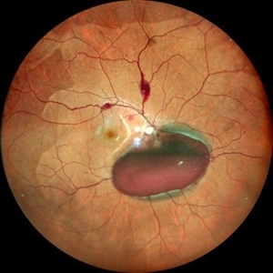
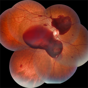
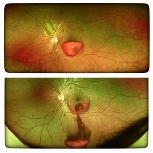
26-year-old male with Valsalva Retinopathy. History of severe cough for 3 days. All hematological investigations were within normal limits.
Photographer: Dr. Unnati Shukla, Consultant, Retina Foundation, Ahmedabad
Imaging device: Nidek Mirante
Condition/keywords: subhyaloid hemorrhage, subretinal hemorrhage, valsalva retinopathy
-
 Valsalva Retinopathy
Nov 18 2022 by Niloofar Piri, MD
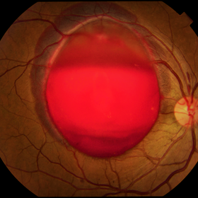
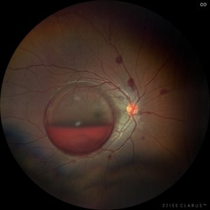
Sudden vision loss immediately after severe vomiting. Color fundus photo demonstrates large sub ILM hemorrhage consistent with valsalva retinopathy.
Photographer: Sean Kelso, Saint Louis University
Condition/keywords: SUB ILM hemorrhage, sub internal limiting membrane haemorrhage, valsalva retinopathy
-
 Preretinal Hemorrhage
May 6 2017 by Mitzy E Torres Soriano, MD
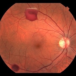
Fundus photograph of a 36-year-old-woman with a preretinal subhyaloid hemorrhage (valsalva retinopathy).
Photographer: Mitzy Torres Soriano
Condition/keywords: macular hemorrhage, premacular hemorrhage, preretinal hemorrhage, subhyaloid hemorrhage, valsalva retinopathy
-
 Valsalva Retinopathy
Apr 5 2018 by Mohamed Tawfik, MD
Fundus photography of 30-year-old male presented with sudden drop of vision .. diagnosed as valsalva retinopathy .. we do YAG hylodotomy ... vision is improved from HM to 1.0.
Photographer: Mohamed A.Tawfik MD,FRCSed
Condition/keywords: valsalva retinopathy
-
 Valsalva retinopathy progression
Oct 4 2023 by Niloofar Piri, MD
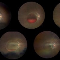
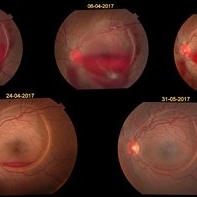
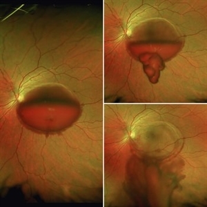
Progression fundus photograph images of a 22 yo female with Valsalva retinopathy secondary to violent emesis. Note the sub ILM layered hemorrhage and gradual decrease then disappearance over time. Last image is at 6 month follow up with 20/20 vision. Of note the silhouette of ILM separation is still visible.
Condition/keywords: valsalva retinopathy
-
---thumb.jpg/image-square;max$300,300.ImageHandler) Primary Subhyaloid Hemorrhage Due to Valsalva Retinopathy
Primary Subhyaloid Hemorrhage Due to Valsalva Retinopathy
Nov 13 2013 by Hamid Ahmadieh, MD
Early venous phase angiogram of the left eye of a 25-year-old man with primary subhyaloid hemorrhage due to Valsalva retinopathy.
Photographer: Nayereh Hadipour, Negah Eye Center, Tehran
Condition/keywords: subhyaloid hemorrhage, valsalva retinopathy
-
 Retinopathy Valsalva
Jan 26 2017 by JEFFERSON R SOUSA, Tecg.º (Biomedical Systems Technology)
Male patient, 23-year-old, with low visual acuity in the right eye. In the ocular examination of the retinography, intense sub hyaloidal hemorrhage.
Photographer: JEFFERSON R SOUSA
Imaging device: Retinografo Topcon TRC-50 DX, Imaginet, campo de 35 graus. Flash 36
Condition/keywords: valsalva retinopathy
-
 Subhyaloid Hemorrhage
Apr 29 2015 by Neha Goel, MS DNB FRCS (Glasg)
Fundus photograph of the right eye of a 25-year-old male with complaints of loss of vision following a bout of coughing.
Photographer: Neha Goel
Imaging device: Zeiss visucam
Condition/keywords: subhyaloid hemorrhage, valsalva retinopathy
-
 Subhyaloid Hemorrhage - Post Hyaloidotomy
Apr 29 2015 by Neha Goel, MS DNB FRCS (Glasg)
Fundus photograph immediately following Nd:YAG laser hyaloidotomy.
Photographer: Neha Goel
Imaging device: Zeiss visucam
Condition/keywords: hyaloidotomy, subhyaloid hemorrhage, valsalva retinopathy
-
 Valsalva Retinopathy
Nov 18 2022 by Niloofar Piri, MD
21 yo female presented with decaresed central vision and scotoma immediately after severe vomiting. Color fundus phtograph demonstrates large sub ILM layered hemorrhage in the macula consistent with valsalva retinopathy. Notice the sacttered blot retinal hemorrhages in mid-periphery.
Photographer: Rocio Bentivegna, MD, Saint Louis University
Condition/keywords: sub ILM hemorrhage, valsalva retinopathy
-
 valsalva retinopathy
Feb 6 2018 by JEFFERSON R SOUSA, Tecg.º (Biomedical Systems Technology)
A 28-year-old female patient, at the 28th week of gestation, presented low vision. Retinal mapping and retinography examination revealed extensive subhaloidal haemorrhage suggestive of RETINOPATHY VALSALVA in pregnancy. In periodic follow-up, spontaneous reabsorption of the hemorrhage was observed.
Photographer: JEFFERSON R SOUSA - Study Center and Ophthalmological Research Dr. Andre M V Gomes, Institute Dr. Suel Abujamra São Paulo-Brazil
Imaging device: Fundus camera Topcon TRC-50 DX, Imaginet, 50 degree field. Flash 50
Condition/keywords: valsalva retinopathy
-
 Valsalva Retinopathy
Dec 28 2020 by Lucas Zago Ribeiro, MD
Fundus image of 34-year-old man with Valsalva mimicking Terson's Syndrome.
Photographer: Lucas Zago Ribeiro, UNIFESP / EPM
Imaging device: Visucam 524
Condition/keywords: valsalva retinopathy
-
 VALSALVA RETINOPATHY
Jun 6 2023 by Akansha Sharma
COLOUR FUNDUS PHOTOGRAPH OF A 21 YEAR OLD MALE WITH VALSALVA RETINOPATHY
Photographer: Dr. Urmil Shah, Dr. Denish Patel, Dr. Akansha Sharma, Bharati Eye Clinic, Ahmedabad, Gujarat
Condition/keywords: SUB RETINAL HEMORRHAGE, valsalva retinopathy
-
 Valsalva retinopathy 2
Jan 11 2013 by Alex P. Hunyor, MD
Valsalva retinopathy, right eye - massive preretinal haemorrhage.
Condition/keywords: valsalva retinopathy
-
 YAG Laser Hyaloidotomy
Aug 31 2025 by Giriraj Vibhute
A 24-year-old young man presented with sudden loss of vision in left eye following history of rigorous coughing. Visual acuity in RE was 6/6, LE was 6/60p. Fundoscopy showed bilateral multiple small intraretinal hemorrhages with LE large premacular subhyaloid hemorrhage just covering the fovea suggestive of bilateral valsalva retinopathy changes. Nd:YAG laser hyaloidotomy was performed to left eye the same day (A250; 2mJ;6 SHOTS). Visual acuity improved to 6/9 immediately following the procedure. After 1 week, the subhyaloid hemorrhage had completely cleared with dispersed intragel hemorrhage in the inferior vitreous cavity with visual acuity of 6/6 in left eye
Photographer: Dr Vani S. MM Joshi eye institute, Hubli
Condition/keywords: valsalva retinopathy, YAG HYALOIDOTOMY
-
 Valsalva Retinopathy
Feb 23 2021 by RAFAEL REIS PEREIRA, MD
Valsalva retinopathy is a specific form of retinopathy characterized by pre-retinal hemorrhages secondary to raised intrathoracic pressure. This is a 31-year-old female who had breast implant surgery and complained of low VA in her left eye since the procedure. The patient had a large subhyaloid hemorrhage and we performed Nd YAG laser restoring 20/20 vision in the 4th-day post-treatment.
Condition/keywords: retina, valsalva retinopathy
-
 A Motor Vehicle Accident Causing Valsalva Retinopathy OD, While Racing A Side By Side 4 Wheel Off-Road Vehicle
Apr 29 2020 by John S. King, MD
43-year-old white male who was injured while racing a side by side 4-wheel off-road vehicle (see Video: https://imagebank.asrs.org/file/53854/sxs-crash-during-a-race-causing-valsalva-retinopathy-od). He presented about three weeks after the injury. He was being seen by his local eye doctor who wanted an evaluation for the retinal heme and scotoma. His main complaint was a central/parcentral scotoma described as a greyish area in vision. Va 20/50 OD, nomotensive, no APD (by technician), anterior segment u/r; see picture for the fundus exam - of note there are superficial/preretinal heme, with layering of the heme superiorly, and small superficial heme at nasal edge of the optic disc; in the parafoveal region nasally there is some mottling of the RPE that may indicate an area of prior commotio retinae (also possible to have TON), which may account for his scotoma. Really bad accident (video), and amazingly, he had no LOC or injuries other than the right retina. Helmet and racing harness seat belt were used.
Photographer: Asli Ahmed
Imaging device: Topcon 50
Condition/keywords: valsalva retinopathy
-
 Subhyaloid Hemorrhage, Right Eye
Jan 28 2015 by Kathy Karsten, COT
Fundus photograph of 38-year-old woman who presented with sudden onset of flashes and a large black floater obscuring vision in the right eye. Recent history of severe coughing and emesis. Diagnosed with subhyaloid hemorrhage and valsalva retinopathy OD.
Photographer: Kathy Karsten, COT
Imaging device: Topcon TRC-50DX
Condition/keywords: subhyaloid hemorrhage
-
 Valsalva Retinopathy
Apr 6 2018 by Jun Dong Dong
A 45-year-old male with no definite history of activities which can suddenly increase the intrathoracic pressure refused the laser treatment and chose the observation.
Photographer: Jun Dong, Shanghai Aier Eye Hospital
Condition/keywords: subhyaloid hemorrhage
-
 A Motor Vehicle Accident Causing Valsalva Retinopathy OD, While Racing A Side By Side 4 Wheel Off-Road Vehicle
May 5 2020 by John S. King, MD
A 43-year-old white male who was injured while racing his side by side 4 wheel off-road vehicle (this is a video he showed me on his phone). He presented about three weeks after the injury. He was being seen by his local eye doctor who wanted an evaluation for the retinal heme and scotoma. His main complaint was a central/parcentral scotoma described as a greyish area in vision. Va 20/50 OD, nomotensive, no APD (by technician), anterior segment u/r; see {https://imagebank.asrs.org/file/53828/sxs-crash-during-a-race-causing-valsalva-retinopathy-od} for the fundus exam - of note there are superficial/preretinal heme, with layering of the heme superiorly; in the parafoveal region nasally there is some mottling of the RPE that may indicate an area of prior commotio retinae (also possible to have TON), which may account for his scotoma. Really bad accident, and amazingly, he had no LOC or injuries other than the right retina. Helmet and racing harness seat belt were used.
Condition/keywords: motor vehicle accident, trauma, valsalva retinopathy
-
 Valsalva Retinopathy
Jul 30 2025 by Akansha Sharma
Color fundus photograph of a 33 year old male with subhyaloid hemorrhage suggestive of valsalva retinopathy.
Photographer: DR. AKANSHA SHARMA
Condition/keywords: subhyaloid hemorrhage, valsalva retinopathy
-
 Valsalva retinopathy mild
Oct 22 2012 by Ronald C. Gentile, MD
A 26-year-old man noticed a central and para-central scotoma in his left eye following strenuous exercise. He was doing push-up while standing on his head. The scotoma and retinal hemorrhage resolved in 2 months with observation.
Photographer: The New York Eye & Ear Infirmary Department of Medical Imaging
Condition/keywords: valsalva retinopathy
-
 YAG-Hyaloidotomy for Subhyaloid Hemorrhage
Apr 3 2018 by Andres Emanuelli, MD
YAG-laser hyaloidotomy in 21- year-old female patient with subhyaloid hemorrhage secondary to valsalva retinopathy.
Photographer: Darimar Peña, Retina Care, Arecibo, Puerto Rico
Imaging device: Visucam Pro NM
Condition/keywords: hyaloidotomy, subhyaloid hemorrhage, valsalva retinopathy
-
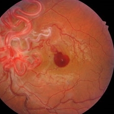 AV malformation with Valsalva retinopathy
Sep 20 2020 by Rajiv M Gandhi, MD,FVRS
Fundus of a 34-year-male who presented with sudden blurring of vision after a bout of severe cough. After examination he was diagnosed with left eye AV malformation with subhyaloid hemorrhage. After 1 month the hemorrhage resolved without any treatment. MRI brain was done to rule out AV malformations in brain but was normal.
Photographer: Dr Rajiv M Gandhi
Imaging device: Kowa 10 alpha
Condition/keywords: Wyburn-Mason
 Loading…