Search results (76 results)
-
 Sickle Cell Retinopathy
Nov 5 2022 by Mateus Queiroz Corrêa, MD
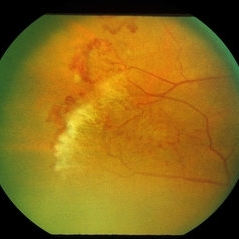
19 -year-old young man with combined rhegmatogenous and tractional retinal detachment secondary to a proliferative sickle retinopathy ( stage V)
Photographer: Mateus Corrêa, Sorocaba Eye Bank Hospital
Imaging device: Optos California
Condition/keywords: Retinal detachment, sickle cell retinopathy
-
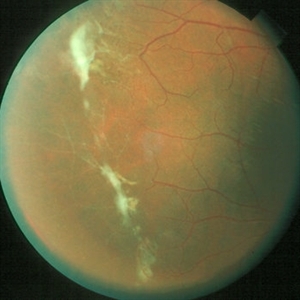
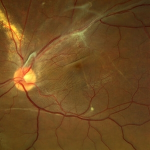 Displaced & folded macula
Oct 10 2022 by Ricardo Leitão Guerra
Tractional retinal detachment due to sickle cell retinopathy leading to a displaced and folded appearance of the macula in this 36-yo male. Subretinal bands are also noticed crossing the macula towards inferior retinal detachment area.
Photographer: Ricardo Leitão Guerra
Imaging device: Clarus 700 - Zeiss
Condition/keywords: folds, sickle cell retinopathy, subretinal bands, tractional retinal detachment
-
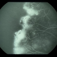
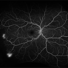 Proliferative Sickle Cell Retinopathy
Feb 1 2023 by Olivia Rainey
Ultra-widefield fluorescein angiography of a 25-year old male with Proliferative Sickle Cell Retinopathy affecting his right eye. Patient stated that he was born with Sickle disease (SC), and has yearly eye exams. He noted no vision concerns over the last year but has typically experienced sickle attacks about 1-2 per year. The physician noted that the fluorescein obtained showed peripheral nonperfusion affecting the patient's nasal and temporal retina as well as neovascularization affecting his left eye more than his right. He recommended pan retinal photocoagulation in his left eye for his temporal and nasal retina, as as well as his right eye following.
Photographer: Olivia Rainey, OCT-C, COA
Imaging device: Optos California
Condition/keywords: early phase, fluorescein angiogram (FA), fluorescein leakage, neovascularization (NV), non-perfusion, proliferative retinopathy, right eye, sickle cell retinopathy, ultra-wide field imaging, ultra-widefield image
-
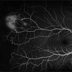
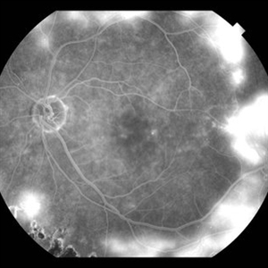
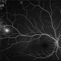 Proliferative Sickle Cell Retinopathy
Jan 29 2021 by Olivia Rainey
Ultra-widefield fluorescein angiogram of a 24-year-old female with proliferative sickle cell retinopathy affecting her right eye. The physician's interpretation of the fluorescein shows seafan neovascularization superotemporally, AV anastomeses, and good peripheral laser. He performed scatter PRP OD on 12/2/2020 to nonperfusion in temporal far periphery. The patient's 12/2020 Hb electrophoresis came back showing Hb SC (rather than sickle cell trait). Patient was born at full term, but she reports that her mother used drugs while pregnant with the patient. The patient also mentioned that her niece has full sickle cell disease and her grandmother, mother, and sibling all have condition on the sickle cell spectrum.
Photographer: Olivia Rainey, OCT-C, COA
Imaging device: Optos California
Condition/keywords: fluorescein angiogram (FA), fluorescein leakage, neovascularization (NV), neovascularization elsewhere (NVE), Optos, sea fan, sickle cell retinopathy
-
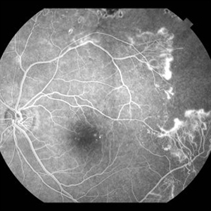
 Retinal Hemorrhages
Mar 10 2021 by Kachelle Brown
Ultra widefield Fluorescein Angiography of a 48-year-old female with retinal hemorrhages affecting her right eye. Physician suspect sickle cell due to family history, and has ordered labs to rule out.
Photographer: Kachelle Brown
Imaging device: Optos California
Condition/keywords: fluorescein angiogram (FA), fluorescein leakage, Optos, retinal hemorrhage, sickle cell retinopathy, ultra-wide field imaging
-
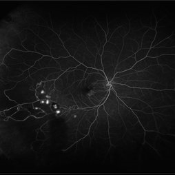
 Sickle Cell Retinopathy
Feb 15 2021 by Kim Barrett
24-year-old female with Sickle Cell Retinopathy, stage 3. She confirms she has the trait as well as her grandmother, mother and a sibling. She has seafan neovascularization superotemporal OD. Current VA is 20/20. Photo is pre-PRP laser with areas of non-profusion temporally.
Photographer: Kim Barrett C.O.A. Retina Specialist of Michigan, Grand Rapids, MI
Imaging device: Optos California
Condition/keywords: neovascularization (NV), pan-retinal photocoagulation (PRP), sickle cell retinopathy, stage 3, trait
-
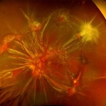
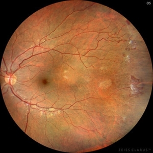
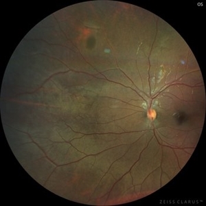 Black Sunburst in Proliferative Sickle Cell Retinopathy
Jul 25 2023 by Kamal Kishore, MD, MBBS
A 17-year-old male with a black sunburst lesion at superonasal periphery.
Photographer: Jessi Wright
Imaging device: Zeiss Clarus
Condition/keywords: Black Sunburst, sickle cell retinopathy
-
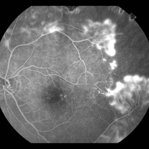
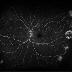
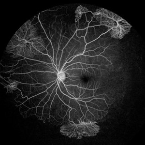 Proliferative Sickle Cell Retinopathy
Jul 8 2025 by Niloofar Piri, MD
Mid AV phase fluorescein angiogram of a 13 yo AA male with SC disease demonstrating multiple classic sea fan neovascularization with peripheral capillary non perfusion (CNP). CNP is more obvious in this image involving the temporal retina and inferonasal retina.
Photographer: Stefan Raev, COT, Saint Louis University
Condition/keywords: Proliferative sickle cell retinopathy, proliferative sickle retinopathy, sickle cell retinopathy
-
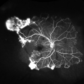 Proliferative Sickle Retinopathy
Jun 13 2025 by Brandon I Fram, MD
30 year-old with HbSC sickle retinopathy found to have profound retinal ischemia and florid peripheral neovascularization.
Imaging device: Fluorescein Angiography
Condition/keywords: proliferative sickle retinopathy, retinal ischemia, sea fan, sickle cell retinopathy
-
 Proliferative Sickle Retinopathy Stage 3
Oct 9 2012 by Alan D. Letson, MD
21-year-old man with Hgb SC Disease and stage 3 PSR with autoinfarction of sea fans
Photographer: Beverly Radcliffe
Condition/keywords: autoinfarction, sea fan, sickle cell, sickle cell retinopathy
-
 Sickle Cell Retinopathy
Sep 28 2012 by Michael P. Kelly, FOPS
Peripheral non-perfusion in sickle cell retinopathy.
Photographer: Michael P. Kelly, FOPS Director, Duke Eye Center Labs, Duke University Hospital
Imaging device: Zeiss FF3C
Condition/keywords: non-perfusion, sickle cell retinopathy
-
 Sickle Cell Retinopathy (Proliferative) - Color
Oct 27 2023 by Ricardo Leitão Guerra
Proliferative sickle cell retinopathy.
Photographer: Ricardo Luz Leitão Guerra, Leitão Guerra - Oftalmologia, Salvador - Brazil
Imaging device: Clarus 700
Condition/keywords: Iridescent spots, non-perfusion, sea fan, sickle cell retinopathy
-
 Sickle cell retinopathy (Proliferative) - FA
Oct 27 2023 by Ricardo Leitão Guerra
Fluorescein angiography in a case of proliferative sickle cell retinopathy.
Photographer: Ricardo Luz Leitão Guerra, Leitão Guerra - Oftalmologia, Salvador - Brazil
Imaging device: Clarus 700
Condition/keywords: Iridescent spots, non-perfusion, sea fan, sickle cell retinopathy
-
 Sickle Cell Retinopathy with Sea Fans (angiogram)
Aug 24 2012 by Geoffrey G. Emerson, MD, PhD, FASRS
Fluorescein angiography (mid phase) of a 40-year-old man with African heritage and sickle SC disease. Sea fans are present temporal to the macula.
Photographer: Geoffrey Emerson, MD, PhD, Retina Center, Minneapolis
Condition/keywords: sea fan, sickle cell retinopathy
-
 Sickle Cell Sea Fan Retinopathy
Jun 4 2014 by Henry J. Kaplan, MD
Sea fan peripheral retinal neovascularization in sickle cell anemia.
Condition/keywords: sea fan, sickle cell retinopathy
-
 Sickle Salmon-Patch Hemorrhage
Oct 23 2012 by Larry Halperin, MD
Salmon-patch hemorrhage in sickle cell
Condition/keywords: salmon patch, sickle cell retinopathy
-
---thumb.JPG/image-square;max$300,300.ImageHandler) Sickle Cell
Sickle Cell
Jul 11 2013 by Jason S. Calhoun
Black female with sickle cell retinopathy in the left eye, temporally.
Photographer: Jason S. Calhoun, Department of Ophthalmology, Mayo Clinic Jacksonville, Florida
Condition/keywords: sickle cell retinopathy
-
 Sickle Cell Retinopathy with Sea Fans (angiogram)
Aug 24 2012 by Geoffrey G. Emerson, MD, PhD, FASRS
Fluorescein angiography (late phase) of a 40-year-old man with African heritage and sickle SC disease. Sea fans are present around the macula (profusely leaking fluorescein dye).
Photographer: Geoffrey Emerson, MD, PhD, Retina Center, Minneapolis
Condition/keywords: sea fan
-
 Sickle Cell Retinopathy with Sea Fans (angiography)
Aug 24 2012 by Geoffrey G. Emerson, MD, PhD, FASRS
Fluorescein angiography (early/mid phase) of a 40-year-old man with African heritage and sickle SC disease. Sea fans are present temporal to the macula (leaking fluorescein).
Photographer: Geoffrey G. Emerson, MD, PhD, Retina Center, Minneapolis
Condition/keywords: sea fan, sickle cell retinopathy
-
 Sickle Cell Sea Fan Retinopathy
Jun 4 2014 by Henry J. Kaplan, MD
Fluorescein angiogram of the same patient shows extensive capillary non-perfusion anterior to the neovascularization area. #2
Condition/keywords: sea fan, sickle cell retinopathy
-
 Sickle Cell Retinopathy
Sep 28 2012 by Raj K. Maturi, MD
Sickle Cell retinopathy
Photographer: Tom Steele, CRA
Imaging device: Optos
Condition/keywords: sickle cell retinopathy
-
 Sickle Cell Retinopathy
Sep 14 2012 by Michael P. Kelly, FOPS
Fluorescein angiogram image of an individual with sickle cell retinopathy using an Optos P200MA ultra-wide field imaging device.
Photographer: Michael P. Kelly, FOPS Director, Duke Eye Center Labs, Duke University Hospital
Imaging device: Optos P200MA
Condition/keywords: Optos, sea fan, sickle cell retinopathy, ultra-wide field imaging
-
 Proliferative Diabetic Retinopathy and SC Disease
Aug 27 2021 by Caesar K. Luo, MD, FASRS
53 year-old male with SC disease complicated by proliferative diabetic retinopathy with severe peripheral non perfusion and vascular sclerosis.
Photographer: Fred Hanamoto, Bay Area Retina Associates
Imaging device: Optos California
Condition/keywords: ischemia, peripheral ischemia, proliferative diabetic retinopathy (PDR), sickle cell retinopathy
-
 Proliferative Diabetic Retinopathy and SC Disease
Aug 27 2021 by Caesar K. Luo, MD, FASRS
53 year-old male with SC disease complicated by proliferative diabetic retinopathy with severe peripheral non perfusion and small, central retained island.
Photographer: Fred Hanamoto, Bay Area Retina Associates
Imaging device: Optos California
Condition/keywords: capillary nonperfusion, peripheral ischemia, proliferative diabetic retinopathy (PDR), retinal ischemia, sickle cell retinopathy
-
 Proliferative Sickle Cell Retinopathy
Feb 1 2023 by Olivia Rainey
Ultra-widefield fluorescein angiography of a 25-year old male with Proliferative Sickle Cell Retinopathy affecting his left eye. Patient stated that he was born with Sickle disease (SC), and has yearly eye exams. He noted no vision concerns over the last year but has typically experienced sickle attacks about 1-2 per year. The physician noted that the fluorescein obtained showed peripheral nonperfusion affecting the patient's nasal and temporal retina as well as neovascularization affecting his left eye more than his right. He recommended pan retinal photocoagulation in his left eye for his temporal and nasal retina, as as well as his right eye following.
Photographer: Olivia Rainey, OCT-C, COA
Imaging device: Optos California
Condition/keywords: early phase, fluorescein angiogram (FA), fluorescein leakage, left eye, neovascularization (NV), proliferative retinopathy, sickle cell retinopathy, ultra-wide field imaging, ultra-widefield image
 Loading…