Search results (134 results)
-
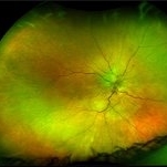
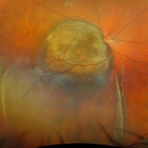 Choroidal Melanoma with Serous Retinal Detachment
Dec 20 2024 by Daniel Davis, OCT-C
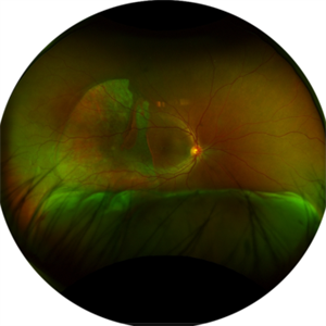
67 year old male presenting with large pigmented choroidal mass with serous retinal detachment.
Photographer: Daniel Davis, OCT-C, The Retina Institute
Imaging device: Optos California
Condition/keywords: Retina detachment
-
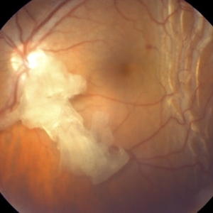 Retained Lens Fragment
Mar 2 2014 by Homayoun Tabandeh, MD, FASRS
Retained lens fragment, choroidal detachment, and serous retinal detachment post cataract surgery
Condition/keywords: retained lens fragments
-
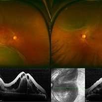
 Serpiginous Choroidopathy
Jun 23 2025 by César Adrián Gómez Valdivia, MD
Fundus photograph of a 29 year-old female patient diagnosed with Serpiginous Choroidopathy. Finings were bilateral. The most common complication of SC is choroidal neovascularization affecting up to 35% of patients. Other reported complications are subretinal fibrosis, cystoid macular edema, branch vein occlusion, serous retinal detachment, optic disc neovascularization ,and anterior uveitis.
Photographer: @eyemissu2
Imaging device: TOPCON TRC-50DX
Condition/keywords: serpiginous choroiditis
-
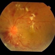
 Hypertensive Retinopathy
Aug 24 2012 by Geoffrey G. Emerson, MD, PhD, FASRS
A 35-year-old man has headaches and decreased vision. The right eye measures 20/25 and the left eye measures 3/200. The blood pressure measures 180/110.
Photographer: Geoffrey Emerson, MD, PhD, Retina Center, Minneapolis
Condition/keywords: hypertensive retinopathy, papilledema, serous retinal detachment
-
 Hypertensive Retinopathy Pre
Mar 10 2014 by Dong Yoon Kim, MD
20-year-old women who had severe preeclamsia visited our clinic for decreased visual acuity on her both eyes. Her visual acuity was 20/100 on both eyes. Fundus examination revealed serous retinal detachment on both eyes. OCT examination revealed subretinal and intraretinal fluid.
Photographer: Sun Tae Kim, University of Ulsan, Asan Medical Center
Imaging device: Optos C200 MA scanning laser ophthalmoscope
Condition/keywords: hypertensive retinopathy
-
 Hypertensive Retinopathy With Bilateral Serous Retinal Detachment at Macula
Jul 29 2014 by Mallika Goyal, MD
Left eye fundus of a 36-year-male with sudden vision drop shows grade 4 hypertensive retinopathy with retinal hemorrhages, exudates and ischaemic disc edema. OCT revealed serous retinal detachment at macula.
Photographer: Mallika Goyal, MD, Apollo Health City, Jubilee Hills, Hyderabad-500033
Condition/keywords: hypertensive retinopathy
-
 Optic Pit
Jul 13 2016 by PAVEL FLORES-MORENO
OCT of a 56-year-old male with 7 days of low visual acuity.
Photographer: Flores-Moreno Pavel
Condition/keywords: optic pit, serous retinal detachment
-
 Serous Retinal Detachment and Retinal Infiltrate due to B. Hensele, Cat-Scratch Disease
Dec 19 2020 by John S. King, MD
64-year-old female had at least a two week history of blurry vision in the right eye. She was being followed for a CRVO in the right eye, and as vision worsened, was referred to our clinic, and saw Dr. Zocchi. Vision in the right eye was CF; there was 1+ cell in the A/C; 1+ vitreous cell was present; disc edema with surrounding SRF as well as a small, white, retinal infiltrate just superior to the optic disc; vessel tortuosity was present as well as a few IRHs (left eye was u/r). There was sub-foveal and PP SRF on OCT. FA in the early to mid phase showed optic disc hyperfluorescence and early filling into the subretinal space. In the later frames there was disc leakage, staining/leakage of the retinal infiltrate, and filling into the subretinal space (See Image). Multiple tests were done, she was started on doxycycline 100 mg BID, and Bartonella serology test came back positive. One week later vision improved to 20/100, a/c cell present, disc edema improved and the SRF was resolving. (will add more photos next visit)
Photographer: Shelly Blair
Imaging device: Optos CA
Condition/keywords: cat scratch retinitis
-
 Serous Retinal Detachment in Advanced Proliferative Diabetic Retinopathy
Feb 15 2024 by Annaka Gooding
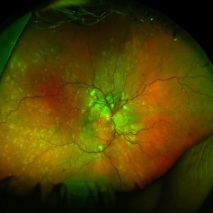
Ultra-Wide fundus photograph of a 29 year old female with a Serous Retinal Detachment in Advanced PDR. Patient present to clinic with LP vision following PPV and fill in PRP. Physician recommended oral prednisone treatment and to reassess at their following visit.
Photographer: Annaka Gooding, CPO
Imaging device: Optos California RGB
Condition/keywords: Diabetes, diabetic macular edema, fundus photography, OPTOS CALIFORNIA, pan-retinal photocoagulation (PRP), pars plana vitrectomy (PPV), proliferative diabetic retinopathy (PDR), serous retinal detachment, ultra-wide field imaging
-
 Von Hippel-Lindau
Aug 23 2012 by Gabriela Lopezcarasa Hernandez, MD
29-year-old woman with decrease in visual acuity secondary to serous retinal detachment in Von Hippel-Lindau.
Photographer: Gabriela Lopezcarasa Hernandez, Hospital Angeles Lomas
Imaging device: FF4
Condition/keywords: serous retinal detachment
-
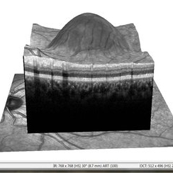 APMPPE With Serous Macular Detachment 3D SD-OCT
Jun 2 2014 by Rameez N Hussain, MD
3D SD-OCT of acute posterior multifocal placoid pigment epitheliopathy (APMPPE) with serous macular detachment.
Photographer: Rameez N Hussain MD, Vitreo Retinal Services, Giridhar Eye Institute, Cochin, India
Imaging device: Heidelberg Spectralis
Condition/keywords: acute posterior multifocal placoid pigment epitheliopathy (APMPPE), serous retinal detachment
-
 Choroidal Melanoma
Jul 4 2012 by John T. Thompson, MD
Amelanotic choroidal melanoma with serous retinal detachment
Condition/keywords: choroidal tumor, exudative retinal detachment, melanoma
-
 Von Hippel-Lindau
Oct 25 2012 by Gabriela Lopezcarasa Hernandez, MD
22 -year-old female with serous and tractional retinal detachment secondary to Von Hippel-Lindau dissease
Photographer: Araceli Rojas, Hospital Angeles Lomas Mexico
Imaging device: Zeiss FF4
Condition/keywords: serous retinal detachment, Von Hippel-Lindau
-
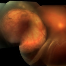 Choroidal Melanoma With a Serous Retinal Detachment
Aug 23 2018 by Nichole Lewis
63-year-old male with a large choroidal melanoma and a serous macula off retinal detachment. Vision is count fingers.
Photographer: Nichole Lewis
Condition/keywords: serous retinal detachment
-
 Collar Button Choroidal Melanoma
Oct 25 2015 by Dwain G. Fuller, MD, JD
Fundus photograph of collar button choroidal melanoma with associated serous retinal detachment and pre-retinal hemorrhages.
Condition/keywords: collar button, malignant melanoma
-
 Exudative Detachment of the Macula in Vogt-Koyanagi-Harada Syndrome
Jan 10 2018 by Peter H. Tang, MD, PhD
SD-OCT imaging of an exudative detachment of the macula in a 27-year-old male diagnosed with Vogt-Koyanagi-Harada Syndrome.
Imaging device: Zeiss Cirrus HD-OCT
Condition/keywords: exudative macula detachment, serous retinal detachment, uveitis, Vogt-Koyanagi-Harada
-
 Giant RPE Rip with Serous Retinal Detachment
Apr 30 2025 by Amber Dubey
A 50 year-old man with sudden onset diminution of vision since 3 days. Optos Image showing a fovea-sparing giant RPE rip temporally with associated serous retinal detachment inferiorly without ocular and systemic co-morbidity or antecedent history of trauma.
Photographer: Dr. Amber Dubey, Sri Sankaradeva Nethralaya, Guwahati, India
Imaging device: Optos imaging system
Condition/keywords: Optos, RPE Rip, serous retinal detachment
-
 Hypertensive Retinopathy, Right
Feb 23 2017 by Alla Goldberg, MD
Fundus photograph of 35-year-old man with severe hypertension (182/128).
Photographer: Sofia Rutiaga, UT Health McGovern Medical School, Cizik Eye Clinic
Condition/keywords: cotton wool spots, Elschnig's spots, hypertensive choroidopathy, hypertensive retinopathy, serous retinal detachment
-
 acute posterior multifocal placoid pigment epitheliopathy
Sep 23 2022 by Jaideep sharma
A 50-year old woman presented to us with unilateral progressive and painless visual blurring. She was diagnosed as a case of CSCR and started on topical dorzolamide with no improvement in VA. Her best-corrected visual acuity (BCVA) was RE 6/6 and LE 6/60 . Eye examination revealed vitritis (grade1) with optic disc hyperemia and multiple serous retinal detachments with choroidal striae in the left eye and a normal right eye. She is k/c/o diabetes. Her past ocular and drug histories were unremarkable. Retinal imaging revealed characteristic features of APMPPE in the left eye. All laboratory testing results were inconclusive. VA and OCT findings significantly improved following the treatment with LE posterior sub tenon’s triamcinolone (40 mg/ml). 1 month post injection VA of the left eye reached 6/6 with resolved serous retinal detachments in this eye. This case is unique as it was managed via PST injection rather than conventional steroid therapy
Photographer: jaideep sharma jaipur calgary eye hospital rajasthan india
Condition/keywords: acute posterior multifocal placoid pigment epitheliopathy (APMPPE), FFA
-
 Acute posterior multifocal placoid pigment epitheliopathy (APMPPE)
Sep 23 2022 by Jaideep sharma
A 50-year old woman presented to us with unilateral progressive and painless visual blurring. She was diagnosed as a case of CSCR and started on topical dorzolamide with no improvement in VA. Her best-corrected visual acuity (BCVA) was RE 6/6 and LE 6/60 . Eye examination revealed vitritis (grade1) with optic disc hyperemia and multiple serous retinal detachments with choroidal striae in the left eye and a normal right eye. She is k/c/o diabetes. Her past ocular and drug histories were unremarkable. Retinal imaging revealed characteristic features of APMPPE in the left eye. All laboratory testing results were inconclusive. VA and OCT findings significantly improved following the treatment with LE posterior sub tenon’s triamcinolone (40 mg/ml). 1 month post injection VA of the left eye reached 6/6 with resolved serous retinal detachments in this eye. This case is unique as it was managed via PST injection rather than conventional steroid therapy
Photographer: jaideep sharma jaipur calgary eye hospital rajasthan india
Condition/keywords: acute posterior multifocal placoid pigment epitheliopathy (APMPPE), FFA
-
 APMPPE With Serous Macular Detachment
Jun 2 2014 by Rameez N Hussain, MD
Acute posterior multifocal placoid pigment epitheliopathy (APMPPE) with serous macular detachment.
Photographer: Rameez N Hussain MD, Vitreo Retinal Services, Giridhar Eye Institute, Cochin, India
Imaging device: Zeiss FF4
Condition/keywords: acute posterior multifocal placoid pigment epitheliopathy (APMPPE), serous retinal detachment
-
 APMPPE With Serous Macular Detachment SD-OCT
Jun 2 2014 by Rameez N Hussain, MD
SD OCT image of acute posterior multifocal placoid pigment epitheliopathy (APMPPE) with serous macular detachment.
Photographer: Rameez N Hussain MD, Vitreo Retinal Services, Giridhar Eye Institute, Cochin, India
Imaging device: Heidelberg Spectralis
Condition/keywords: acute posterior multifocal placoid pigment epitheliopathy (APMPPE), serous retinal detachment
-
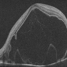
 B-scan Ultrasound of Choroidal Melanoma with Serous Retinal Detachment
Sep 5 2025 by Kristen Wagner
B-scan ultrasound of a choriodal melanoma with serous retinal detachment.
Photographer: Kristen Wagner, COT Tennessee Retina
Condition/keywords: B scan ultrasound, Choroidal melanoma, serous retinal detachment
-
 Central Serous Retinopathy-OCT
Jun 14 2018 by Mitzy E Torres Soriano, MD
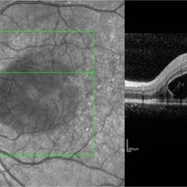
62-year-old male patient with chronic central serous chorioretinopathy in his right eye. OCT shows serous neurosensory retinal detachment and retinal pigment epithelial detachment.
Condition/keywords: central serous chorioretinopathy (CSCR), central serous retinopathy (CSR), optical coherence tomography (OCT), pigment epithelial detachment (PED), serous retinal detachment
-
 Choroid hemangioma
Sep 7 2022 by JEFFERSON R SOUSA, Tecg.º (Biomedical Systems Technology)
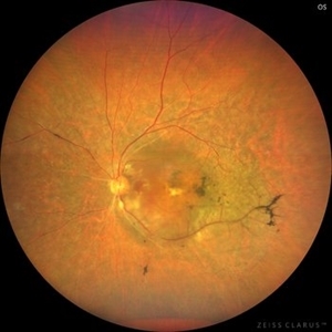
Patient 54 years old, Female, progressive loss of vision. In the multimodal evaluation of the retina showed important retinal alterations. A discreet opacity of the media impairs the quality of the images. In the Autofluorescent Background Image with a green filter, because it reaches a depth in the retinal tissue, it is able to show changes that affect the retinal pigment epithelium, it was better in this case than with the green filter. WF retinography shows an elevated, slightly reddish lesion, probable serous retinal detachment, mobilization of pigments and phantom vessels.
Photographer: JEFFERSON ROCHA DE SOUSA - Retinal Department at Instituto Dr. Suel Abujamra Sao Paulo-Brazil
Imaging device: Clarus 700 - Zeiss 135 degree images. Multimodal Evaluation
Condition/keywords: elevated retinal lesion, hemangioma, melanoma, serous retinal detachment
 Loading…