 Initializing download.
-
 By Adrienne W. Scott, MD, FASRS
Wilmer Eye Institute, Johns Hopkins University School of Medicine - Uploaded on May 28, 2014.
- Last modified by Caroline Bozell on May 29, 2014.
- Rating
- Appears in
- 28-May-2014
- Photographer
- David Emmert, Wilmer Eye Institute, Baltimore, MD
- Imaging device
- Fundus camera
- Description
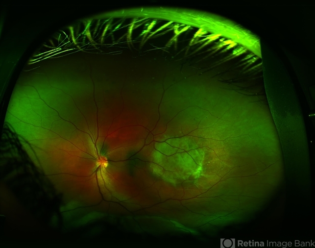
- Fundus photograph of an 11-year-old boy with a choroidal hemangioma and associated serous retinal detachment.