Search results (18 results)
-
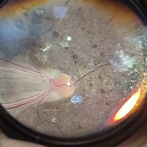
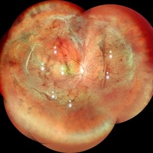 Tractional Retinal Detachment
Mar 22 2024 by Anjana Mirajkar, MS Ophthalmology
A widefield color photo of RE of a 17 year old male showing tractional retinal detachment likely a ROP sequelae.
Photographer: Dr. Anjana Mirajkar -Retina Foundation, Ahmedabad
Imaging device: Mirante-Nidek
Condition/keywords: ROP sequelae
-
 Syphilis Neuroretinopathy
Apr 2 2018 by JEFFERSON R SOUSA, Tecg.º (Biomedical Systems Technology)
Female patient, 21-years-old, with complaint of low vision in the right eye for 3 years. According to information from the patient's history, at the time she noticed the low vision, it also coincided with a picture of a strong urinary infection as well as episodes of constant tonsillitis. Yes, the patient did not seek medical attention and self-medicated with antibiotics. In ophthalmologic evaluation, as well as examinations of color retinography and ocular fundus autofluorescence, important pigmentary alterations were observed following vascular arches with pigment mobilization in osteoclasts (aspect of a unilateral pigmentary retinitis secondary to the inflammatory process). Which suggested inflammatory process sequelae. Through the laboratory tests, he had positive (+) confirmation for SYPHILIS NEURORETINOPATHY .
Photographer: JEFFERSON R SOUSA - Study Center and Ophthalmological Research Dr. Andre M V Gomes, Institute Dr. Suel Abujamra São Paulo-Brazil
Imaging device: Fundus camera Topcon TRC-50 DX, Imaginet 5.0, angle de 50 graus. Flash 36 / Mosaic with 10 images.
Condition/keywords: neurosyphilitic optic atrophy, retinitis pigmentosa, syphilis, syphilis neuroretinopathy
-
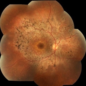
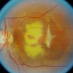
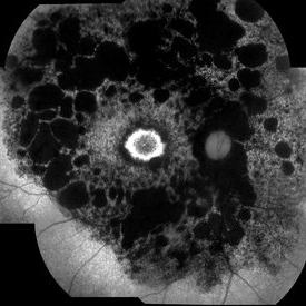
 Atypical RP with Typhoid Retinitis Sequelae with Old CRAO
Dec 5 2024 by Tejaswita Verma
FAF of a 20 year old female who presented with 2 months history of sudden painless vision loss, bilaterally light perception vision, s/o presumed atypical RP, bilateral old CRAO with typhoid retinitis sequelae.
Photographer: DR. TEJASWITA VERMA
Imaging device: MIRANTE
Condition/keywords: CRAO, retinitis pigmentosa, typhoid fever
-
 Branch Retinal Vein Occlusion (BRVO)
Sep 12 2023 by Ben Serar
Fundus Photograph of RE showing exudates at the macula with macular edema, with collaterals and peri-venous sheathing along the inferotemporal vessel arcade, as a sequelae of branch retinal vein occlusion.
Condition/keywords: branch retinal vein occlusion (BRVO), macular edema
-
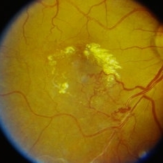
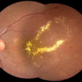 Leaking Aneurysms in Diabetic Retinopathy
Mar 22 2024 by Vaidehi Sathaye
Fundus photograph of LE of a 50 year old female with leaking aneurysms encircled by hard exudates, as a sequelae of Diabetic Retinopathy.
Photographer: Dr. Vaidehi Sathaye
Imaging device: Topcon
Condition/keywords: aneurysm, diabetic retinopathy, hard exudates
-
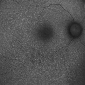
 Left Inferior Quadrantopia - Microperimetry
Jan 17 2024 by Francisco Fraga Santini Canto
Microperimetry of a 63-year-old male with sequelae of ischemic stroke in the right parietal lobe years ago. In the right eye, the patient also had a central scotoma secondary to silicone oil toxicity.
Photographer: Leonardo Hideki Nomachi Naito
Imaging device: Navis-EX Microperimeter
Condition/keywords: ischemic stroke, microperimeter, neuro-ophtalmolgy, silicone oil toxicity, visual field defect
-
 Left Inferior Quadrantopia - Microperimetry
Jan 17 2024 by Francisco Fraga Santini Canto
Microperimetry of a 63-year-old male with sequelae of ischemic stroke in the right parietal lobe years ago.
Photographer: Leonardo Hideki Nomachi Naito
Imaging device: Navis-EX Microperimeter
Condition/keywords: ischemic stroke, microperimetry, neuro, neuro-ophtalmology, visual field defect
-
 ROP SEQUELAE
Apr 26 2023 by Kalyan Singh
Fundus photograph of an young boy with history of premature birth.
Photographer: Dr Kalyan Singh, Junior resident , Department of ophthalmology, GSVM MEDICAL COLLEGE KANPUR
Imaging device: One plus 10 R ( smartphone)
Condition/keywords: ROP MACULAR DRAGGING
-
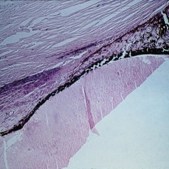
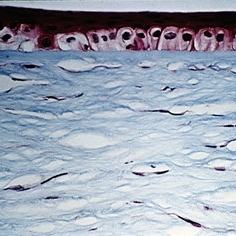 Slide 12-24
Feb 27 2019 by Lancaster Course in Ophthalmology
Sequelae. Basal layer of the corneal epithelium is pale and swollen by edema fluid. Further accumulation of edema fluid will lift the epithelium off of Bowman's membrane, resulting in bullous keratopathy (see Slide 12-16) (Masson's tri chromex250).
Condition/keywords: edema, epithelium, sequelae
-
 Slide 12-25
Feb 27 2019 by Lancaster Course in Ophthalmology
Sequelae. The anterior chamber is deep, and the pupillary iris shows ectropion uveae resulting from shrinkage of new fibrovascular tissue (rubeosis iridis) on the anterior surface of the iris. The lens is dislocated posteriorly into the vitreous. All changes are the result of blunt trauma.
Condition/keywords: ectropion uveae, rubeosis, sequelae
-
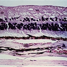
 Slide 12-26
Feb 27 2019 by Lancaster Course in Ophthalmology
Sequelae. Massive peripheral anterior synechia and ectropion uveae have resulted from blunt trauma. Note the postcontusion deformity of the anterior chamber angle (H&E x40).
Condition/keywords: anterior synechiae, ectropion uveae, sequelae
-
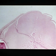 Slide 12-27
Feb 27 2019 by Lancaster Course in Ophthalmology
Sequelae. Increased intraocular pressure has resulted in stretching and thinning of the sclera in the equatorial region, resulting in an equatorial staphyloma (H&E x3).
Condition/keywords: sclera, sequelae, staphyloma
-
 Slide 12-28
Feb 27 2019 by Lancaster Course in Ophthalmology
Sequelae. The inner retinal layers are atrophic in this case of long standing glaucoma (H&E x101).
Condition/keywords: sequelae
-
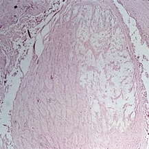 Slide 12-29
Feb 27 2019 by Lancaster Course in Ophthalmology
Sequelae. Cavernous degeneration of the optic nerve (Schnabel's cavernous atrophy) shows cystic spaces which appear clear in hematoxylin and eosin stained sections (H&E x21).
Condition/keywords: optic atrophy, sequelae
-
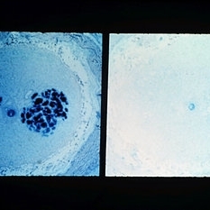 Slide 12-30
Feb 27 2019 by Lancaster Course in Ophthalmology
Sequelae. Stains for acid mucopolysaccharides (left) show spaces to contain AMP. The AMP disappears if the tissue on the slide is first digested with hyaluronidase (right), showing that the material is hyaluronidase-sensitive AMP, presumably hyaluronic acid (left view, AMPxl6; right view, hyaluronidase-AMPx16).
Condition/keywords: sequelae
-
 Subretinal haemorrhage
Sep 26 2023 by Ben Serar
Fundus photograph showing subretinal bleed with subretinal scarring and fibrosis as sequelae.
Condition/keywords: Subretinal haemorrhage
-
 Syphilis Neuroretinopathy
Apr 2 2018 by JEFFERSON R SOUSA, Tecg.º (Biomedical Systems Technology)
Female patient, 21-years-old, with complaint of low vision in the right eye for 3 years. According to information from the patient's history, at the time she noticed the low vision, it also coincided with a picture of a strong urinary infection as well as episodes of constant tonsillitis. Yes, the patient did not seek medical attention and self-medicated with antibiotics. In ophthalmologic evaluation, as well as examinations of color retinography and ocular fundus autofluorescence, important pigmentary alterations were observed following vascular arches with pigment mobilization in osteoclasts (aspect of a unilateral pigmentary retinitis secondary to the inflammatory process). Which suggested inflammatory process sequelae. Through the laboratory tests, he had positive (+) confirmation for SYPHILIS NEURORETINOPATHY .
Photographer: JEFFERSON R SOUSA - Study Center and Ophthalmological Research Dr. Andre M V Gomes, Institute Dr. Suel Abujamra São Paulo-Brazil
Imaging device: Fundus camera Topcon TRC-50 DX, Imaginet 5.0, angle de 50 graus. Flash 100 / Mosaic with 10 images.
Condition/keywords: autofluorescence imaging, neurosyphilitic optic atrophy, retinitis pigmentosa, syphilis, syphilis neuroretinopathy
-
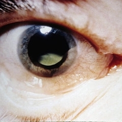
 Wegener's Disease
Feb 20 2015 by H. Michael Lambert, MD
Thinning and vascularization of the peripheral cornea, a sequelae of peripheral ulcerative keratitis and scleritis.
Condition/keywords: corneal furrow, Wegener's granulomatosis
 Loading…