Search results (50 results)
-
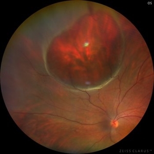 Giant Retinal Cyst
Sep 20 2025 by JORGE SOBERANES
Fundus photograph of a 45-year-old-man with a large cyst on the nasal superior side of the retina. The patient had a history of a pneumatic retinopexy two years ago and the cyst has been there since that.
Photographer: Dr. Jorge Soberanes, Asociación para Evitar la Ceguera en México (APEC), UNAM
Condition/keywords: abnormal retina, pneumatic retinopexy, retinal cyst
-
 Macrocyst in the Fovea
Feb 2 2021 by Peter J Belin, MD
36-year-old male with a white cataract and a chronic total retinal detachment for 1 year presented with a recurrent PVR detachment after primary repair 2 weeks prior. This OCT- EDI demonstrates a large retinal cyst through the fovea.
Photographer: Holly Cheshier, CRA, OCT-C, COT
Imaging device: Heidelberg Spectralis
Condition/keywords: chronic retinal detachment, proliferative vitreoretinopathy (PVR), retinal cyst, retinal macrocyst
-
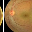
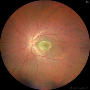
 Macular fold posterior microphthalmos OS
Apr 24 2022 by Mariam Cernichiaro-Espinosa, MD
Macular OCT of 6-year-old girl with posterior microphthalmos, showing macular fold and one intraretinal cyst OS.
Photographer: Mariam Cernichiaro-Espinosa, Asociación para Evitar la Ceguera, I.A.P. Mexico City, Mexico.
Imaging device: Zeiss Clarus
Condition/keywords: posterior microphthalmos
-
 Retinal Cyst
Aug 14 2020 by Noy Ashkenazy, MD, MS
Fundus photograph of a 13-year-old male with a chronic retinal detachment following a penetrating ocular trauma. There is a retinal cyst and proliferative vitreoretinopathy.
Photographer: Giselle DeOliveira
Imaging device: Retcam III
Condition/keywords: chronic retinal detachment, proliferative vitreoretinopathy (PVR), retinal cyst
-
 Retinal Detachment
Nov 9 2012 by Norman Byer
This 18-year-old girl gave the history of having been hit in this eye three years before with a fist and of having retinal surgery nine months previously, which was temporarily successful. When the photograph was taken, she had a total left retinal detachment with a small nasal dialysis which had not been treated. She also had two prominent intraretinal cysts, one of which is shown here. The retina promptly reattached following further surgery and the next slide shows an interesting change in this cyst.
Condition/keywords: intraretinal cyst, small nasal dialysis
-
 Retinal Lesion
Nov 9 2012 by Norman Byer
This 30-year-old man sustained a severe blow to his brow region which resulted in a variety of injuries including hyphema and vitreous and retinal hemorrhages. This photograph shows a retinal lesion, which is either a tiny area of elevated full thickness retina or a traumatic retinal cyst.
Condition/keywords: elevated retinal lesion, retinal hemorrhage, traumatic retinal cyst
-
 Subretinal Cysticercosis Post Vitrectomy
Sep 10 2020 by Anamika Dwivedi
Fundus photograph of a 22-year-old male, case of subretinal cysticercosis, after PPV and cyst removal showing laser scar at the site of the previous cyst.
Photographer: Dr Anamika Dwivedi
Imaging device: topcon
Condition/keywords: bilateral subretinal cysticercosis
-
 Cystercercosis
Sep 17 2012 by Prema Abraham, MD
Fundus photograph of 25-year-old male with subretinal cystercercosis.
Photographer: Dan Parks, Black Hills Regional Eye Institute, Rapid City South Dakota
Imaging device: Topcon 50 EX
Condition/keywords: cysticercosis
-
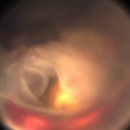
 Documented Retinal Pars Plana Cysts
Mar 14 2018 by Asaf Friehmann
Photograph taken during indentation of a 74-year-old patient who underwent a 25G pars plana vitrectomy (PPV) for repair of dislocated IOL, when this rarely documented peripheral retinal cyst which was found.
Photographer: Alexander Rubowitz
Condition/keywords: peripheral retinal cyst
-
 Intraretinal Cysts in Chronic Retinal Detachment
Dec 8 2020 by Alice Kim
B-scan ultrasound showing multiple intraretinal cysts in the setting of chronic retinal detachment and proliferative vitreoretinopathy.
Condition/keywords: chronic retinal detachment, intraretinal cyst, proliferative vitreoretinopathy (PVR)
-
 Chronic Retinal Detachment
Oct 12 2012 by Jeffrey G. Gross, MD, FASRS
Chronic RD with multiple retinal cysts, B scan ultrasound.
Condition/keywords: B scan ultrasound, chronic retinal detachment, retinal cyst
-
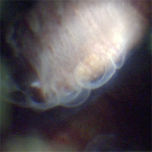
 Old RRD With Retinal Cysts and High Watermark
Apr 10 2020 by Dipak Nag, MBBS, FCPS, MSc, FRF
Intra-operative fundus picture of a 20-year-old boy showing multiple retinal cysts and high watermark in a case of old inferior retinal detachment OD.
Photographer: Dipak
Condition/keywords: high watermark, retinal cyst
-
 Cystercercosis
Sep 17 2012 by Prema Abraham, MD
Fundus photograph of 25-year-old male with subretinal cystercercosis.
Photographer: Dan Parks, Black Hills Regional Eye Institute, Rapid City South Dakota
Imaging device: Topcon 50EX
Condition/keywords: cysticercosis
-
 Cystercercosis
Sep 17 2012 by Prema Abraham, MD
Fundus photgraph of 25-year-old male with subretinal cystercercosis.
Photographer: Dan Parks, Black Hills Regional Eye Institute, Rapid City South Dakota
Imaging device: Topcon 50EX
Condition/keywords: cysticercosis
-
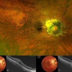 Bow-Tie Macular Hemorrhage With Cyst- Atypical Presentation of Myopic Choroidal Neovascularization
Mar 26 2021 by RUSHIK PATEL
The image of right eye of 51-year-old lady with high myopia show " Bow-Tie" macular hemorrhage (A). Optical coherence tomography (B) scan passing through hemorrhage showed intraretinal cystic lesion. During the course of intravitreal anti-VEGF injection treatment, the lesion converted into typical myopic choroidal neovascularization (C).
Photographer: Rushik Patel, Netralaya Super Speciality Eye Hospital
Condition/keywords: cyst, macular hemorrhage, myopic choroidal neovascularization (CNV)
-
 Chronic retinal detachment changes
Apr 29 2022 by Otakar Dušek, M.D. Ph.D.
Colour fundus photo of 22-year-old woman with bulous retinal detachment number 5-9, old demarcation lines and inferotemporal periheral secondary retinal cyst.
Photographer: Otakar Dušek, Charles University, Prague
Imaging device: Zeiss Clarus
Condition/keywords: chronic retinal detachment, demarcation line, peripheral retinal cyst
-
 Chronic Retinal Detachment: Features Slide 1
Oct 22 2012 by Ronald C. Gentile, MD
Chronic retinal detachments can be associated with demarcation lines (tidemarks), subretinal bands or sheets, and retinal cysts. Fundus photo of a chronic inferior retinal detachment reveals multiple demarcation lines inferior to the center of the fovea as a result of an inferior temporal dialysis.
Photographer: The New York Eye & Ear Infirmary Department of Medical Imaging
Condition/keywords: chronic retinal detachment, demarcation line
-
 Chronic Retinal Detachment: Features Slide 2
Oct 22 2012 by Ronald C. Gentile, MD
Chronic retinal detachments can be associated with demarcation lines (tidemarks), subretinal bands or sheets, and retinal cysts. Fundus photo of a chronic retinal detachment reveals a branching subretinal band superior nasal to the macula with a portion extending to the inferior margin of the optic disc.
Photographer: The New York Eye & Ear Infirmary Department of Medical Imaging
Condition/keywords: chronic retinal detachment, subretinal bands
-
 Chronic Retinal Detachment: Features Slide 3
Oct 22 2012 by Ronald C. Gentile, MD
Chronic retinal detachments can be associated with demarcation lines (tidemarks), subretinal bands or sheets, and retinal cysts. Fundus photo of a chronic retinal detachment reveals a retinal cyst within the peripherally detached temporal retina.
Condition/keywords: chronic retinal detachment
-
 CSR with Fibrin
Jan 28 2025 by Vishal Agrawal, MD, FRCS,FACS,FASRS
A 31-year-old female was referred with a diagnosis of subretinal cysticercosis. BCVA was 20/200 OS. OCT showed a large subfoveal bacillary layer detachment (BALAD) without any scolex. FFA revealed a smoke-stack appearance. A final diagnosis of CSR with Fibrin was made and was managed conservatively. BCVA at final visit was 20/20.
Photographer: Dr Ayushi Gupta
Imaging device: Clarus 700
Condition/keywords: central serous chorioretinopathy (CSCR)
-
 CSR with Fibrin-FFA
Jan 29 2025 by Vishal Agrawal, MD, FRCS,FACS,FASRS
A 31-year-old female was referred with a diagnosis of subretinal cysticercosis. BCVA was 20/200 OS. OCT showed a large subfoveal bacillary layer detachment (BALAD) without any scolex. FFA revealed a smoke-stack appearance. A final diagnosis of CSR with Fibrin was made and was managed conservatively. BCVA at final visit was 20/20.
Photographer: Dr Ayushi Gupta
Imaging device: Clarus 700
Condition/keywords: central serous chorioretinopathy (CSCR)
-
 Diabetic Macular Edema
Feb 12 2025 by Kimberly Wakester
Horizontal OCT scan of a 63-year-old woman with diabetic macular edema in the right eye. When reviewing the scan, one of the intraretinal cyst (IRC) appears heart shaped. A fun scan to see just a few day's before Valentine's day.
Photographer: Kimberly Wakester, COA
Imaging device: Heidelberg
Condition/keywords: diabetic macular edema, intraretinal cyst
-
 Eye Finally Got the Ring... But the Retina Was Too Detached to Care
Nov 5 2025 by SHRADDHA RAJ SHRIVASTAVA
Left Eye B-scan ultrasound of a patient with old retinal detachment shows open funnel shaped hyperechoic membranous echoes, with high amplitude spikes on A-scan and a poor after-movement on dynamic B-scan, suggestive of retinal detachment. We can see a round echogenicity in sub-retinal location, with clear contents within, suggestive of a retinal cyst. This B-scan image is indicative of a long-standing chronic retinal detachment with secondary retinal cyst.
Photographer: Dr. Shraddha Raj Shrivastava
Condition/keywords: B scan ultrasound, chronic retinal detachment, OLD RD, open funnel RD, retinal cyst
-
 Intraretinal cysts
Nov 15 2021 by Marcelo Zas, MD PhD
Left eye from a young patient with a chronic rhegmatogenous retinal detachment presenting intraretinal cysts.
Photographer: Zas Marcelo MD PhD
Condition/keywords: chronic retinal detachment, intraretinal cyst
-
 Intraretinal cysts
Nov 15 2021 by Marcelo Zas, MD PhD
Left eye from a young patient with a chronic rhegmatogenous retinal detachment presenting intraretinal cysts.
Photographer: Zas Marcelo MD PhD
Condition/keywords: chronic retinal detachment, intraretinal cyst
 Loading…