Search results (625 results)
-
 Hypertensive Retinopathy
Feb 25 2013 by Suber S. Huang, MD, MBA, FASRS
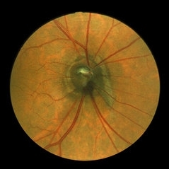
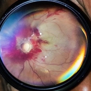
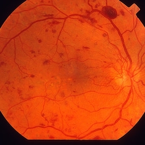
32-year-old African American male with Grade IV hypertensive retinopathy and acute renal failure. Vision OD 20/70, OS 20/25. Creatine 7.1. BP: 250/150.
Photographer: Geoffrey Pankhurst, University Hospitals, Eye Institute/Dept. Ophthalmology and Visual Sciences Case Western Reserve University Cleveland, OH
Imaging device: Topcon TRC 50x
Condition/keywords: acute renal failure, disc edema, exudate, hypertension, hypertensive retinopathy, ischemia, macular edema, macular ischemia, optic disc edema
-
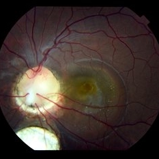
 Optic Disc Coloboma`
Mar 26 2018 by Purva Patwari
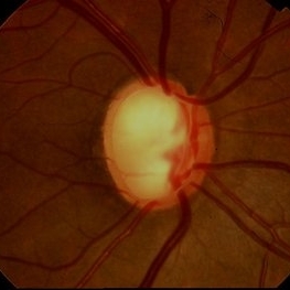
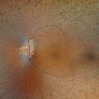
16-year-old female patient with vision of 6/60 presented with diminished vison. Other eye was normal.She had a normal birth history and developmental milestone. Look at the optic disc coloboma extending upto the macula. Intercalary membrane looks normal.
Photographer: Dr Purva Patwari, Patwari Retina Center, Ahmedabad, Gujarat , India
Imaging device: ZEISS VISU 500
Condition/keywords: coloboma, coloboma of optic disc, optic disc
-
 Optic Disc Melanocytoma
Jun 4 2014 by Henry J. Kaplan, MD
Optic disc melanocytoma with jet black pigmentation . #1
Condition/keywords: melanocytoma, optic disc melanocytoma
-
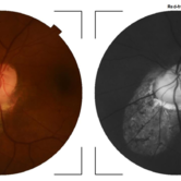
 Optic Nerve Head Drusen With Idiopathic CNV
Feb 17 2017 by Kristen Wagner
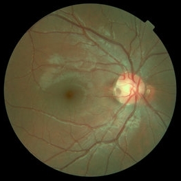
22-year-old female fundus photograph of a right eye with Optic Nerve Drusen with Idiopathic CNV.
Photographer: Kristen Wagner, COT, OSC Ophthalmic Photographer, Tennessee Retina, Nashville TN
Condition/keywords: choroidal neovascularization (CNV), drusen of optic disc, optic disc drusen
-
 Cat Eye Syndrome
Feb 11 2020 by Sophia El Hamichi, MD
A 3-year-old female with cat eye syndrome including iris, chorioretinal and optic nerve colobomas. Note the CNV temporally to the optic nerve coloboma (blue arrows)
Photographer: Giselle De Oliveira, Bascom Palmer Eye Institute, Miami
Imaging device: RetCam
Condition/keywords: cat eye syndrome, chorioretinal coloboma, choroidal neovascularization (CNV), coloboma, coloboma of optic disc, optic nerve coloboma
-
 Congenital Prepapillary Arterial Loop With a Figure-of-Eight Configuration
Mar 27 2019 by Tammy Mclaughlin
Congenital prepapillary arterial loop with a figure-of-eight configuration. OD. There is a twisted anomalous vessel eminating from the optic disc into the vitreous. Likely a congenital anomaly. Does not require treatment and should not be a vision threat.
Photographer: Tammy Mclaughlin, Carolina Retina Center 645 W. Wesmark Blvd. Sumter, Sc 29150
Condition/keywords: congenital anomaly, congenital prepapillary vascular loop
-
 Glaucoma
Feb 9 2015 by Govindarajan Venkatesan
Glaucoma.
Photographer: Govindarajan Venkatesan
Condition/keywords: optic disc
-
---thumb.jpg/image-square;max$300,300.ImageHandler) Melanocytoma
Melanocytoma
Feb 13 2013 by From the Collections of Thomas M. Aaberg, MD and Thomas M. Aaberg Jr., MD
Melanocytoma, color fundus photo, optic disc.
Condition/keywords: melanocytoma, optic disc
-
 Melanocytoma of the Optic Nerve
Apr 6 2024 by Hector Gabriel Moreno Solano, MD, MHA
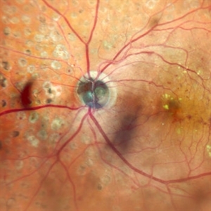
Fundus photograph of a 57-year-old male presented for an ophthalmological evaluation with a chief complaint of progressive visual loss. Indirect ophthalmoscopy revealed proliferative diabetic retinopathy, without macular edema, and a hyperpigmented lesion at the optic disc which corresponds to a melanocytoma.
Photographer: Héctor Gabriel Moreno-Solano
Imaging device: Clarus 700
Condition/keywords: diabetic retinopathy, intraocular tumor, melanocytoma, optic nerve
-
 Melanocytoma of the Optic Nerve
Apr 6 2024 by Hector Gabriel Moreno Solano, MD, MHA
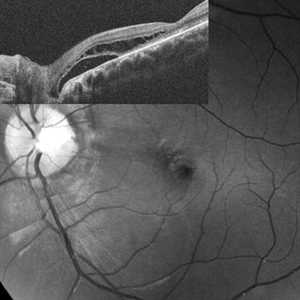
Optic Nerve laser scan image reconstruction of a 57-year-old male presented for an ophthalmological evaluation with a chief complaint of progressive visual loss. Indirect ophthalmoscopy revealed proliferative diabetic retinopathy, without macular edema, and a hyperpigmented lesion at the optic disc which corresponds to a melanocytoma.
Photographer: Héctor Gabriel Moreno-Solano, MD, MHA
Imaging device: Mirante
Condition/keywords: intraocular tumor, macular edema, melanocytoma, optic nerve
-
 Optic disc avulsion
Mar 5 2023 by Kalyan Singh
Case of post traumatic optic disc avulsion of young male adult.
Photographer: Kalyan Singh, GSVM medical college, Kanpur
Imaging device: Smartphone (1 plus 10R)
Condition/keywords: optic disc, trauma
-
 Optic Disc Drusen, RP
Apr 21 2025 by Virginia Gebhart
28 year old male with stable retinitis pigmentosa and optic disc drusen OU. Bardet-Biedl variant identified in previous genetic testing. BCVA 20/50 OD, 20/30 OS
Photographer: Virginia Gebhart, Retina Consultants of Carolina
Imaging device: Optos California
Condition/keywords: Drusen, optic disc drusen, retinitis pigmentosa
-
 Optic Disc Edema and Hemorrhages with Subdural Hematoma
Oct 1 2012 by Jeffrey G. Gross, MD, FASRS
Optic disc edema and hemorrhages with subdural hematoma.
Condition/keywords: optic disc edema, subdural hematoma
-
 Optic Disc Pit
Nov 27 2016 by Rita Couceiro, MD, MS
15-year-old boy with an optic disc pit of the right eye (incidental finding during routine fundoscopy).
Photographer: Andreia Rocha
Condition/keywords: optic disc pit
-
 Optic Disc Pit Schisis RD
Apr 29 2013 by Michael Colucciello, MD, FASRS
Optic disc pit with peripapillary RD and macular schisis, fundus photograph and SD-OCT overlay.
Condition/keywords: optic disc pit
-
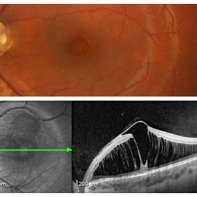 Optic Disc Pit with Maculopathy
Feb 25 2021 by Niloofar Piri, MD
Color fundus photograph and SD OCT of a 6-year-old patient with optic disc pit associated with large retinoschisis involving the entire macula. SD OCT demonstrating large retinoschisis with ILM draping centrally giving it the appearance of pseudohole on the corresponding central area of color photo. Vision 20/80
Photographer: Lisa Breeding, St Louis University
Condition/keywords: maculopathy, optic disc
-
 Serpiginous Choroidopathy
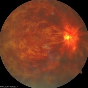
Jun 23 2025 by César Adrián Gómez Valdivia, MD
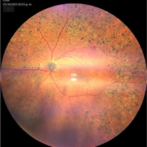
Fundus photograph of a 29 year-old female patient diagnosed with Serpiginous Choroidopathy. Finings were bilateral. The most common complication of SC is choroidal neovascularization affecting up to 35% of patients. Other reported complications are subretinal fibrosis, cystoid macular edema, branch vein occlusion, serous retinal detachment, optic disc neovascularization ,and anterior uveitis.
Photographer: @eyemissu2
Imaging device: TOPCON TRC-50DX
Condition/keywords: serpiginous choroiditis
-
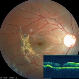 Choroidal Rupture
Apr 7 2021 by Priya Rasipuram Chandrasekaran, MBBS, DO, DNB, FRCS
The fundus photo of a 24-year-old male shows crescent shaped choroidal rupture away from fovea and concentric to the optic disc following cricket ball injury. The corresponding optical coherence tomography shows disruption of the choriocapillaris, retinal pigment epithelium and Bruch’s membrane while the neurosensory retina remains intact. The fovea is not involved.
Condition/keywords: choroidal rupture
-
 Coloboma involving the Optic nerve, Retina, and Choroid
Dec 6 2021 by Jesus Lozano, MD
78-year-old woman after prophylactic laser photocoagulation (PLP) for her RE Coloboma involving the optic nerve, retina, and choroid. At 6 month follow up, patient preserved her FC vision as it was before the procedure. Retina attached.
Photographer: Yair Bet Yosef, Hadassah Medical Center. Israel
Imaging device: Optos Silverstone fundus image
Condition/keywords: coloboma, coloboma of choroid, coloboma of macula, coloboma of optic disc, PLP, prophylactic photocoagulation
-
 Coloboma of Optic Disc
Apr 28 2019 by Bastián Schmidt Arias
Fundus photograph of an 63-year-old woman with retinal coloboma.
Photographer: Bastian Schmidt
Condition/keywords: coloboma of optic disc
-
 Colobomatous Optic Disc Maculopathy
Feb 13 2020 by Yoshihiro Yonekawa, MD, FASRS
Beautifully focused fundus photograph of a teenage girl with submacular fluid from a colobomatous optic disc.
Photographer: Netanya Lerner, COA, Wills Eye Hospital/Mid Atlantic Retina
Imaging device: Topcon
Condition/keywords: chorioretinal coloboma, coloboma of optic disc, congenital optic nerve pit, subretinal fluid
-
 Colobomatous Optic Disc Maculopathy
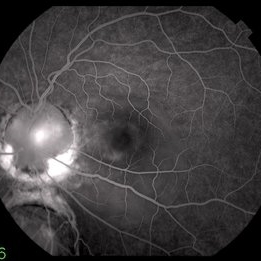
Feb 13 2020 by Yoshihiro Yonekawa, MD, FASRS
Fluorescein angiography, late frame, of a teenage girl with submacular fluid from a colobomatous optic disc. The camera is focused is on the elevated macula, and the disc is subtly defocused.
Photographer: Netanya Lerner, COA, Wills Eye Hospital/Mid Atlantic Retina
Imaging device: Topcon
Condition/keywords: chorioretinal coloboma, coloboma of optic disc, congenital optic nerve pit, subretinal fluid
-
 CRVO
CRVO
Nov 26 2020 by Priya Rasipuram Chandrasekaran, MBBS, DO, DNB, FRCS
A 44-year-old male patient presented with no underlying systemic illness presented with this picture showing extensive scattered superficial and deep retinal haemorrhages to confluent retinal hemorrhages extending to all the quadrants associated with marked dilatation and tortuosity of vessels and associated with optic disc edema, macular edema and retinal thickening giving the appearance of blood and thunder retina.
Condition/keywords: central retinal vein occlusion (CRVO)
-
 Diabetes HRC-PDR
Mar 29 2013 by Henry J. Kaplan, MD
NVD on the optic disc extended superiorly and temporally.
Condition/keywords: neovascularization of the disc (NVD)
-
 Early Phase FA of Optic Disc Capillary Hemangioblastoma
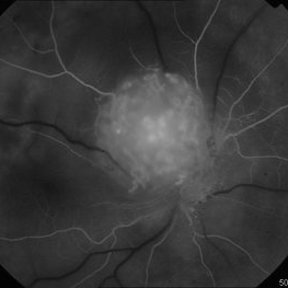
Mar 18 2014 by Arwa Azmeh, MD, PhD
FA showed early hyperfluorescent spots over the mass.
Condition/keywords: retinal hemangioblastoma
 Loading…