Search results (27 results)
-
 Macular Telangiectasis
May 13 2019 by Hashim Ali Khan, OD, FAAO
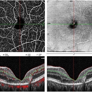
OCT-angio of superficial vascular network and structural OCT of a 60-years-old female demonstrating macular TEL showing alterations in FAZ and vascular remodeling and increased the intercapillary distance.
Imaging device: Optical Coherence Tomography Angiography
Condition/keywords: idiopathic macular telangiectasia, macular telangiectasia, macular telangiectasia type 2
-
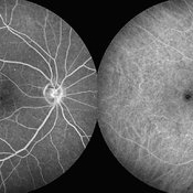
 Macular Telangiectasia Type 2 & CNV
Sep 22 2012 by Hamid Ahmadieh, MD
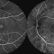
FA and ICG angiography imagings of the left eye of a 70-year-old man with idiopathic macular telangiectasia type 2 and CNV.
Photographer: Hamid Ahmadieh, MD, Ophthalmic Research Center, Labbafinejad Medical Center, Shahid Beheshti University of Medical Sciences
Imaging device: HRA
Condition/keywords: choroidal neovascularization (CNV), idiopathic macular telangiectasia, indocyanine green (ICG) angiography
-
 Macular Telangiectasia Type 2
Sep 22 2012 by Hamid Ahmadieh, MD
Autofluorescence imagings of both eyes of a 70-year-old man with idiopathic macular telangiectasia type 2.
Photographer: Hamid Ahmadieh, MD, Ophthalmic Research Center, Labbafinejad Medical Center, Shahid Beheshti University of Medical Sciences
Imaging device: HRA
Condition/keywords: autofluorescence imaging, idiopathic macular telangiectasia
-
 Macular Telangiectasia Type 2
Sep 22 2012 by Hamid Ahmadieh, MD
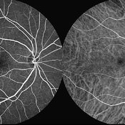
FA and ICG angiography imagings of the right eye of a 70-year-old man with idiopathic macular telangiectasia type 2.
Photographer: Hamid Ahmadieh, MD, Ophthalmic Research Center, Labbafinejad Medical Center, Shahid Beheshti University of Medical Sciences
Imaging device: HRA
Condition/keywords: idiopathic macular telangiectasia, indocyanine green (ICG) angiography
-
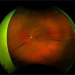
 End Point of Macular Telangiectasia (Mac Tel) Type 2
Oct 31 2024 by JULIAN VILLARREAL, MD
60 year old female with an end-stage proliferative macular telangiectasia type 2 with right-angle retinal vessels, manifested as blunted arterioles and venules that connect the superficial and deeper retinal plexus, chorioretinal anastomosis with a fibrovascular scar and a typical retinal pigment hyperplasia , fellow eye showed a focal discontinuity in the ellipsoid zone with a loss of the outer and a disorganization of the inner retinal layers, not involving the foveal center and a non exudative neovascularization
Photographer: Julián Villarreal MD
Imaging device: Zeiss Clarus 700
Condition/keywords: Mac Tel type 2, macular telangiectasia type 2
-
 Lipemia Retinalis
Dec 10 2019 by Jane S Myung, MD
Fundus photograph of a 58-year-old male with a past ocular history of macular telangiectasia type 2 who came in for routine follow-up with no vision changes and was found to have nearly complete retinal vessel whitening with a creamy appearance in both eyes. His past medical history includes diabetes, hypertension and hyperlipidemia, and he admits to stopping all his systemic medications several months prior.
Photographer: Puna Vongdara
Imaging device: Optos Ultrawidefield Camera
Condition/keywords: lipemia retinalis
-
 MACTEL
Mar 7 2025 by T. P . VIGNESH, MBBS,MS
Fundus photograph of the right eye of an 62-year-old woman with macular telangiectasia type 2.
Photographer: Sivanath
Imaging device: EIDON
Condition/keywords: IJT
-
 MACTEL
Mar 7 2025 by T. P . VIGNESH, MBBS,MS
Fundus photograph of the left eye of an 62-year-old woman with macular telangiectasia type 2.
Photographer: Sivanath
Imaging device: EIDON
Condition/keywords: macular telangiectasia type 2
-
 Mactel Type 2
Jul 1 2022 by T. P . VIGNESH, MBBS,MS
Fundus photo of a 70 year old man with Mactel with scarred subretinal neovascular membrane.
Photographer: Bharathi Singaravel
Imaging device: Zeiss Clarus
Condition/keywords: macular telangiectasia type 2
-
 Macular Telangectasia Type 2 FA OS
Apr 12 2018 by Aaron P. Appiah, MD
45-year-old African American, female with type 2 diabetes mellitus, showing bilateral, temporal macular telangectasis and peripheral microaneurysms.
Imaging device: Optos California
Condition/keywords: macular telangiectasia type 2
-
 Macular Telangectasia Type 2 OCT OD
Apr 12 2018 by Aaron P. Appiah, MD
45-year-old African American female, with type 2 diabetes mellitus, bilateral macular telangiectasia, hyper- fluorescence on FA, and cystoid changes on OCT.
Imaging device: Cirrus HD-OCT
Condition/keywords: macular telangiectasia type 2
-
 Macular Telangectasia Type 2 OCT OS
Apr 12 2018 by Aaron P. Appiah, MD
45-year-old African American female, with type 2 diabetes mellitus, bilateral macular telangiectasia, hyper- fluorescence on FA, and cystoid changes on OCT.
Imaging device: Cirrus HD-OCT
Condition/keywords: macular telangiectasia type 2
-
 Macular TelangectasiaType 2 FA OD
Apr 12 2018 by Aaron P. Appiah, MD
45-year-old African American, female with type 2 diabetes mellitus, showing bilateral, temporal macular telangectasis & peripheral microaneurysms
Imaging device: Optos California
Condition/keywords: macular telangiectasia type 2
-
 Macular Telangiectasia Type 2
Sep 22 2012 by Hamid Ahmadieh, MD
Late phase FA and ICG angiography imagings of the right eye of a 70-year-old man with idiopathic macular telangiectasia type 2.
Photographer: Hamid Ahmadieh, MD, Ophthalmic Research Center, Labbafinejad Medical Center, Shahid Beheshti University of Medical Sciences
Imaging device: HRA
Condition/keywords: idiopathic macular telangiectasia, indocyanine green (ICG) angiography
-
 Macular Telangiectasia Type 2
Mar 29 2024 by Lucy V Cobbs, M.D.
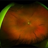
Color fundus photograph of a right eye depicts fundus changes typically seen early in disease onset of MacTel type 2. The first clinical sign of disease is subtle retinal opacification parafoveally. RPE changes initially occur temporally along dilated capillaries and then later involve the rest of the parafoveal region as disease progresses.
Condition/keywords: Mac Tel type 2, retina
-
 Macular Telangiectasia Type 2
Mar 29 2024 by Lucy V Cobbs, M.D.
Color fundus photograph of the right eye of a 61-year-old Caucasian female shows that as disease progresses, parafoveal capillaries become dilated temporally and then circumferentially around the fovea. The vessels also appear to make “right angle” turns as they plunge into deep retina. Punctate crystals may form at the vitreoretinal interface in almost half of MacTel type 2 patients and do not correlate with disease progression. This patient had bilateral asymmetric disease involvement, which is typical for MacTel type 2.
Condition/keywords: fundus photograph
-
 Macular Telangiectasia Type 2
Mar 29 2024 by Lucy V Cobbs, M.D.
Fundus autofluorescence photograph of both eyes of a patient with MacTel type 2. Fundus autofluorescence can aid in early diagnosis of disease, showing development of foveal hyperautofluorescence corresponding to deterioration of macular pigment and possible damage to Muller cells. As the disease progresses, RPE hyperplasia may develop and manifests as hypoautofluorescent regions.
Condition/keywords: Mac Tel type 2, retina
-
 Macular Telangiectasia type 2
Mar 31 2023 by Niloofar Piri, MD
Fundus autofluorescence of both eyes in a diabetic patient with Mac tel type 2 demonstrating classic temporal foveal hyperAF.
Condition/keywords: idiopathic macular telangiectasia, Mac Tel type 2, macular telangiectasia type 2
-
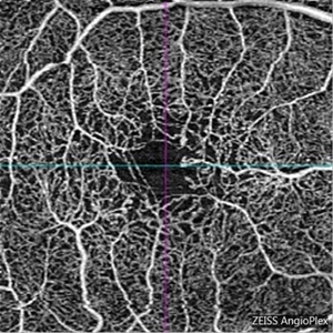 Macular Telangiectasia Type 2
Mar 8 2018 by Daniel R Agarwal, MD
OCT Angiography image in a 51-year-old male with fogging of vision and leaking on fluorescein angiography.
Photographer: Jen Welsh
Imaging device: Zeiss Angioplex OCTA
Condition/keywords: macular telangiectasia, macular telangiectasia type 2
-
 Macular Telangiectasia Type 2
Mar 29 2024 by Lucy V Cobbs, M.D.
Color fundus photograph of the left eye of a 70-year-old male with a disciform scar resulting from a neovascular membrane. A minority of MacTel type 2 patients develop neovascular disease, and the gold standard treatment is anti-VEGF intravitreal therapy. Without treatment, membranes may progress to severe central macular scarring. Late stages of proliferative MacTel type 2 may be confused with AMD, and a differentiating aspect is that MacTel type 2 typically lacks pigment epithelial detachments and drusen.
Condition/keywords: Mac Tel type 2
-
 Macular Telangiectasia Type 2 & CNV
Sep 22 2012 by Hamid Ahmadieh, MD
Color fundus photograph & OCT imagings of the left eye of a 70-year-old man with idiopathic macular telangiectasia type 2 and CNV.
Photographer: Hamid Ahmadieh, MD, Ophthalmic Research Center, Labbafinejad Medical Center, Shahid Beheshti University of Medical Sciences
Imaging device: Topcon Fundus Camera & Topcon OCT
Condition/keywords: choroidal neovascularization (CNV), idiopathic macular telangiectasia, optical coherence tomography (OCT)
-
 Macular Telangiectasia Type 2 & CNV
Sep 22 2012 by Hamid Ahmadieh, MD
Late phase FA and ICG angiography imagings of the left eye of a 70-year-old man with idiopathic macular telangiectasia type 2 and CNV.
Photographer: Hamid Ahmadieh, MD, Ophthalmic Research Center, Labbafinejad Medical Center, Shahid Beheshti University of Medical Sciences
Imaging device: HRA
Condition/keywords: choroidal neovascularization (CNV), idiopathic macular telangiectasia, indocyanine green (ICG) angiography
-
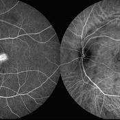
 Macular Telangiectasia Type 2 Fluorescein Angiography
Mar 29 2024 by Lucy V Cobbs, M.D.
Fluorescein angiography of the left eye of a 45-year-old African American female with MacTel type 2 and Type 2 Diabetes. This angiogram demonstrates peripheral microaneurysms characteristic of mild non proliferative diabetic retinopathy and temporal foveal leakage with telangiectatic macular capillaries classic for MacTel type 2. There is a well-established association between the two conditions.
Condition/keywords: Mac Tel type 2
-
 Macular Telangiectasia Type 2 OCT
Mar 29 2024 by Lucy V Cobbs, M.D.
Optical coherence tomography demonstrates a cavitation involving the inner retina with a thin ILM drape over the region of tissue loss. In addition, there is underlying focal disruption of the ellipsoid zone. These hyporreflective cavitations do not correlate with leakage on fluorescein angiography and are distinct from cysts in that they are not thought to be fluid filled.
Condition/keywords: Mac Tel type 2
 Loading…