 Initializing download.
-
 By Lucy V Cobbs, M.D.
NYU Langone Health - Uploaded on Mar 29, 2024.
- Last modified by Joshua Friedman on Apr 1, 2024.
- Rating
- Appears in
- Miscellaneous
- Condition/keywords
- fundus photograph
- Description
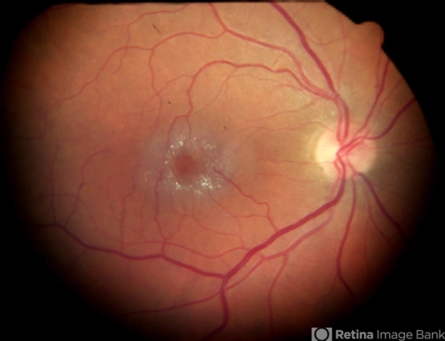
- Color fundus photograph of the right eye of a 61-year-old Caucasian female shows that as disease progresses, parafoveal capillaries become dilated temporally and then circumferentially around the fovea. The vessels also appear to make “right angle” turns as they plunge into deep retina. Punctate crystals may form at the vitreoretinal interface in almost half of MacTel type 2 patients and do not correlate with disease progression. This patient had bilateral asymmetric disease involvement, which is typical for MacTel type 2.
")
")
")


---thumb.JPG/image-square;max$79,0.ImageHandler "Welding arc maculopathy")


---thumb.jpg/image-square;max$79,0.ImageHandler "Normal Fundus Photo")