Search results (48 results)
-
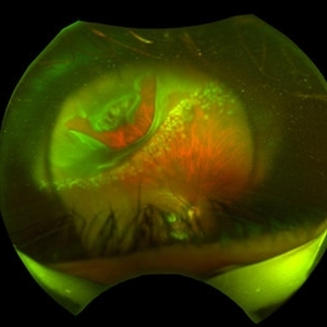
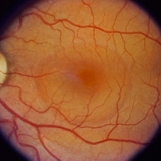
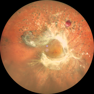 Annular Tractional Retinal Detachment
Jul 4 2024 by Hector Gabriel Moreno Solano, MD, MHA
52-year-old Hispanic female patient with a diagnosis of type II diabetes mellitus of 15 years of evolution, comes to the retina service for progressive visual loss in the right eye (single functional eye) with visual acuity of 20/100, Fundus examination reveals laser-modified proliferative diabetic retinopathy with activity + annular tractional retinal detachment with macular involvement.
Photographer: Hector Gabriel Moreno Solano, MD, MHA, HGZ #20 IMSS Puebla.
Imaging device: Mirante
Condition/keywords: macular detachment, proliferative diabetic retinopathy (PDR), tractional retinal detachment
-
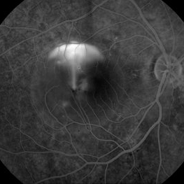 CSCR Mushroom Cloud
Feb 23 2015 by James J. Bedrick, MD
Late transit FA of a large active sub-foveal CSCR leak. You may view this pair in stereo to appreciate the plume of leakage within this large serous RD of the macula. This patient presented with a BCVA of 20/200 and fluorescein and historic evidence of prior episodes of leakage. After discussion of known treatment options including observation, he elected to be treated initially with oral rifampin and BCVA improved to 20/40 with persistent metamorphosis and a shallower persistent macular detachment over several visits. Rifampin was discontinued and he then received sub-threshold micro-pulse laser photocoagulation with an 810 diode which resulted in the patient reporting full restoration of his vision subjectively within a month. He failed to keep his follow-up appointment.
Photographer: Diana Bodnar, COT
Imaging device: Topcon 50X with Merge capture station
Condition/keywords: CSCR subfoveal leak
-
 Exudative Macular Detachment After Intensive Laser Photocoagulation
Mar 12 2016 by Sjakon G Tahija, MD
Fundus photograph of 44-year-old man with exudative detachment of the macula after vitrectomy and ILM peeling for proliferative diabetic retinopathy combined with intensive endolaser photocagulation.
Photographer: Avris Siahaan, Klinik Mata Nusantara
Condition/keywords: exudative detachment, pan-retinal photocoagulation (PRP)
-
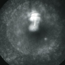
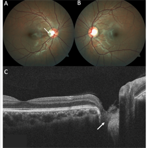 Optic Disc Pit
Nov 8 2021 by Michael Grinton
Optic disc pits are rare congenital abnormalities of the optic nerve head. Colour fundus image of an asymptomatic 18-year old male shows an optic disc pit in the right eye (A, white arrow); a small, grey, oval shaped excavation in the temporal segment of the optic disc. These pits are usually unilateral (B shows normal colour fundus of left eye) and asymptomatic. Imaging with optical coherence tomography (C) shows the optic disc pit in cross section (white arrow) and normal macular structure. In some patients with the condition, fluid can accumulate underneath the macular (serous macular detachment).
Condition/keywords: Optic disc pit, Optic nerve pit, Optic pit
-
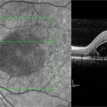
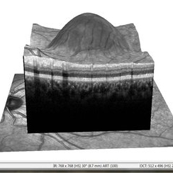 APMPPE With Serous Macular Detachment 3D SD-OCT
Jun 2 2014 by Rameez N Hussain, MD
3D SD-OCT of acute posterior multifocal placoid pigment epitheliopathy (APMPPE) with serous macular detachment.
Photographer: Rameez N Hussain MD, Vitreo Retinal Services, Giridhar Eye Institute, Cochin, India
Imaging device: Heidelberg Spectralis
Condition/keywords: acute posterior multifocal placoid pigment epitheliopathy (APMPPE), serous retinal detachment
-
 "Hang in There"
Apr 20 2021 by Tomas Minelli, MD
Fundus wide field photograph of a 50-year-old man with a macular detachment associated with a big temporal superior tear. The laser is firmly holding the progression of the tear in the 14th day post- laser. BCVA 20/20
Photographer: Livia Conci, Universtity of São Paulo
Imaging device: Optos Daytona
Condition/keywords: giant retinal tear
-
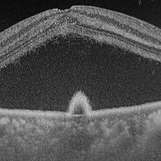
 Choroidal Rupture, Subretinal and Vitreous Hemorrhage Secondary to Blunt Trauma
Dec 29 2012 by Humberto Ruiz-Garcia, MD
SD-OCT obtained at 72 hours which shows neurosensory macular detachment and severe thinning (impending macular hole).
Photographer: Humberto Ruiz-Garcia
Imaging device: Cirrus HD OCT
Condition/keywords: traumatic macular hole
-
 APMPPE With Serous Macular Detachment
Jun 2 2014 by Rameez N Hussain, MD
Acute posterior multifocal placoid pigment epitheliopathy (APMPPE) with serous macular detachment.
Photographer: Rameez N Hussain MD, Vitreo Retinal Services, Giridhar Eye Institute, Cochin, India
Imaging device: Zeiss FF4
Condition/keywords: acute posterior multifocal placoid pigment epitheliopathy (APMPPE), serous retinal detachment
-
 APMPPE With Serous Macular Detachment SD-OCT
Jun 2 2014 by Rameez N Hussain, MD
SD OCT image of acute posterior multifocal placoid pigment epitheliopathy (APMPPE) with serous macular detachment.
Photographer: Rameez N Hussain MD, Vitreo Retinal Services, Giridhar Eye Institute, Cochin, India
Imaging device: Heidelberg Spectralis
Condition/keywords: acute posterior multifocal placoid pigment epitheliopathy (APMPPE), serous retinal detachment
-
 Bilateral Central Serous Retinopathy
Mar 26 2019 by Gary R. Cook, MD, FACS
Late-phase frame of FA of 37-year-old white male with acute CSR OD showing pooling of dye beneath the small central RPED centrally, a smokestack-type leak from the RPE defect just above it, and mild late pooling of dye outlining the large neurosensory macular detachment; VA = 20/80-1.
Imaging device: Topcon VT-50
Condition/keywords: central serous retinopathy (CSR), FA late phase, FA late phase leakage, neurosensory detachment of retina
-
 Central serous chorioretinopathy
Nov 18 2022 by T. P . VIGNESH, MBBS,MS
OCT of a 45 year old man revealing serous macular detachment with a subfoveal PED .
Photographer: Priyanka
Imaging device: Topcon Triton
Condition/keywords: central serous chorioretinopathy (CSCR)
-
 Central Serous Chorioretinopathy (CSR)
Sep 26 2023 by Ben Serar
Fundus photograph of RE showing serous macular detachment in a case of Central Serous Chorioretinopathy.
Condition/keywords: Central Serous Chorioretinopathy (CSR)
-
 Central Serous Chorioretinopathy (CSR)
Sep 21 2023 by Ben Serar
Fundus photograph of LE showing serous macular detachment in a case of Central Serous Chorioretinopathy.
Condition/keywords: Central Serous Chorioretinopathy (CSR)
-
 Central Serous Chorioretinopathy (CSR)
Sep 21 2023 by Ben Serar
Fundus photograph of RE showing serous macular detachment in a case of Central Serous Chorioretinopathy.
Condition/keywords: Central Serous Chorioretinopathy (CSR)
-
 Central Serous Chorioretinopathy (CSR)
Sep 21 2023 by Ben Serar
Fundus photograph showing increased cup-disc ratio with nasalisation of vessels , with thinning of Neuroretinal rim and bayonetting of vessels in a case of Glaucomatous Optic Atrophy (GOA) Fundus photograph of LE showing serous macular detachment in a case of Central Serous Chorioretinopathy (CSR).
Condition/keywords: Central Serous Chorioretinopathy (CSR)
-
 Central Serous Chorioretinopathy (CSR)
Sep 14 2023 by Ben Serar
Fundus photograph of LE showing serous macular detachment in a case of Central Serous Chorioretinopathy.
Condition/keywords: Central Serous Chorioretinopathy (CSR)
-
 Central Serous Chorioretinopathy (CSR)
Sep 14 2023 by Ben Serar
Fundus photograph of LE showing serous macular detachment in a case of Central Serous Chorioretinopathy.
Condition/keywords: Central Serous Chorioretinopathy (CSR)
-
 Central Serous Chorioretinopathy (CSR)
Sep 12 2023 by Ben Serar
Fundus photograph of LE showing serous macular detachment in a case of Central Serous Chorioretinopathy.
Condition/keywords: Central Serous Chorioretinopathy (CSR)
-
 Central Serous Chorioretinopathy (CSR)
Sep 12 2023 by Ben Serar
Fundus photograph of RE showing serous macular detachment in a case of Central Serous Chorioretinopathy.
Condition/keywords: Central Serous Chorioretinopathy (CSR)
-
 Central Serous Chorioretinopathy (CSR)
Sep 12 2023 by Ben Serar
Fundus photograph of LE showing serous macular detachment in a case of Central Serous Chorioretinopathy.
Condition/keywords: Central Serous Chorioretinopathy (CSR)
-
 Central Serous Chorioretinopathy (CSR)
Sep 12 2023 by Ben Serar
Fundus photograph of LE showing serous macular detachment in a case of Central Serous Chorioretinopathy
Condition/keywords: Central Serous Chorioretinopathy (CSR)
-
 Central Serous Retinopathy
Mar 26 2019 by Gary R. Cook, MD, FACS
43-year-old white male with acute CSR OD showing a large neurosensory macular detachment in his right eye.
Imaging device: Topcon VT-50
Condition/keywords: central serous retinopathy (CSR), neurosensory detachment of retina
-
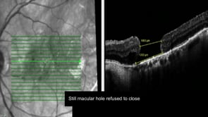 Closing the Persistent Macular Hole: Third Time’s the Charm
May 7 2019 by Srinivas Joshi, MD, FASRS
Patient was operated for macular hole with vitrectomy and conventional ILM peeling following which macular hole did not close. In second step, macular detachment induction with subretinal BSS injection using 42G needle was done but macular hole did not close. In third attempt a neurosensory retinal patch graft surgery was done and finally the macular hole was closed successfully with type 1 closure.
Condition/keywords: macular detachment, macular hole, neurosensory retinal patch graft, subretinal BSS
-
 CSR with large RPED
Mar 26 2019 by Gary R. Cook, MD, FACS
Mid-phase FA showing large RPED inferonasal to optic disc with overlying cruciate pigment figures (black lines) and neurosensory macular detachment OD.
Imaging device: Topcon VT-50
Condition/keywords: central serous retinopathy (CSR), neurosensory detachment of retina, retinal pigment epithelium (RPE) detachment
-
 FA 40 Seconds - Large Hemorrhage With Macular Detachment Due to AMD
Nov 7 2019 by John S. King, MD
81-year-old white female with three day history of seeing a "dark blob" nasally OD; no blood thinners; vision was 20/100- J16 with 2+NSC OD; OCT (not shown) had large SRF that included the fovea and extended out temporally. Posterior segment showed a large amount of SRF in the macula with some SRH in the inferior portion of the macula, hemorrhagic PEDs temporally with some RPE scarring and SRH in the periphery. On the FA there is blockage by the SRH and SRPE heme; there is staining peripherally; there is a wave of leakage that extends out into the macula and pools into to subretinal space.
Photographer: Brandon Peter
Condition/keywords: retinal pigment epithelium, subretinal hemorrhage, wet age-related macular degeneration (wet AMD)
 Loading…