Search results (241 results)
-
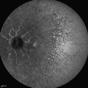
 Acute Posterior Multifocal Placoid Pigment Epitheliopathy
Feb 20 2024 by Soobien Lee
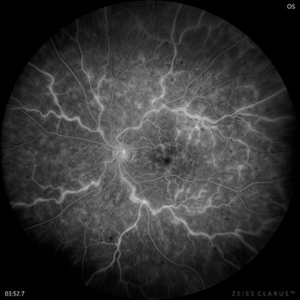
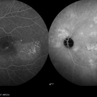
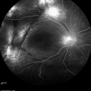
Fluorescein angiogram of a 20-year-old caucasian female with viral prodrome and vision loss OS>OD secondary to Acute Posterior Multifocal Placoid Pigment Epitheliopathy (APPME). Early blockage with late hyperfluorescent leakage can be seen on fluorescein angiography of the left eye.
Photographer: Ashley Metzger, Elman Retina Group
Imaging device: Optos Ultra-Widefield Fluorescein Angiography
Condition/keywords: acute posterior multifocal placoid pigment epitheliopathy (APMPPE), bacilliary layer detachment, FA, FA late phase, FA late phase leakage, fluorescein angiogram (FA), Optos, uveitis, white dot syndrome
-
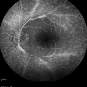
 Central Retinal Artery Occlusion
Aug 23 2012 by Gerardo Garcia-Aguirre, MD
Fluorescein angiogram, late phase, of a central retinal artery occlusion, showing very delayed filling and wide areas of capillary nonperfusion.
Photographer: Noemí Hernández, Asociación para Evitar la Ceguera en México
Condition/keywords: capillary nonperfusion, central retinal artery occlusion (CRAO), vessel sheathing
-
 Retinal Lightning
Apr 24 2022 by Mariam Cernichiaro-Espinosa, MD
Late phase venous leakage on IV fluorescein angiography from a 32-year-old male with central retinal vein occlusion (CRVO).
Photographer: Mariam Cernichiaro-Espinosa, Asociación para Evitar la Ceguera, I.A.P. Mexico City, Mexico.
Imaging device: Zeiss Clarus
Condition/keywords: central retinal vein occlusion (CRVO)
-
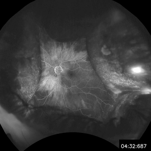
 Central Retinal Artery Occlusion & Cilioretinal Artery Sparing
Dec 22 2012 by Hamid Ahmadieh, MD
Late phase FA image of the right eye of a 34-year-old man with sudden drop of vision due to CRAO. The macula is involved despite cilioretinal artery sparing .
Photographer: Zohre Salimi; Labbafinejad Medical Center, Shahid Beheshti University of Medical Sciences , Tehran
Imaging device: Heidelberg HRA
Condition/keywords: central retinal artery occlusion (CRAO), cilioretinal sparing
-
 Central Retinal Artery Occlusion & Cilioretinal Artery Sparing
Dec 22 2012 by Hamid Ahmadieh, MD
Late phase FA image of the right eye of a 34-year-old man with sudden drop of vision due to CRAO. The macula is involved despite cilioretinal artery sparing .
Photographer: Zohre Salimi; Labbafinejad Medical Center, Shahid Beheshti University of Medical Sciences
Imaging device: Heidelberg HRA
Condition/keywords: central retinal artery occlusion (CRAO), cilioretinal sparing
-
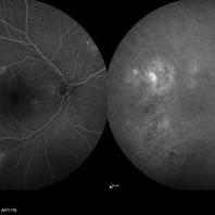
 Central Retinal Vein Occlusion with Severe Retinal Ischemia
Jan 19 2022 by Olivia Rainey
Ultra-widefield fluorescein angiogram of a 56-year-old male with a Central Retinal Vein Occlusion with Severe Retinal Ischemia affecting his right eye. The patient presented on 1/19/2022, sc20/20-2 vision in the right eye. The patient has had a good response to Eylea with complete resolution of edema. The physician is considering PRP to ischemic periphery in the future and given the degree of ischemia in both eyes, she recommends that the patient's PCP check carotid Doppler US.
Photographer: Olivia Rainey, OCT-C, COA
Imaging device: Optos California
Condition/keywords: central retinal vein occlusion (CRVO), FA late phase, fluorescein angiogram (FA), ischemic CRVO, Optos, retinal ischemia, ultra-wide field imaging
-
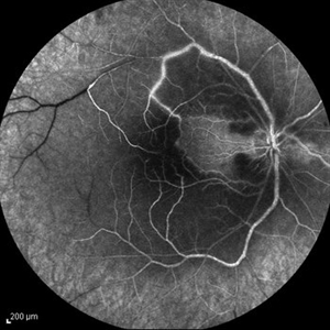
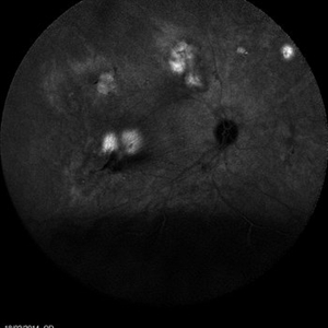
 Central Serous Chorioretinopathy
Jan 25 2022 by Olivia Rainey
Late phase widefield fluorescein angiography of a 60-year-old male with Central Serous Chorioretinopathy. Chronic history of CSR followed with observation without treatment prior to presenting at our office. The physician noted subfoveal subretinal fluid with recent visual decline. FA shows multifocal leakage and ICG shows hypercyanescence. OCTA, ICG, and FA consistent with CSR, and without concern for CNVM thus will observe without anti-VEGF at this time. PDT therapy recommended.
Photographer: Olivia Rainey, OCT-C, COA
Imaging device: Heidelberg Spectralis
Condition/keywords: 55-degrees, central serous chorioretinopathy (CSCR), central serous retinopathy (CSR), chronic central serous chorioretinopathy (CSCR), fluorescein angiogram (FA), heidelberg spectralis, indocyanine green (ICG) angiography, left eye
-
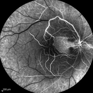
 Central Serous Chorioretinopathy
Jan 25 2022 by Olivia Rainey
Late phase widefield fluorescein angiography of a 60-year-old male with Central Serous Chorioretinopathy. Chronic history of CSR followed with observation without treatment prior to presenting at our office. The physician noted significant findings on exam and imaging with multifocal areas of inactive and active changes OD. FA shows superotemporal macular leakage, subtle inferonasal macular leakage and staining as well as multifocal hypercyanescence on ICG. Fortunately foveal sparing and thus observation is recommended at this time OD.
Photographer: Olivia Rainey, OCT-C, COA
Imaging device: Heidelberg Spectralis
Condition/keywords: 55-degrees, central serous chorioretinopathy (CSCR), central serous retinopathy (CSR), chronic central serous chorioretinopathy (CSCR), fluorescein angiogram (FA), fluorescein leakage, heidelberg spectralis, indocyanine green (ICG) angiography, late phase
-
 Central Serous Choroidopathy - Angiography
Jun 27 2018 by Gabriel Costa Andrade, PhD
46-year-old male with central serous choroidopathy in left eye. VA OS cc 20/40. Late phase FA photo shows multiple foci of leakage.
Photographer: Gabriel Andrade, RETINA CLINIC, São Paulo, BRAZIL
Imaging device: Optos California
Condition/keywords: central serous retinopathy (CSR)
-
 Central Serous Choroidopathy, CSR, with Foci of Leakage
Oct 9 2012 by James B. Soque, CRA, OCT-C, COA, FOPS
50 y/o WM with Central Serous Choroidopathy Left eye. VA OS cc 20/80. Topcon 3D 1000 SD OCT image reveals Sub RPE detachment in several locations, and subretinal fluid blister. Color, Early, and Late phase FA photos enclosed. FA shows obvious ‘smoke stack’ appearance of leakage in superonasal fovea, and 3 other foci of leakage. Late FA Photo shown.
Photographer: James Soque CRA COA
Imaging device: Topcon TRC 50 EX, with OIS V 10.5.74 Software. 5 MP Camera
Condition/keywords: central serous chorioretinopathy (CSCR), central serous retinopathy (CSR)
-
 Combined hamartoma - FA 2
Jan 11 2013 by Alex P. Hunyor, MD
Combined hamartoma of retina and RPE, right eye - late phase fluorescein angiogram. Note: scanned negative film FA
Condition/keywords: combined hamartoma
-
 Cystoid Macular Degeneration
Feb 1 2023 by Kachelle Brown
Fluorescein Angiogram of a 56 year old woman with bilateral Cystoid Macular Degeneration. Patient vision was 20/60 OU.
Photographer: Kachelle Brown OMA, Retina Specialist of Michigan
Condition/keywords: cystoid macular degeneration, cystoid macular edema (CME), FA late phase, fluorescein angiogram (FA)
-
 Multifocal Exudative Detachments Due to VKH
May 14 2014 by Avris Romario Diparaja Siahaan
FA (Late Phase) a 38-year-old man with multifocal CSR and inferior exudative retinal detachment on both eyes (Harada Syndrome).
Photographer: Avris Romario Diparaja Siahaan, Klinik Mata Nusantara
Imaging device: Heidelberg HRA + OCT Spectralis
Condition/keywords: multifocal central serous chorioretinopathy (CSCR)
-
 Multifocal Exudative Detachments Due to VKH
May 14 2014 by Avris Romario Diparaja Siahaan
ICG (Late Phase) a 38-year-old man with multifocal CSR and inferior exudative retinal detachment on both eyes (Harada Syndrome).
Photographer: Avris Romario Diparaja Siahaan, Klinik Mata Nusantara
Imaging device: Heidelberg HRA + OCT Spectralis
Condition/keywords: indocyanine green (ICG) angiography, multifocal central serous chorioretinopathy (CSCR)
-
 Retinal Fold Angiography
Feb 9 2017 by Dominic M Buzzacco, MD
Late phase angiogram of 10-month-old male with congenital retinal fold. Contralateral eye had normal angiography.
Photographer: Dominic M Buzzacco MD, Midwest Retina
Imaging device: Retcam 3
Condition/keywords: retinal fold
-
 Vogt Koyanagi Harada
Oct 7 2015 by Avris Romario Diparaja Siahaan
Simultaneous FA + ICG (Late Phase) of a 42-year-old woman with Harada Syndrome in both eyes.
Photographer: Yohanes Harry Purwanto, Klinik Mata Nusantara
Imaging device: Heidelberg HRA + OCT
Condition/keywords: indocyanine green (ICG) angiography, late phase, Vogt-Koyanagi-Harada
-
 ROP FA OD
Apr 27 2018 by Brenda Fallas
4-month-old baby with regressed ROP post-Avastin.
Photographer: Brenda Fallas, Bascom Palmer Eye Institute, Miami, FL
Imaging device: RETCAM III 130 degree lens mongtage
Condition/keywords: FA late phase leakage, fluorescein angiogram (FA), retina, retinopathy of prematurity (ROP)
-
 ROP FA OS
Apr 27 2018 by Brenda Fallas
4-month-old baby with regressed ROP post-Avastin.
Photographer: Brenda Fallas, Bascom Palmer Eye Institute, Miami, FL
Imaging device: RETCAM III 130 degree lens montage
Condition/keywords: FA late phase leakage, fluorescein angiogram (FA), retinopathy of prematurity (ROP)
-
 Central Serous Chorioretinopathy, Fluorescein Angiogram
Aug 23 2012 by Gerardo Garcia-Aguirre, MD
Fluorescein angiogram, late phase, showing hyperfluorescent spot, larger than earlier phases.
Photographer: Noemí Hernández, Asociación para Evitar la Ceguera en México
Imaging device: Zeiss FF4
Condition/keywords: central serous chorioretinopathy (CSCR)
-
 Angioid Streaks
Sep 29 2012 by Hamid Ahmadieh, MD
Late phase ICG angiography image of the right eye of a 59-year-old man with angioid streaks.
Photographer: Hamid Ahmadieh, MD; Ophthalmic Research Center, Labbafinejad Medical Center, Shahid Beheshti University of Medical Sciences
Imaging device: Heidelberg Spectralis
Condition/keywords: angioid streaks, indocyanine green (ICG) angiography
-
 Angioid Streaks
Sep 29 2012 by Hamid Ahmadieh, MD
Late phase ICG angiography image of the left eye of a 59-year-old man with angioid streaks.
Photographer: Hamid Ahmadieh, MD; Ophthalmic Research Center, Labbafinejad Medical Center, Shahid Beheshti University of Medical Sciences
Imaging device: Heidelberg Spectralis
Condition/keywords: angioid streaks, indocyanine green (ICG) angiography
-
 Angioid Streaks & CNV (Fig 4)
Aug 25 2012 by Hamid Ahmadieh, MD
Late phase ICG angiography imaging of a 53-year-old woman with a juxtafoveal CNV secondary to angioid streaks.
Photographer: Hamid Ahmadieh, Ophthalmic Research Center, Labbafinejad Medical Center
Imaging device: Heidelberg Spectralis
-
 Behcet's Disease
Mar 13 2013 by Hamid Ahmadieh, MD
Late phase FA of the left eye of a 23-year-old man with retinal vasculitis due to Behcet's disease .
Photographer: Solmaz Shahmohammad, Negah Eye Center, Tehran
Imaging device: Heidelberg Spectralis
Condition/keywords: retinal vasculitis
-
 Bruch’s membrane rupture
Jan 11 2013 by Hyung-Woo Kwak, MD
An area of Bruch’s membrane rupture involving the fovea is seen on indocyanine green angiography: late phase (right).
Photographer: Misook Lee, Kyung Hee Univsersity Hospital, Seoul
Imaging device: Zeiss f 450 plus
Condition/keywords: Bruch's membrane, myopic choroidal neovascularization (CNV)
-
 Choroidal Detachment
Jan 6 2020 by Sarah Oelrich
Choroidal detachment
Photographer: Sarah Oelrich CRA COT OCT-C
Imaging device: Optos
Condition/keywords: choroidal detachment, detachment, FA late phase
 Loading…