Search results (73 results)
-
 Commotio Retinae
Aug 7 2025 by Gabriel Costa Andrade, PhD
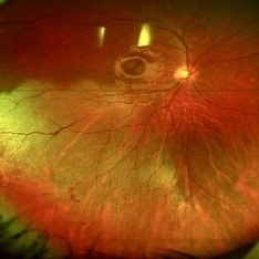
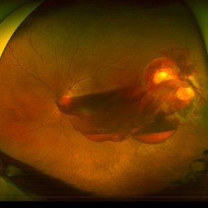
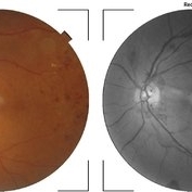
Color fundus photograph of a 13-year-old girl who was hit by accidental discharge of gel bullet in her right eye. She presented with retinal whitening with intraretinal hemorrhages in temporal inferior area of the peripheral retina.
Photographer: Gabriel Andrade
Condition/keywords: macula, Retina, Trauma
-
 Congenital Retinal Macrovessel
Oct 13 2023 by Jacob D. Grodsky, MD
41 y/o male who presented with acute onset of blurred vision OD. Visual acuity was 20/200 OD; 20/25 OS. Examination was consistent with congenital retinal macrovessel through the macula with intraretinal hemorrhage as seen in the fundus photo. Intravitreal bevacizumab was injected, and visual acuity improved to 20/40 at 4-week follow-up. MRA head and neck was ordered to rule out other vascular anomalies.
Condition/keywords: congenital retinal macrovessel, RETINAL MACROVESSEL
-
 Diabetic Retinopathy
Jun 4 2025 by Paulina Araujo
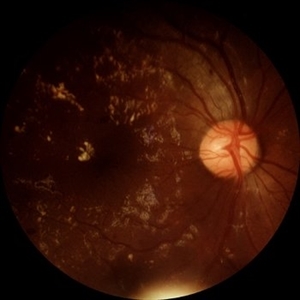
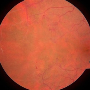
The 55-degree central fundus photograph of the right eye demonstrates numerous hard exudates, dot intraretinal hemorrhages, and microaneurysms.
Photographer: Paulina D.Araujo Martínez, Asociación para Evitar la Ceguera en México I.A.P., Hospital Dr Luis Sánchez Bulnes.
Condition/keywords: diabetic retinopathy
-
 Macular Edema
Jun 4 2025 by Paulina Araujo
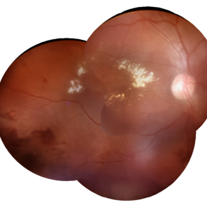
The composite fundus photograph of the right eye demonstrates circinate hard exudates in the thickened macular area, along with flame-shaped intraretinal hemorrhages along the inferior temporal arcade.
Photographer: Paulina D.Araujo Martínez, Asociación para Evitar la Ceguera en México I.A.P., Hospital Dr Luis Sánchez Bulnes.
Condition/keywords: macular edema
-
 YAG Laser Hyaloidotomy
Aug 31 2025 by Giriraj Vibhute
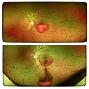
A 24-year-old young man presented with sudden loss of vision in left eye following history of rigorous coughing. Visual acuity in RE was 6/6, LE was 6/60p. Fundoscopy showed bilateral multiple small intraretinal hemorrhages with LE large premacular subhyaloid hemorrhage just covering the fovea suggestive of bilateral valsalva retinopathy changes. Nd:YAG laser hyaloidotomy was performed to left eye the same day (A250; 2mJ;6 SHOTS). Visual acuity improved to 6/9 immediately following the procedure. After 1 week, the subhyaloid hemorrhage had completely cleared with dispersed intragel hemorrhage in the inferior vitreous cavity with visual acuity of 6/6 in left eye
Photographer: Dr Vani S. MM Joshi eye institute, Hubli
Condition/keywords: valsalva retinopathy, YAG HYALOIDOTOMY
-
 Ruptured Macroaneurysm
May 22 2019 by Nichole Lewis
FA of a 91-year-old woman with a ruptured macroaneurysm, intraretinal hemorrhage and subretinal hemorrhage. VA 20/400.
Photographer: Nichole Lewis
Condition/keywords: intraretinal hemorrhage, ruptured macroaneurysm, subretinal hemorrhage
-
 Diabetic Retinopahty
Nov 2 2022 by pedro fernandes souza neto
Fundus photograph of a 40-year-old man with diabetes and hypertension shows hard exudates, difuse intraretinal hemorrhages and splinter hemorrhages.
Photographer: Pedro Fernandes, Universidade Federal da Bahia, Brazil.
Condition/keywords: diabetic mellitus, hypertensive retinopathy, retinopathy
-
 Penetrating Trauma with Retinal Detachment
Apr 30 2019 by Olivia Rainey
Ultra-wide field pseudocolor image of a 39-year-old female with penetrating trauma resulting in a retinal detachment with an intraretinal hemorrhage affecting the left eye. Patient was struck with a champagne glass in October of 2018, which lacerated the eyelid and globe. Patient was "seeing red" when she first came to the office and after multiple surgeries she was seeing 20/20 at her last check in April 2019.
Photographer: Olivia Rainey
Imaging device: Optos
Condition/keywords: hemorrhage, left eye, Optos, penetrating trauma, ruptured globe, ultra-wide field imaging
-
 Lyme Disease
Feb 13 2013 by From the Collections of Thomas M. Aaberg, MD and Thomas M. Aaberg Jr., MD
Papilledema, intra-retinal hemorrhage, periopticneuritis.
Condition/keywords: intraretinal hemorrhage, Lyme disease, periopticneuritis
-
 Worsening of Diabetic Retinopathy Severity Level Associated with Initiation of Tight Glucose Control
Feb 11 2013 by Neil M. Bressler, MD
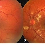
Tight control of systemic blood sugar in juvenile-onset diabetes may lead to worsen diabetic retinopathy for a temporary period, and will lead to better retinopathy condition in longer term of the disease1. A 20-year-old man with Type 1 diabetes presented with severe non-proliferative diabetic retinopathy in both eyes at the initiation of intensive diabetes treatment (Fig A, C). 3 months after initiating tight blood sugar control, his fundus shows substantial worsening of this diabetic retinopathy severity level with new neovascularization of the disc, numerous new nerve fiber layer infarcts surrounding the optic nerve (in the absence of any systemic hypertension) and a marked increase in intraretinal hemorrhages in both eyes (Fig B, D). Reference: 1. The Diabetes Control and Complications Trial Research Group. The effect of intensive treatment of diabetes on the development and progression of long-term complications in insulin-dependent diabetes mellitus. N Engl J Med 1993; 329:977-986.
-
---thumb.jpg/image-square;max$300,300.ImageHandler) Acute retinal necrosis
Acute retinal necrosis
Feb 15 2013 by From the Collections of Thomas M. Aaberg, MD and Thomas M. Aaberg Jr., MD
Diffuse intraretinal hemorrhages and whitening in the posterior pole consistent with acute retinal necrosis.
Condition/keywords: macular edema, microangiopathy, retinal necrosis, retinal whitening
-
 Angiographic Storm: Fluorescein Leakage in Retinal Vasculitis
Nov 17 2025 by SHRADDHA RAJ SHRIVASTAVA
This left eye montage fundus fluorescein angiography (FFA) image of a 19 year old male with idiopathic retinal vasculitis, having skip vasculitic lesions predominantly involving retinal veins. There are areas of blocked fluorescence due to intraretinal hemorrhages, the involved veins have filling defects and occlusions, leading to formation of numerous collateral channels. The inflamed vessels also show perivascular fuzzy hyperfluorescent stain due to leakage of dye. We can also see multiple peripheral capillary non perfusion (CNP) areas, with a 'hot disc', suggestive of ongoing inflammation.
Photographer: Dr. Shraddha Raj Shrivastava
Imaging device: Nidek Mirante SLO/OCT (Confocal scanning/Spectral domain OCT)
Condition/keywords: FA late phase leakage, Fundus Fluorescein Angiography, idiopathic retinal vasculitis, optic disc leakage, VASCULITIS
-
---thumb.jpg/image-square;max$300,300.ImageHandler) Binder3 P12 Slide82
Binder3 P12 Slide82
Feb 15 2013 by From the Collections of Thomas M. Aaberg, MD and Thomas M. Aaberg Jr., MD
Color fundus photograph showing peripheral retinal nonperfusion, retinal neovascularization elsewhere (NVE), venous beading and dilatation, retinal microaneurysms, and intraretinal hemorrhage.
Condition/keywords: peripheral retinal nonperfusion, proliferative retinopathy, retinal neovascularization
-
 Branch Retinal Vein Occlusion with Macular Edema
Aug 23 2012 by Gerardo Garcia-Aguirre, MD
Fluorescein angiogram composition of the left eye, showing hypofluorescent areas corresponding to intraretinal hemorrhages.
Photographer: Noemí Hernández, Asociación para Evitar la Ceguera en México
Condition/keywords: branch retinal vein occlusion (BRVO), macular edema
-
 Broken macroaneurysm
Nov 27 2022 by Nassim Alejandro Abreu Arbaje, MD
Fundus video frame of a 58 year old male who had a PPV on his left eye because a retinal macroaneurysm that broke a bled on all 3 retinal planes.
Photographer: Nassim Abreu, Hospital Dr. Elías Santana
Imaging device: NGenuity 3D system
Condition/keywords: broken macroanerysm, intraretinal hemorrhage, macroaneurysm, subretinal hemorrhage, vitreous hemorrhage
-
 BRVO With Decreased Vision from Ruptured Aneurysm in Area of Collateral Vessels
Dec 27 2017 by John S. King, MD
BRVO with decreased vision from ruptured aneurysm in area of collateral vessels; central IRH.
Imaging device: Cirrus
Condition/keywords: arteriolar macroaneurysm, branch retinal vein occlusion (BRVO), collateral retinal vessel, intraretinal hemorrhage
-
 Central Retinal Vein Occlusion
Dec 22 2018 by FELIPE PEREIRA
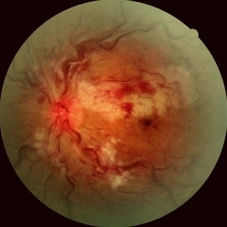
25-year-old male patient with acute and painless vision loss of left eye. The fundus examination demonstrate optic disc swelling, venous tortuosity, diffuse intraretinal hemorrhage and severe macular edema. There is also extensive exudative retinal detachment with lipid deposits in the posterior pole, mainly around the vessels. The systemic work up was negative, including serologies, rheumatologic and hematological markers and cholesterol and triglycerides within normal limits.
Photographer: Felipe Pereira
Imaging device: Vizucan, Zeiss
Condition/keywords: central vein occlusion, ischemic CRVO
-
 Central Retinal Vein Occlusion - After 3 Consecutive Ranibizumab Injections
Jan 26 2013 by Ratimir Lazic, MD, PhD
Color fundus photography of a 67-year-old female. Intraretinal hemorrhages in posterior pole, tortuous and dilated veins with optociliary shunts visible on optic nerve head. No macular edema can be noticed.
Photographer: Marko Lukic, MD
Imaging device: Zeis Visucam Lite 2
Condition/keywords: central retinal vein occlusion (CRVO)
-
 Choroidal MRSA Abscess
Apr 15 2021 by Rui Zhang, BA
A 14-year-old boy receiving induction chemotherapy for acute lymphocytic leukemia (ALL) complained of floaters and central scotoma in his left eye. (A) Fundus photography showed sub-macular choroidal abscess with intraretinal hemorrhage and edema. (B) OCT confirmed that the abscess had not penetrated the retinal pigment epithelium (RPE). Due to systemic septic signs (fever, tachycardia, tachypnea, new-onset papules), blood cultures were drawn and they came back positive for methicillin-resistant staphylococcus aureus (MRSA). Patient was promptly treated with both IV and intravitreal antibiotics. This is a case of sub-macular choroidal MRSA abscess in the setting of systemic bacteremia in an immunocompromised host.
Photographer: Raymond Mok, CRA COMT (Dartmouth-Hitchcock Medical Center)
Imaging device: Optical coherence tomography
Condition/keywords: abscess, acute leukemia, MRSA sepsis
-
 Chronic Retinal Vein Occlusion
Jul 8 2012 by Jeffrey S. Heier, MD
Chronic RVO with vascular changes, intraretinal hemorrhages
Imaging device: Zeiss
Condition/keywords: chronic retinal vein occlusion, intraretinal hemorrhage
-
 CMV Retinitis
Jan 10 2019 by Rahul Komati, MD
63-year-old male with history of plasma cell leukemia, presenting with photopsias and 20/25 vision. Fundus photograph shows superior area of retinitis, intraretinal hemorrhage, and vessel sclerosis. Retinitis regressed with systemic valganciclovir and 5 intravitreal foscarnet injections over 3 weeks.
Photographer: Pamela Hulvey, University of Chicago
Imaging device: Optos
Condition/keywords: CMV retinitis
-
---thumb.jpg/image-square;max$300,300.ImageHandler) CMV with leukemia
CMV with leukemia
Feb 15 2013 by From the Collections of Thomas M. Aaberg, MD and Thomas M. Aaberg Jr., MD
color fundus photograph of a patient with leukemia complicated by CMV retinitis, manifesting as intraretinal hemorrhage, nerve fiber layer infarction, and retinal exudation
Condition/keywords: leukemia
-
 Coat's Disease with Exudative RD
Feb 12 2025 by Tejaswita Verma
Fundus photo of a 7 year old boy with vision Counting fingers close to face in the right eye and intermittent outward deviation of the right eye observed by parents. Fundus examination shows subretinal exudates, telengiectatic vessels in superotemporal quadrant, intraretinal hemorrhages, macular scar, NVD.
Photographer: DR. TEJASWITA VERMA
Imaging device: MIRANTE
Condition/keywords: Coats' disease, exudative retinal detachment
-
 Combined Cilioretinal Artery and Central Retinal Vein Occlusion
May 14 2016 by Ines Leal
Combined cilioretinal artery and central retinal vein occlusion in an otherwise 49-year-old healthy female patient. Color fundus photography shows whitening of the retina in the distribution of the cilioretinal artery and intraretinal hemorrhages with tortuous and engorged veins.
Photographer: Inês Leal, MD, Department of Ophthalmology, Faculty of Medicine, Universidade de Lisboa,
Condition/keywords: cilioretinal artery occlusion, venous occlusion
-
 Diabetic Retinopathy
Apr 5 2018 by JYOTI PATIL, Ph.D.
A FAG image of a 74-year-old female. Diabetic changes of the posterior pole and midperipheral retina can be seen. dots (microaneurisms) and hypoflorescent areas (intraretinal hemorrhages) can be seen.
Photographer: Dr.Aditya Kelkar
Condition/keywords: diabetic retinopathy
 Loading…