 Initializing download.
-
 By JYOTI PATIL, Ph.D.
Savitribai Phule Pune Unviversity
Co-author(s): Dr.Aditya Kelkar - Uploaded on Apr 5, 2018.
- Last modified by Caroline Bozell on Apr 6, 2018.
- Rating
- Appears in
- Diabetic Retinopathy
- Condition/keywords
- diabetic retinopathy
- Photographer
- Dr.Aditya Kelkar
- Imaging device
- Optical coherence tomography system
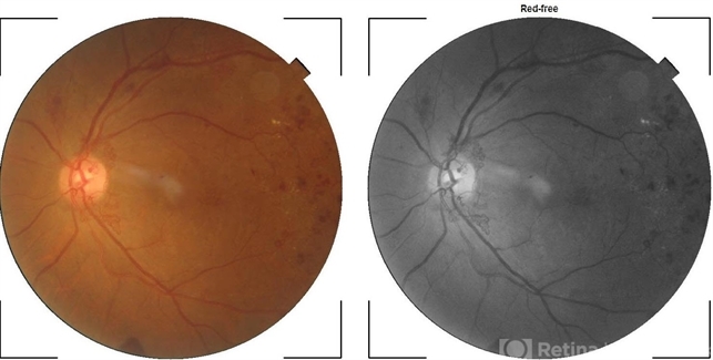
- Description
- A FAG image of a 74-year-old female. Diabetic changes of the posterior pole and midperipheral retina can be seen. dots (microaneurisms) and hypoflorescent areas (intraretinal hemorrhages) can be seen.


