Search results (111 results)
-
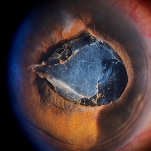 Inflammatory pupillary membrane in patient with endophthalmitis
Jan 28 2023 by Kingston Rodolfo Ureña-Wong, MD, Opht, MSc
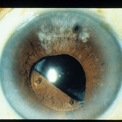
Anterior segment photography of a 54-year-old woman with post phacoemulsification endophthalmitis. She did not improve after first intravitreal antibiotics injection and develop an inflammatory pupillary membrane. After two vitrectomies, and a complete three intravitreal injections scheme, we decided to remove the intraocular lens and capsules.
Photographer: Marco Antonio Rubio-Atonal,UNAM, Asociación para evitar la ceguera en México
Imaging device: Zeiss Clarus 700
Condition/keywords: endophthalmitis, pupillary membranes
-
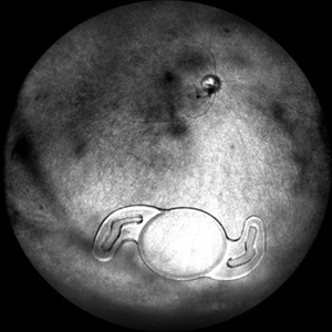
 Dropped IOL
Apr 5 2018 by Mohamed Tawfik, MD
Intra operative photo of a case of dropped IOL after phaco+IOL.
Photographer: Mohamed A,Tawfik
Imaging device: Intra operative Photography Screen shoot
Condition/keywords: dropped intraocular lens (IOL)
-
 Fraternal Twins
May 22 2023 by Gustavo M. Hüning, MD, MBA, FASRS
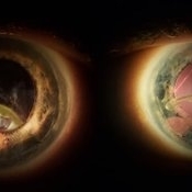
Intrasurgical photograph using a non-contact system and 3D visualization system of a 65-year-old woman who suffered an ocular trauma.
Photographer: Gustavo M. Hüning, Hüning Clínica do Olhar, Santa Maria - Brazil
Imaging device: Alcon Luxor combined with Alcon nGenuity
Condition/keywords: dislocated intraocular lens (IOL), implant, pars plana vitrectomy (PPV)
-
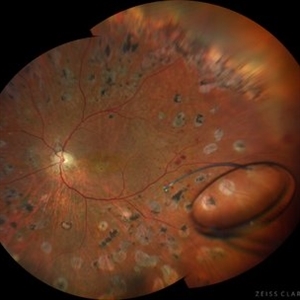 Intraocular lens luxated to the vitreous cavity
Jun 24 2023 by Mariam Cernichiaro-Espinosa, MD
Three-piece intraocular lens luxated to the vitreous cavity in a patient with photocoagulated diabetic retinopathy after blunt trauma.
Photographer: Mariam Cernichiaro-Espinosa, Asociación para Evitar la Ceguera en México, I.A.P. Mexico City, Mexico.
Imaging device: Zeiss Clarus
Condition/keywords: diabetic retinopathy, intraocular lense in vitreous, lens luxation
-
 Eye of the Hurricane
Apr 8 2025 by Gustavo Uriel Fonseca Aguirre
Ultrasound biomicroscopy of a post-operative eye (status post trabeculectomy and phacoemulsification) reveals a patent ostium on the right side, along with an intraocular lens in position. A hyphema is observed displaying small convection currents, creating a circular motion pattern due to the temperature gradient between the iris and cornea. Notably, the blood flow can be seen circulating toward the trabeculectomy site.
Photographer: Gustavo U. Fonseca Aguirre, Hospital Conde de Valenciana, Ciudad de México
Condition/keywords: Hyphema, trabeculectomy
-
Eye of the Hurricane
Apr 9 2025 by Gustavo Uriel Fonseca Aguirre
Ultrasound biomicroscopy of a post-operative eye (status post trabeculectomy and phacoemulsification) reveals a patent ostium on the right side, along with an intraocular lens in position. A hyphema is observed displaying small convection currents, creating a circular motion pattern due to the temperature gradient between the iris and cornea. Notably, the blood flow can be seen circulating toward the trabeculectomy site.
Condition/keywords: hyphema, trabeculectomy
-
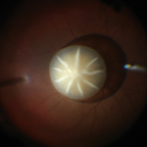 Dislocated Cataractous Lens
Jun 19 2025 by Mrinali Gupta, MD, FASRS
Intraoperative image of a chronically dislocated cataractous lens. The patient underwent pars plana vitrectomy, lensectomy, and placement of an anterior chamber intraocular lens, with improvement in vision from Count Fingers to 20/20 without correction.
Photographer: Mrinali Gupta, MD
Imaging device: Intraoperative surgical video (Zeiss Lumera scope, Resight lens)
Condition/keywords: dislocated crystalline lens
-
 Dislocated Intraocular Lens
Nov 15 2024 by Tejaswita Verma
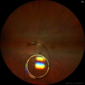
Fundus image of a spontaneously posteriorly dislocated IOL 10 years following surgery. Other eye had a subluxated opacified IOL.
Photographer: DR. TEJASWITA VERMA
Imaging device: MIRANTE
Condition/keywords: dislocated intraocular lens (IOL)
-
 Dislocated Intraocular Lens (IOL)
Aug 2 2019 by JEFFERSON R SOUSA, Tecg.º (Biomedical Systems Technology)
A 53-year-old male patient suffered blunt trauma 15 days after cataract surgery. Note total dislocation of the intraocular lens. No glass reaction.
Photographer: JEFFERSON R SOUSA - Study Center and Ophthalmological Research Dr. Andre M V Gomes, Institute Dr. Suel Abujamra São Paulo-Brazil
Imaging device: Topcon TRC-50 DX, Imaginet 4.0, angle de 50 graus. Flash 18w-s
Condition/keywords: dislocated intraocular lens (IOL)
-
 Dislocated IOL
Jan 13 2014 by David Callanan, MD
Dislocated IOL, 63-year-old female.
Condition/keywords: dislocated intraocular lens (IOL)
-
 Dislocated IOL
Jun 4 2024 by Marlee Curnutt
Slit lamp photo of a 64 year old woman presenting with worsening vision and depth perception after trauma induced by a dog, which dislocated her IOL. The patient's IOL haptic was seen in the AC, and almost abutting cornea. Patient's vision upon presentation was DCC CF@1 feet. Patient was counseled and underwent an IOL exchange.
Photographer: Marlee Curnutt, COA
Imaging device: Galaxy A42
Condition/keywords: dislocated intraocular lens (IOL), haptic, IOL, right eye, slit lamp photo, slit lamp photography, trauma
-
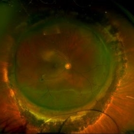
 Dislocated IOL
Sep 20 2025 by JORGE SOBERANES
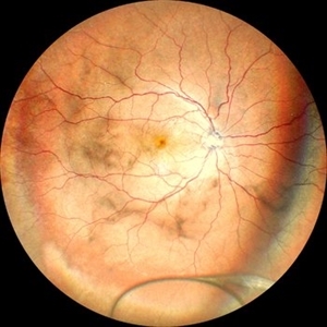
Fundus photograph of a 65-year-old man with a history of cataract surgery one year ago and bad vision since that.
Photographer: Dr. Jorge Soberanes, APEC, Universidad Nacional Autónoma México
Condition/keywords: dislocated lens, intraocular lens dislocation
-
 Dislocated IOL
May 15 2018 by Morgan Benton
Ultra-wide field pseudocolor image of a 68-year-old male with a dislocated IOL after cataract surgery in the left eye. Patient was only able to count fingers at one foot and could pinhole to 20/60.
Photographer: Morgan Benton
Imaging device: Optos
Condition/keywords: color fundus photograph, dislocated intraocular lens (IOL), left eye, Optos, ultra-wide field imaging
-
 Dislocated Lens
Jun 29 2013 by Jason S. Calhoun
84-year-old female comes in with blurred vision in the left eye. VA was 20/30, right eye and count fingers in the left eye. Fundus examination reveals dislocation of the IOL into the vitreous inferiorily at 6-o'clock. Suggest surgery to fix the problem.
Photographer: Jason S. Calhoun, Mayo Clinic Jacksonville, Florida
Imaging device: TOPCON TRC 50-EX
Condition/keywords: dislocated posterior chamber intraocular lens (PCIOL)
-
 Dislocated-P/C IOL Bag Complex
Nov 27 2018 by Maria H. Berrocal, MD
85-year-old who underwent phaco IOL 15 years prior, who noticed loss of vision OS.
Photographer: Thaylan Calderon, Berrocal & Associates, San Juan, PR
Imaging device: Optos
Condition/keywords: dislocated intraocular lens (IOL)
-
 Dropped Lens and IOLs
Apr 5 2018 by Mohamed Tawfik, MD
Group photo of dropped crystalline lens and IOLs.
Photographer: Mohamed A.Tawfik MD , FRCSed
Imaging device: intra opeative Photography Screen shoot
Condition/keywords: dropped intraocular lens (IOL), dropped nucleus
-
 IOL
IOL
Jan 17 2018 by Emily Cooper
Optos image of 47-year-old man with a now worsening retinal detachment that had been treated by pneumatic retinopexy.
Photographer: Emily Cooper, Retina Specialists of Michigan
Imaging device: Optos Ultra Wide Field
Condition/keywords: chronic retinal detachment, intraocular lens (IOL)
-
 IOL in Vitreous Cavity Right Eye
May 22 2021 by Akansha Sharma
Fundus infra-red image of a 47-year-old male with IOL in vitreous cavity right eye.
Photographer: Dr. Akansha Sharma-Retina Foundation, Ahmedabad
Condition/keywords: intraocular lens (IOL)
-
 Kissing Choroidals
Dec 18 2025 by Talhah - Zubair, MD
72 year old woman developed suprachoroidal hemorrhage during Yamane scleral intraocular lens fixation. At clinic follow up they were found to be appositional. Suprachoroidal tissue plasminogen activator was injected in clinic and an emergent choroidal cutdown drainage was performed the following day with subsequent resolution of apposition. Appositional choroidals were managed urgently to avoid retinal adhesion/membrane formation.
Condition/keywords: appositional choroidals, kissing choroidals, suprachoroidal hemorrhage
-
 Metastatic Breast Carcinoma
Jan 21 2021 by Jamin S. Brown, MD
This anterior segment photograph was taken with a smartphone camera attached to a regular Haag Streit slit lamp ocular demonstrates unusual clustering of white cells on the posterior surface of the intraocular lens. The clinical diagnosis is metastatic breast carcinoma to the vitreous, which is very rare.
Photographer: Stefanie Palmer CRA, Retina Vitreous Surgeons of CNY
Imaging device: Cell phone camera
Condition/keywords: anterior segment, breast cancer, cell phone camera, slit lamp photo
-
---thumb.JPG/image-square;max$300,300.ImageHandler) Retinal Detachment With Dislocated IOL Lens
Retinal Detachment With Dislocated IOL Lens
Jun 30 2013 by Jason S. Calhoun
47-year-old male who had trauma to the right eye. Patient had retinal detachment surgery in the past (scleral buckle), to the right eye. Patient came in with another retinal detachment with dislocated PC IOL lens. Notice the haptics tearing the retina. Patient underwent vitrectomy with gas exchange. VA was hand motion 1 day post-op.
Photographer: Jason S. Calhoun, Mayo Clinic Jacksonville, Florida
Condition/keywords: dislocated posterior chamber intraocular lens (PCIOL), retinal tear
-
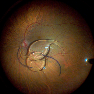
 Rhegmatogenous retinal detachment with dislocated IOL in a Morning Glory anomaly
Jul 27 2023 by Gustavo Aguirre-Suarez
Fundus photograph of a 13-year-old male with a history of congenital cataract surgery in his right eye in 2019. The patient presents with sudden visual loss. Upon examination, a dislocated IOL is observed in the posterior segment, accompanied by a rhegmatogenous retinal detachment featuring peripheral retinal tears and horseshoe breaks. Additionally, a morning glory disc anomaly is also present in this patient.
Photographer: Gustavo Aguirre-Suarez
Imaging device: Mirante, NIDEK
Condition/keywords: dislocated posterior chamber intraocular lens (PCIOL), Morning Glory Anomaly, rhegmatogenous retinal detachment
-
 Scleral Buckling IOL Drop
Aug 6 2023 by Dr.Sheetal Divate
A 27 year old female with an old history of trauma and operated with scleral buckling and cataract surgery in the past came recently with complaints of DOV . Findings noted where IOL drop, inferior retinal detachment and old scleral buckle indent.
Photographer: Dr.Sheetal Divate
Imaging device: Optos Advance
Condition/keywords: dislocated intraocular lens (IOL), Retinal Detachment, scleral buckle
-
 Subconjuntival IOL After Blunt Trauma
Jun 27 2018 by Gabriel Costa Andrade, PhD
A 73-year-old male patient was referred to our ophthalmic emergency department with complaints of redness, pain, and diminution of vision in his left eye, after fall from height. The patient underwent small incision cataract surgery with polymethylmethacrylate (PMMA) IOL implantation in both the eyes 15 years back through superior sclerocorneal incision under local anesthesia. His best-corrected visual acuity was perception of light in the left eye. Ophthalmic examination using slit lamp biomicroscopy of the left eye revealed diffuse subconjunctival hemorrhage with no conjunctival laceration and inferior bulbar conjunctiva showed traumatic pseudophacocele with a sign “golden half ring,” suggesting the presence of PCIOL in subconjunctival space.There was total hyphema obscuring the view of rest of the ocular structures in his left eye.
Photographer: Gabriel Andrade, RETINA CLINIC, São Paulo, BRAZIL
Condition/keywords: dislocated intraocular lens (IOL), trauma
-
 IOL With BAG in Vitreous of Myopic Eye
Apr 14 2017 by Manish Nagpal, MD, FRCS (UK), FASRS
50-year-old male having myopia presented with a IOL in vitreous within its bag.
Photographer: Pooja Barot
Condition/keywords: intraocular lens (IOL), intraocular lense in vitreous, intraocular lense with bag, myopia
 Loading…