Search results (169 results)
-
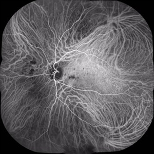
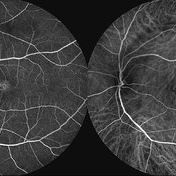
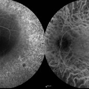
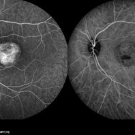 Best Disease
Mar 9 2013 by Hamid Ahmadieh, MD
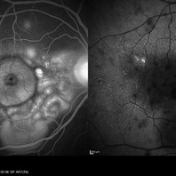
FA and ICG Angiography of the left eye of a 49-year-old man with advanced Best disease.
Photographer: Soodabeh Fooladin, Negah Eye Center, Tehran
Imaging device: Heidelberg Spectralis
Condition/keywords: Best disease, indocyanine green (ICG) angiography
-
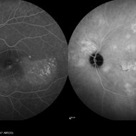 Central Serous Chorioretinopathy
Jan 25 2022 by Olivia Rainey
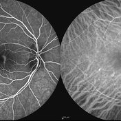
Late phase widefield fluorescein angiography of a 60-year-old male with Central Serous Chorioretinopathy. Chronic history of CSR followed with observation without treatment prior to presenting at our office. The physician noted subfoveal subretinal fluid with recent visual decline. FA shows multifocal leakage and ICG shows hypercyanescence. OCTA, ICG, and FA consistent with CSR, and without concern for CNVM thus will observe without anti-VEGF at this time. PDT therapy recommended.
Photographer: Olivia Rainey, OCT-C, COA
Imaging device: Heidelberg Spectralis
Condition/keywords: 55-degrees, central serous chorioretinopathy (CSCR), central serous retinopathy (CSR), chronic central serous chorioretinopathy (CSCR), fluorescein angiogram (FA), heidelberg spectralis, indocyanine green (ICG) angiography, left eye
-
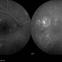 Central Serous Chorioretinopathy
Jan 25 2022 by Olivia Rainey
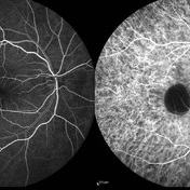
Late phase widefield fluorescein angiography of a 60-year-old male with Central Serous Chorioretinopathy. Chronic history of CSR followed with observation without treatment prior to presenting at our office. The physician noted significant findings on exam and imaging with multifocal areas of inactive and active changes OD. FA shows superotemporal macular leakage, subtle inferonasal macular leakage and staining as well as multifocal hypercyanescence on ICG. Fortunately foveal sparing and thus observation is recommended at this time OD.
Photographer: Olivia Rainey, OCT-C, COA
Imaging device: Heidelberg Spectralis
Condition/keywords: 55-degrees, central serous chorioretinopathy (CSCR), central serous retinopathy (CSR), chronic central serous chorioretinopathy (CSCR), fluorescein angiogram (FA), fluorescein leakage, heidelberg spectralis, indocyanine green (ICG) angiography, late phase
-
 Choroidal Melanoma 3 Ways
Jan 16 2025 by Virginia Gebhart
RGB/FA/ICG of 76 year old female with a new choroidal melanoma. Pt scheduled for plaque radiation. BCVA 20/400
Photographer: Virginia Gebhart, Retina Consultants of Carolina
Imaging device: Optos California
Condition/keywords: fluorescein angiogram (FA), indocyanine green (ICG) angiography, OPTOS CALIFORNIA RGB
-
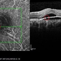 Discontinuity RPE
Oct 17 2014 by Avris Romario Diparaja Siahaan
A simultan ICG angiography + OCT of 56-year-old man that shows a image of discontinuity retinal pigment ephitelial.
Photographer: Harni Christine Damanik, Klinik Mata Nusantara
Imaging device: Heidelberg Spectralis
Condition/keywords: indocyanine green (ICG) angiography, optical coherence tomography (OCT), retinal pigment epithelium
-
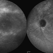
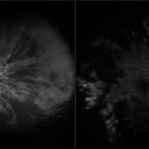
 Indocyanine Green (ICG) of Circumscribed Choroidal Hemangioma (CCH)
Feb 6 2025 by Jack B Margines, MD, MHCI
Peripheral patchy hyperfluorescence is seen on this early image of ICG-A on a 53-year-old asymptomatic with an extramacular circumscribed choroidal hemangioma.
Photographer: W Ryan Miliam, CRA, OCT-C, University of California, Irvine Gavin Herbert Eye Institute
Imaging device: Optos
Condition/keywords: choroidal hemangioma, indocyanine green (ICG) angiography
-
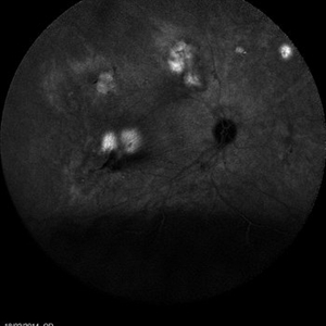
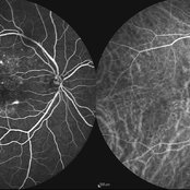
 Multifocal Choroiditis
Aug 16 2018 by FELIPE PEREIRA
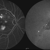
Mid-phase indocyanine green angiography of a 25-year-old woman with sudden central vision loss and photopsias for 7 days. The hypofluorescent lesions in the macula and nasal to the disc correspond to the yellow-white deep lesions in the fundus examination. No leakage is observed at any stage of the exam
Photographer: Claudio Zett Lobos
Imaging device: HEIDELBERG SPECTRALIS HRA
Condition/keywords: indocyanine green (ICG) angiography, multifocal choroiditis, white dot syndrome
-
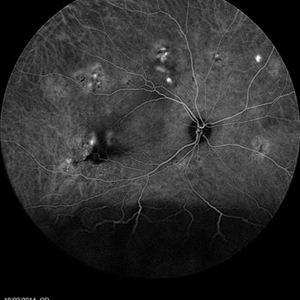
 Multifocal Exudative Detachments Due to VKH
May 14 2014 by Avris Romario Diparaja Siahaan
ICG (Late Phase) a 38-year-old man with multifocal CSR and inferior exudative retinal detachment on both eyes (Harada Syndrome).
Photographer: Avris Romario Diparaja Siahaan, Klinik Mata Nusantara
Imaging device: Heidelberg HRA + OCT Spectralis
Condition/keywords: indocyanine green (ICG) angiography, multifocal central serous chorioretinopathy (CSCR)
-
 Multifocal Exudative Detachments Due to VKH
May 14 2014 by Avris Romario Diparaja Siahaan
ICG (Mid Phase) a 38-year-old man with multifocal CSR and inferior exudative retinal detachment on both eyes (Harada Syndrome).
Photographer: Avris Romario Diparaja Siahaan, Klinik Mata Nusantara
Imaging device: Heidelberg HRA + OCT Spectralis
Condition/keywords: indocyanine green (ICG) angiography, multifocal central serous chorioretinopathy (CSCR)
-
 PCV
PCV
Jul 26 2014 by Avris Romario Diparaja Siahaan
An ICG image of a 65-year-old-woman with PCV on her left eye (mid phase).
Photographer: Avris Romario Diparaja Siahaan, Klinik Mata Nusantara
Imaging device: Heidelberg Spectralis
Condition/keywords: indocyanine green (ICG) angiography, polypoidal choroidal vasculopathy (PCV)
-
 Vogt Koyanagi Harada
Oct 7 2015 by Avris Romario Diparaja Siahaan
Simultaneous FA + ICG (Late Phase) of a 42-year-old woman with Harada Syndrome in both eyes.
Photographer: Yohanes Harry Purwanto, Klinik Mata Nusantara
Imaging device: Heidelberg HRA + OCT
Condition/keywords: indocyanine green (ICG) angiography, late phase, Vogt-Koyanagi-Harada
-
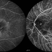
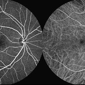
 Acute Central Serous Chorioretinopathy
Sep 15 2012 by Hamid Ahmadieh, MD
Mid-phase FA & ICG angiography images of a 30-year-old man with acute CSCR.
Photographer: Hamid Ahmadieh, MD, Ophthalmic Research Center, Labbafinejad Medical Center, Shahid Beheshti University of Medical Sciences
Imaging device: Heidelberg HRA
Condition/keywords: central serous chorioretinopathy (CSCR), indocyanine green (ICG) angiography
-
 PED due to CSCR 4
Sep 2 2012 by Hamid Ahmadieh, MD
Early phase FA & ICG images of a 37-year-old man with a serous PED secondary to CSCR
Photographer: Hamid Ahmadieh, Ophthalmic Research Center, Labbafinejad Medical Center
Imaging device: Heidelberg Spectralis
Condition/keywords: central serous chorioretinopathy (CSCR), indocyanine green (ICG) angiography, pigment epithelial detachment (PED)
-
 Polypoidal Choroidal Vasculopathy: Case 1 - Image 6of 7
Oct 4 2012 by Gregg T. Kokame, MD, MMM, FASRS
OCT/Indocyanine Green Angiography image of a 57-year-old woman with treatment-naive polypoidal choroidal vasculopathy. Series of images provides an comparative view of the same condition while utilizing a variet of different imaging procedures.
Photographer: Andrew Yuen, Retina Consultants of Hawaii
Imaging device: Heidelberg Spectralis
Condition/keywords: branching vascular network (BVN), indocyanine green (ICG) angiography, optical coherence tomography (OCT), polypoidal choroidal vasculopathy (PCV)
-
 Angioid Streaks
Sep 29 2012 by Hamid Ahmadieh, MD
Late phase ICG angiography image of the right eye of a 59-year-old man with angioid streaks.
Photographer: Hamid Ahmadieh, MD; Ophthalmic Research Center, Labbafinejad Medical Center, Shahid Beheshti University of Medical Sciences
Imaging device: Heidelberg Spectralis
Condition/keywords: angioid streaks, indocyanine green (ICG) angiography
-
 Angioid Streaks
Sep 29 2012 by Hamid Ahmadieh, MD
Late phase ICG angiography image of the left eye of a 59-year-old man with angioid streaks.
Photographer: Hamid Ahmadieh, MD; Ophthalmic Research Center, Labbafinejad Medical Center, Shahid Beheshti University of Medical Sciences
Imaging device: Heidelberg Spectralis
Condition/keywords: angioid streaks, indocyanine green (ICG) angiography
-
 Multifocal CSCR
Sep 2 2012 by Hamid Ahmadieh, MD
Late-phase FA and ICG angiograms of a 36-year-old man with an active multifocal CSCR.
Photographer: Hamid Ahmadieh, Ophthalmic Research Center, Labbafinejad Medical Center
Imaging device: Heidelberg Spectralis
Condition/keywords: central serous chorioretinopathy (CSCR), indocyanine green (ICG) angiography
-
 Macular Telangiectasia Type 2 & CNV
Sep 22 2012 by Hamid Ahmadieh, MD
FA and ICG angiography imagings of the left eye of a 70-year-old man with idiopathic macular telangiectasia type 2 and CNV.
Photographer: Hamid Ahmadieh, MD, Ophthalmic Research Center, Labbafinejad Medical Center, Shahid Beheshti University of Medical Sciences
Imaging device: HRA
Condition/keywords: choroidal neovascularization (CNV), idiopathic macular telangiectasia, indocyanine green (ICG) angiography
-
 Cystoid Macular Edema (CME)
Sep 11 2012 by Hamid Ahmadieh, MD
Late phase FA & ICG angiography imagings of the left eye a 17-year-old boy with CME & retinal periphlebitis secondary to chronic intermediate uveitis.
Photographer: Hamid Ahmadieh, MD, Ophthalmic Research Center, Labbafinejad Medical Center, Shahid Beheshti University of Medical Sciences
Imaging device: Heidelberg Spectralis
Condition/keywords: cystoid macular edema (CME), indocyanine green (ICG) angiography, intermediate uveitis
-
 Multifocal CSCR 2
Sep 2 2012 by Hamid Ahmadieh, MD
Early-phase FA and ICG angiograms of a 36-year-old man with an active multifocal CSCR.
Photographer: Hamid Ahmadieh, Ophthalmic Research Center, Labbafinejad Medical Center
Imaging device: Heidelberg Spectralis
Condition/keywords: central serous chorioretinopathy (CSCR), indocyanine green (ICG) angiography
-
 Polypoidal Choroidal Vasculopathy
Aug 25 2012 by Hamid Ahmadieh, MD
FA & ICG angiography imagings of a 73-year-old man with a peripapillary PCV.
Photographer: Hamid Ahmadieh, Ophthalmic Research Center, Labbafinejad Medical Center
Imaging device: Heidelberg Spectralis
Condition/keywords: indocyanine green (ICG) angiography, polypoidal choroidal vasculopathy (PCV)
-
 Retinitis Pigmentosa
Sep 11 2012 by Hamid Ahmadieh, MD
FA & ICG angiography images of a 40-year-old man with RP.
Photographer: Hamid Ahmadieh, MD, Ophthalmic Research Center, Labbafinejad Medical Center, Shahid Beheshti University of Medical Sciences
Imaging device: Heidelberg Spectralis
Condition/keywords: indocyanine green (ICG) angiography, retinitis pigmentosa
-
 Macular Telangiectasia Type 2
Sep 22 2012 by Hamid Ahmadieh, MD
FA and ICG angiography imagings of the right eye of a 70-year-old man with idiopathic macular telangiectasia type 2.
Photographer: Hamid Ahmadieh, MD, Ophthalmic Research Center, Labbafinejad Medical Center, Shahid Beheshti University of Medical Sciences
Imaging device: HRA
Condition/keywords: idiopathic macular telangiectasia, indocyanine green (ICG) angiography
-
 Acute Central Retinal Artery Occlusion
Jul 27 2022 by Becca Harris
Ultra widefield FA/ICG of a 24 year old female with an acute central retinal artery occlusion affecting the right eye. Patient presented with extreme headaches following DAVF surgery the previous day. Patient has Factor VIII deficiency and had a cerebral venous thrombosis 9 years ago and lost vision in the right eye at that time. Patient has history of optic sheath fenestration OU and craniotomy. On initial evaluation, she had a CRAO as well as diffuse choroidal nonperfusion noted on optos FA. Suspect nonperfusion to third and sixth nerve leading to palsy. Occlusion of vasculature in the setting of recent endovascular embolization of fistulas in the CNS. Discussed diagnosis and poor prognosis with parents and patient. Patient had no light perception at the time of her initial appointment.
Photographer: Becca Harris
Imaging device: Optos California
Condition/keywords: Choroidal non-perfusion, fluorescein angiogram (FA), indocyanine green (ICG) angiography, non-perfusion, Optos, Right Eye, ultra-wide field imaging
-
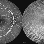 Acute Central Serous Chorioretinopathy
Sep 15 2012 by Hamid Ahmadieh, MD
Early phase FA & ICG angiography images of a 30-year-old man with acute CSCR.
Photographer: Hamid Ahmadieh, MD, Ophthalmic Research Center, Labbafinejad Medical Center, Shahid Beheshti University of Medical Sciences
Imaging device: Heidelberg HRA
Condition/keywords: central serous chorioretinopathy (CSCR), indocyanine green (ICG) angiography
 Loading…