Search results (159 results)
-
 Hypertensive Retinopathy
Feb 25 2013 by Suber S. Huang, MD, MBA, FASRS
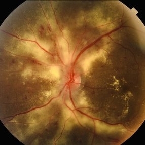
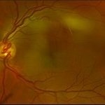
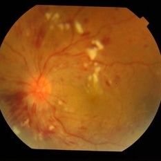
32-year-old African American male with Grade IV hypertensive retinopathy and acute renal failure. Vision OD 20/70, OS 20/25. Creatine 7.1. BP: 250/150.
Photographer: Geoffrey Pankhurst, University Hospitals, Eye Institute/Dept. Ophthalmology and Visual Sciences Case Western Reserve University Cleveland, OH
Imaging device: Topcon TRC 50x
Condition/keywords: acute renal failure, disc edema, exudate, hypertension, hypertensive retinopathy, ischemia, macular edema, macular ischemia, optic disc edema
-
 Hypertensive Retinopathy
Apr 21 2024 by César Adrián Gómez Valdivia, MD
Hypertensive Retinopathy
Photographer: Erika Paulina Ornelas Cazares
Imaging device: TOPCON TRC-50DX
Condition/keywords: hypertensive retinopathy
-
 Hypertensive Retinopathy
May 26 2025 by César Adrián Gómez Valdivia, MD
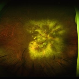
Fundus photograph of a 62 year-old woman with history of untreated hypertension and chronic kidney disease. Findings were bilateral.
Photographer: @eyemissu2
Imaging device: OPTOS
Condition/keywords: hypertensive retinopathy
-
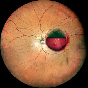 Premacular Subhyaloid Hemorrhage
Jan 20 2021 by Nivesh Gupta
A 41-year-old male patient complaining of diminution of vision in left eye since 6 days. His best corrected visual acuity finger counting at 2 meters.
Photographer: Nivesh Gupta, Retina Fellow, Retina Foundation, Ahmedabad, India
Condition/keywords: hypertensive retinopathy, preretinal hemorrhage, subhyaloid hemorrhage
-
 Prepapillary Vascular Loop
Mar 11 2020 by Asdrubal F Moreno, MD
Fundus color photograph of a 80-year-old woman with a unilateral congenital prepapillary vascular loop and hypertensive retinopathy, focus on the vascular loop end for perception.
Photographer: Asdrubal Moreno, Fundacion AVAO, Universidad de Los Andes, Venezuela
Imaging device: Zeiss Visucam 500
Condition/keywords: congenital prepapillary vascular loop, peripapillary
-
 Anterior Uveitis
May 4 2021 by Hannah Keller
Anterior Uveitis OPTOS on 81-year-old male.
Photographer: Hannah Keller, Retina Specialists of Michigan
Condition/keywords: anterior uveitis, hypertensive retinopathy, OD, Optos
-
 BRVO / Arterial Embolus / Hypertensive Retinopathy
Apr 8 2014 by David Callanan, MD
77-year-old white female, BRVO / arterial Embolus / hypertensive retinopathy.
Condition/keywords: arterial embolus, branch retinal vein occlusion (BRVO), hypertensive retinopathy
-
 HTN Retinopathy with Pre-Papillary Vascular Loop OS
Jun 4 2018 by Hosam Attia, MD
Close-up color fundus photograph of 53-year-old, African American male with history of diabetes, hypertension, depicting chronic hypertensive retinopathy changes and unilateral pre-papillary vascular loop OS.
Imaging device: Optos California
Condition/keywords: congenital prepapillary vascular anomaly, congenital prepapillary vascular loop, prepapillary vascular loop
-
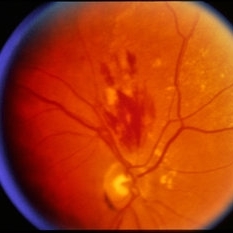
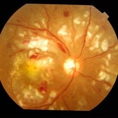
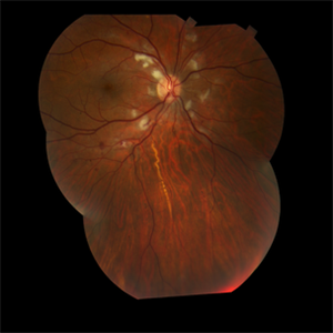
 Hypertensive Retinopathy
Jun 28 2013 by Jason S. Calhoun
Patient came in complaining of spots in vision in both eyes. VA was 20/25 - right eye and 20/20- left eye. Fundus exam reveals little hemorrhages with cotton wool spots due to hypertension and anemia.
Photographer: Jason S. Calhoun, Mayo Clinic Jacksonville, Florida
Imaging device: TOPCON TRC 50-EX
Condition/keywords: hypertensive retinopathy
-
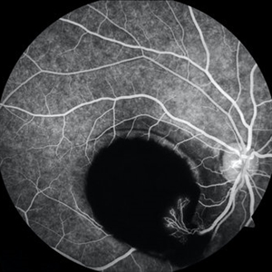
 Hypertensive Retinopathy
Aug 24 2012 by Geoffrey G. Emerson, MD, PhD, FASRS
A 35-year-old man has headaches and decreased vision. The right eye measures 20/25 and the left eye measures 3/200. The blood pressure measures 180/110.This fluorescein angiogram shows leakage of dye from the optic disc (papilledema), ischemia, and dilated capillaries around the foveal avascular zone
Photographer: Geoffrey Emerson, MD, PhD, Retina Center, Minneapolis
Condition/keywords: hypertensive retinopathy, ischemia, papilledema
-
 Hypertensive Retinopathy
Aug 24 2012 by Geoffrey G. Emerson, MD, PhD, FASRS
A 35-year-old man has headaches and decreased vision. The right eye measures 20/25 and the left eye measures 3/200. The blood pressure measures 180/110.
Photographer: Geoffrey Emerson, MD, PhD, Retina Center, Minneapolis
Condition/keywords: hypertensive retinopathy, papilledema, serous retinal detachment
-
 Hypertensive Retinopathy
Oct 20 2012 by Hyung-Woo Kwak, MD
The sudden appearance of cotton wool spots with hypertension retinopathy is known as accelerated hypertension. This patient has acute hypertension of rapid onset and measured systolic blood pressure was more than 200mmhg at this time.
Imaging device: Zeiss F450 plus
Condition/keywords: cotton wool spots, hypertension
-
 Hypertensive Retinopathy
Dec 24 2017 by Purva Patwari
52-year-old female diagnosed of hypertension by retina evaluation.
Photographer: Dr Purva Patwari, Patwari Retina Center, Ahmedabad, Gujarat , India
Imaging device: ZEISS VISU500
Condition/keywords: hypertensive retinopathy, neovascularization elsewhere (NVE), Roth spots
-
 Hypertensive Retinopathy
Feb 10 2016 by Mallika Goyal, MD
Bilateral hypertensive retinopathy in a 19-year-old girl with renal disease, hypertension and anemia.
Photographer: Mallika Goyal, MD, Apollo Health City, Hyderabad, India
Condition/keywords: hypertensive retinopathy
-
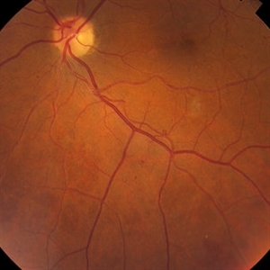
 Hypertensive Retinopathy
Jun 4 2025 by Paulina Araujo
The 55-degree central fundus photograph of the right eye reveals vascular tortuosity, generalized arteriolar narrowing with a vein-to-artery ratio of 3:1, along with Guist and Bonnet signs.
Photographer: Paulina D.Araujo Martínez, Asociación para Evitar la Ceguera en México I.A.P., Hospital Dr Luis Sánchez Bulnes.
Condition/keywords: hypertensive retinopathy
-
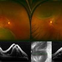 Hypertensive Retinopathy Pre
Mar 10 2014 by Dong Yoon Kim, MD
20-year-old women who had severe preeclamsia visited our clinic for decreased visual acuity on her both eyes. Her visual acuity was 20/100 on both eyes. Fundus examination revealed serous retinal detachment on both eyes. OCT examination revealed subretinal and intraretinal fluid.
Photographer: Sun Tae Kim, University of Ulsan, Asan Medical Center
Imaging device: Optos C200 MA scanning laser ophthalmoscope
Condition/keywords: hypertensive retinopathy
-
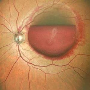
 Hypertensive Retinopathy With Bilateral Serous Retinal Detachment at Macula
Jul 29 2014 by Mallika Goyal, MD
Left eye fundus of a 36-year-male with sudden vision drop shows grade 4 hypertensive retinopathy with retinal hemorrhages, exudates and ischaemic disc edema. OCT revealed serous retinal detachment at macula.
Photographer: Mallika Goyal, MD, Apollo Health City, Jubilee Hills, Hyderabad-500033
Condition/keywords: hypertensive retinopathy
-
 Papilledema With Hypertensive Retinopathy
Apr 14 2014 by Dipankar Barua, M.Sc
Male patient, 25-years-old. On examination his vision of the right eye is 6/6 and left eye is 6/36. It seems to be a case papilloedema with hypertensive retinopathy.
Photographer: Dipankar Barua
Imaging device: Topcon TRC 50 DX (IA)
Condition/keywords: hypertensive retinopathy, papilledema
-
 Pre Macular Subhyaloid Hemorrhage
Jan 20 2021 by Nivesh Gupta
A 41 year old male patient complaining of diminution of vision in left eye since 6 days. His best corrected visual acuity finger counting at 2 meters.
Photographer: Nivesh Gupta, Retina Fellow, Retina Foundation, Ahmedabad, India
Condition/keywords: hypertensive retinopathy, preretinal hemorrhage, subhyaloid hemorrhage
-
 Sub-ILM Hemorrhage with Neovessels
Apr 30 2020 by Saurabh Deshmukh, MBBS, DNB, FVRS, MNAMS
Late arteriovenous phase FA showing a large sub-internal limiting membrane hemorrhage with overlying neovessels. This hypertensive patient presented with a visual acuity of counting fingers at 2 meters. The patient was advised intravitreal anti-VEGF injection, Nd: YAG Membranotomy, and systemic control of hypertension.
Photographer: Saurabh Deshmukh, Sri Sankaradeva Nethralaya, Guwahati, India
Imaging device: Topcon TRC-50 DX
Condition/keywords: hypertensive retinopathy, neovascularization elsewhere (NVE), subILM hemorrhage
-
 Diabetic Retinopahty
Nov 2 2022 by pedro fernandes souza neto
Fundus photograph of a 40-year-old man with diabetes and hypertension shows hard exudates, difuse intraretinal hemorrhages and splinter hemorrhages.
Photographer: Pedro Fernandes, Universidade Federal da Bahia, Brazil.
Condition/keywords: diabetic mellitus, hypertensive retinopathy, retinopathy
-
 Prepapillary Vascular Loop
Mar 11 2020 by Asdrubal F Moreno, MD
Fundus color photograph of a 80-year-old woman with a unilateral congenital prepapillary vascular loop and hypertensive retinopathy, focused on the retinal plane for perception.
Photographer: Asdrubal Moreno, Fundacion AVAO, Universidad de Los Andes, Venezuela
Imaging device: Zeiss Visucam 500
Condition/keywords: congenital prepapillary vascular loop, peripapillary
-
 Siegrist Streak
Nov 6 2012 by F. Ryan Prall, MD
32-year-male with history of hypertension, recent admission for malignant hypertension.
Photographer: Tom Egnatz, Indiana University
Condition/keywords: hypertensive retinopathy, malignant hypertension
-
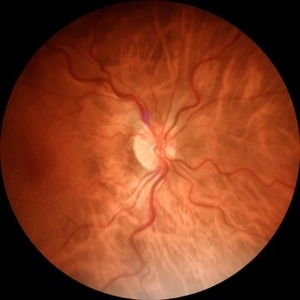
 Copperwire Reflex and AV-Nipping
Mar 1 2014 by Homayoun Tabandeh, MD, FASRS
Copperwire reflex and AV-Nipping in a patient with hypertensive retinopathy.
Condition/keywords: hypertensive retinopathy
-
---thumb.jpg/image-square;max$300,300.ImageHandler) Hypertensive Retinopathy
Hypertensive Retinopathy
Jan 8 2014 by David Callanan, MD
43-year-old male with hypertensive retinopathy.
Condition/keywords: hypertensive retinopathy
 Loading…