Search results (55 results)
-
 Post Subretinal tpa , viterectomy and gas
May 6 2022 by Shobhit Chawla, M.S.
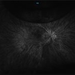
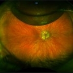
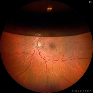
SUBMACULAR HAEMORRHAGE IN A 38YEAR OLD LADY PATIENT CAUSE POLYP BLEED IN PCV. Following viterectomy , subretinal tpa . gas and aflibercept injection. 7 day post operative image.
Photographer: Shobhit Chawla
Imaging device: Zeiss Clarus 500
Condition/keywords: aflibercept, intravitreal gas bubble, submacular hemorrhage, tissue plasminogen activator (tPA), vitrectomy
-
 Anterior Segment Gas Bubble and PFC Interface
Jun 21 2018 by Maria Stephanie R. Jardeleza, MD
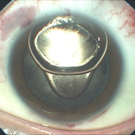
Anterior segment photographs of 30-year-old male who underwent superior rhegmatogenous retinal detachment repair with intraocular gas tamponade. Perfluorocarbon was used to flatten the macula to prevent a macular fold and was removed during PFC/air exchange. Post operative week two visit shows gas migration into the anterior chamber with retained PFC on the posterior aspect of the gas bubble/anterior surface of the lens. Patient had been maintaining face down positioning.
Photographer: Andy Zepeda, COA, Retina Clinic, San Antonio Eye Center, San Antonio, TX
Condition/keywords: retained perfluorocarbon, vitreous substitutes
-
---thumb.jpg/image-square;max$300,300.ImageHandler) C3F8 gas bubble after retinal detachment surgery
C3F8 gas bubble after retinal detachment surgery
Feb 1 2013 by Sharon Fekrat, MD FACS FASRS
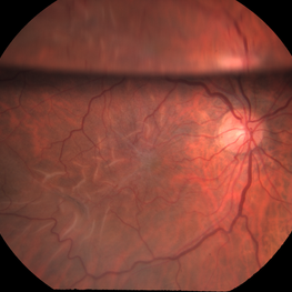
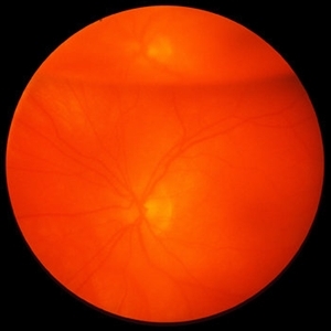
63 year old man s/p encircling scleral buckle and 23g pars plana vitrectomy for a macula off phakic rhegmatogenous retinal detachment. This fundus photograph shows the effect of the encircling buckle and the residual C3F8 intravitreal gas bubble in the right eye.
Photographer: Tiffanie Keaton, Duke Eye Imaging, Duke University Eye Center, Durham, NC
Imaging device: Optos
Condition/keywords: intravitreal gas bubble, vitrectomy
-
 Gas Bubble Extending into Anterior Chamber
Oct 12 2012 by Jeffrey G. Gross, MD, FASRS
Gas bubble extending into anterior chamber in aphakic eye, after PPV.
Condition/keywords: anterior chamber, aphakic eye, gas bubble, pars plana vitrectomy (PPV)
-
 Pseudophakic RRD, S/P Buckle/Vit. w/ Residual Gas Fish Eggs OD
May 23 2018 by Hosam Attia, MD
71-year-old male, s/p combined buckle vitrectomy for recurrent, macula-off, rhegmatogenous retinal detachment, with residual gas fish eggs OD.
Imaging device: Optos California Ultra-Wide Field Fundus Camera
Condition/keywords: encircling scleral buckle, gas bubble, intraocular gas, intravitreal gas bubble
-
 Retinal Detachment with Retinal Hole
Sep 30 2013 by Jason S. Calhoun
Patient in with complaints of floaters in the right eye. VA was 20/40 with no improvement. Fundus exam shows retinal detachment from 9-12 o'clock with hole at 10:30 posteriorly. Pneumatic retinopexy was performed with C3F8 Gas bubble and laser around the retinal tear in the right eye.
Photographer: Jason S. Calhoun, Department of Ophthalmology, Mayo Clinic Jacksonville, Florida
Imaging device: TOPCON TRC 50-EX
Condition/keywords: retinal hole
-
 Anterior Chamber Gas and PFC Migration
Jun 21 2018 by Maria Stephanie R. Jardeleza, MD
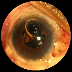
Anterior segment photographs of 30-year-old male who underwent superior rhegmatogenous retinal detachment repair with intraocular gas tamponade. Perfluorocarbon was used to flatten the macula to prevent a macular fold and was removed during PFC/air exchange. Post operative week two visit shows gas migration into the anterior chamber with retained PFC layered in a tear drop shape posterior to the gas bubble and anterior to the lens. Patient had been maintaining face down positioning.
Photographer: Andy Zepeda, COA, Retina Clinic, San Antonio Eye Center, San Antonio, TX
Condition/keywords: retained perfluorocarbon, retina surgery complications, vitreous substitutes
-
 Pseudophakic RRD, S/P Buckle/Vit. w/ Residual Gas Fish Eggs OD
May 23 2018 by Hosam Attia, MD
71-year-old male, s/p combined buckle vitrectomy for recurrent, macula-off, rhegmatogenous retinal detachment, with residual gas fish eggs OD.
Imaging device: Optos California Ultra-Wide Field Fundus Camera
Condition/keywords: encircling scleral buckle, gas bubble, intraocular gas, intravitreal gas bubble
-
 Retained perfluorocarbon in the anterior segment
Dec 19 2012 by Eric A. Postel, MD
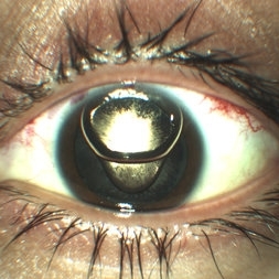
color photograph of the anterior segment of an aphakic eye with retained perfluorocarbon below the intraocular gas bubble
Condition/keywords: retained perfluorocarbon
-
 C3F8 Gas Bubble for Retinal Tear/ Pneumatic Retinopexy
Jun 27 2013 by Jason S. Calhoun
Patient comes in with retinal detachment and a pneumatic retinopexy was performed. Gas bubble is visible with optic nerve reflected.
Photographer: Jason S. Calhoun, Mayo Clinic Jacksonville, Florida
Imaging device: TOPCON TRC 50-EX
Condition/keywords: pneumatic retinopexy
-
 Epiretinal Membrane
Mar 3 2017 by Nichole Lewis
64-year-old female in a post op period with an epiretinal membrane and folds with a gas bubble. S/P repaired partial retinal detachment with multiple tears.
Photographer: Nichole Lewis
Condition/keywords: choroidal folds, epiretinal membrane (ERM)
-
---thumb.JPG/image-square;max$300,300.ImageHandler) Gas Bubble
Gas Bubble
Jul 12 2013 by Jason S. Calhoun
Patient had a pneumatic retinopexy for retinal detachment. Fundus shows C3F8 gas bubble superiorly.
Photographer: Jason S. Calhoun, Department of Ophthalmology, Mayo Clinic Jacksonville, Florida
Condition/keywords: pneumatic retinopexy
-
 Gas Bubble
May 21 2021 by Raj K. Maturi, MD
S/P PPV 73-year-old woman with small retinal detachment. Originally presented with decrease vision and residual vitreous floaters. Upon examination small localized retinal detachment with multiple breaks.
Photographer: Charlotte Harris ,Midwest Eye Institute 10300 N Illinois St, Carmel Indiana 46290
Imaging device: Nikon Optos California P200TDx
Condition/keywords: gas bubble
-
 Gas Bubble
May 21 2021 by Raj K. Maturi, MD
S/P PPV 73-year-old woman with small retinal detachment. Originally presented with decrease vision and residual vitreous floaters. Upon examination small localized retinal detachment with multiple breaks.
Photographer: Charlotte Harris ,Midwest Eye Institute 10300 N Illinois St, Carmel Indiana 46290
Imaging device: Nikon Optos California P200TDx
Condition/keywords: gas bubble
-
 Gas Bubble
May 21 2021 by Raj K. Maturi, MD
S/P PPV 73-year-old woman with small retinal detachment. Originally presented with decrease vision and residual vitreous floaters. Upon examination small localized retinal detachment with multiple breaks.
Photographer: Charlotte Harris ,Midwest Eye Institute 10300 N Illinois St, Carmel Indiana 46290
Imaging device: Nikon Optos California P200TDx
-
 Gas Bubble in Anterior Chamber
Jul 14 2013 by Jason S. Calhoun
Gas bubble injected in anterior chamber to control IOP.
Photographer: Jason S. Calhoun, Department of Ophthalmology, Mayo Clinic Jacksonville, Florida
Imaging device: TOPCON D-90 SL NIKON CAMERA
Condition/keywords: gas bubble
-
 Hydrophilic IOL With Fine Bubble Opacities Post-PPV for RD
Aug 21 2018 by Russell Pokroy, MD
Anterior segment photograph of 68-year-old woman 2 years after PPV for RD shows fine bubble-like opacities in the central part of this hydrophilic intraocular lens (IOL). These opacities are thought to be due to prolonged contact of the IOL with a large gas bubble in the vitreous cavity, particularly hydrophilic acrylic IOLs exposed to C3F8 gas. In addition, perfluorocarbon liquid exposure during surgery may put the IOL at risk of gas-induced opacity development. Minor Elschnig pearls are seen anterior to the wide optic-haptic junction in the capsule bag at 6-o-clock. A large symmetric capsulotomy with a rolled edge is seen behind the IOL. The slightly decreased yet stable vision of 20/30 was thought to be due to the central opacification of the IOL.
Photographer: Russell Pokroy, Assaf Harofe Medical Center, Israel
Condition/keywords: hydrophilic intraocular lens, IOL opacification
-
 Intralenticular Gas
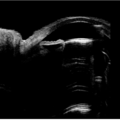
Aug 10 2024 by Varsha Reddy
Ultrasound biomicroscopy image of resolving intralenticular gas at office visit approximately 4 weeks later (B). The lens remained unchanged in thickness at 3.78 mm before and after resolution of the gas bubble, but developed a cataract.
Condition/keywords: intralenticular gas, pneumatic retinopexy, post-op
-
 Intraocular Gas bubble - Wide field Fundus Autofluorescence
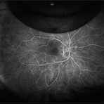
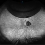
Sep 6 2021 by Ricardo Leitão Guerra
Wide field confocal scanning laser ophthalmoscopy 7 days after vitrectomy to treat a macular hole.
Imaging device: Zeiss Clarus 700
Condition/keywords: gas bubble, macular hole, pars plana vitrectomy (PPV), post-vitrectomy, vitrectomy
-
 Intraocular Gas Bubble - Wide-field True Color CSLO
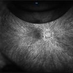
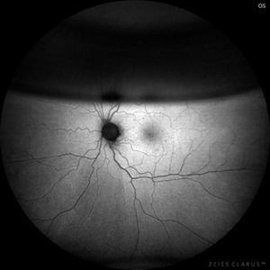
Sep 6 2021 by Ricardo Leitão Guerra
Wide field confocal scanning laser ophthalmoscopy 7 days after vitrectomy to treat a macular hole.
Imaging device: Zeiss Clarus 700
Condition/keywords: gas bubble, macular hole, pars plana vitrectomy (PPV), post-vitrectomy, vitrectomy
-
 Intravitreal Gas Bubble after PPV
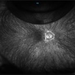
Oct 12 2012 by Jeffrey G. Gross, MD, FASRS
Intravitreal gas bubble after PPV, with mirroring reflection of ON.
Condition/keywords: intravitreal gas bubble, mirroring reflection of ON, pars plana vitrectomy (PPV)
 Loading…