Search results (197 results)
-
 Disseminated Retinitis and Retinochoroiditis, Metastatic
May 16 2017 by Karen Panzegrau
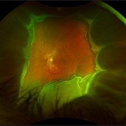
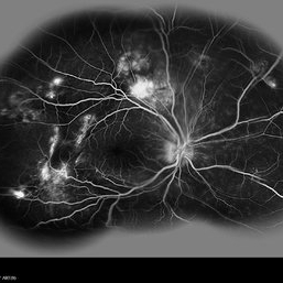
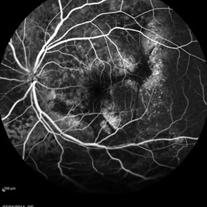
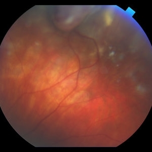
Fundus photograph of 44-year-old male with plasmacytoma infiltation of the choroid confirmed by biopsy, associated with disseminated retinitis, and retinochoroiditis. Vision is LP. Patient treated with intravitreal methotrexate
Photographer: Karen Panzegrau
Imaging device: Optos
Condition/keywords: metastatic lesion, methotrexate, Optos, plasmacytoma, retinitis, retinochoroiditis, unilateral exudative retinal detachment
-
 Choroidal Melanoma with Exudative Retinal Detachment
Mar 2 2023 by Aditya S Kelkar, MS, FRCS, FASRS,FRCOphth
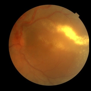
Color fundus photograph of the left eye of a 45 year old male showing choroidal melanoma with exudative retinal detachment.
Photographer: Dr. Pranali Surawase, National Institute of Ophthalmology, Pune, India.
Imaging device: Zeiss Clarus 500
Condition/keywords: choroidal mass, exudative retinal detachment, Retinal detachment
-
 Coats' Disease With Exudative Retinal Detachment and Retinal Macrocyst
Dec 9 2019 by Sophia El Hamichi, MD
A 3-year-old male with a presentation of a complex Coats' disease in the left eye with exudative retinal detachment, abnormal telangiectatic vasculature, and inferotemporal retinal macrocyst/retinoschisis.
Photographer: Abby Orcutt-Hayes, Murray Ocular Oncology and Retina
Imaging device: RetCam
Condition/keywords: Coats' disease, exudative detachment, montage, retinal macrocyst
-
 Choroidal metastasis case 3 image 1
Jan 11 2013 by Alex P. Hunyor, MD
Large choroidal metastasis from breast carcinoma, with exudative retinal detachment.
Condition/keywords: choroidal metastasis
-
---thumb.JPG/image-square;max$300,300.ImageHandler) Coats Disease
Coats Disease
Oct 11 2012 by Anat Loewenstein, MD
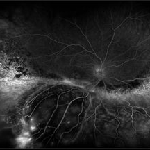
Fluorescein angiography of 6 -year-old girl whose parents have noticed leukocoria in her right eye. On examination severe exudative retinal detachment was diagnosed. On FA of the right eye peripehral capillary non perfusion and peripheral capillary dilatations were seen.
Photographer: Galit Yair-Pur
-
 Exudative Retinal Detachment and Branch Retinal Vein Occulsion
Oct 29 2020 by Olivia Rainey
Ultra-widefield fluorescein anigogram of a 51-year-old female with an exudative retinal detachment and branch retinal vein occlusion with retinal neovascularization affecting her right eye. The physician stated that the multiple aneurysmal dilations noted in the inferior periphery are responsible for the exudative RD seen on exam. He is considering Coat's vs FEVR given family history of aneurysms/congenital heart pathology per patient. He encouraged the patient to control their blood pressure, cholesterol, blood sugar, and co-morbidities which may have promoted the BRVO. He recommended antiVEGF injections to control the vascular leakage. Given the severe presentation and imminent threat to her vision, he recommended Eylea as first line therapy.
Photographer: Olivia Rainey, OCT-C, COA
Imaging device: Optos California
Condition/keywords: branch retinal vein occlusion (BRVO), chronic retinal detachment, fluorescein angiogram (FA), fluorescein leakage, inferior retina, inferior retinal detachment, Optos, ultra-wide field imaging
-
 Fluorescein Angiogram of Coats's Disease With Exudative Retinal Detachment
Dec 9 2019 by Sophia El Hamichi, MD
A 3-year-old male presenting a complex Coats' disease of the left eye with exudative retinal detachment, abnormal telangiectatic vasculature with peripheral nonperfusion and leakage.
Photographer: Abby Orcutt-Hayes, Murray Ocular Oncology and Retina
Condition/keywords: Coats' disease, exudative detachment, fluorescein angiogram (FA), montage, retinal macrocyst
-
---thumb.jpg/image-square;max$300,300.ImageHandler) Harada's with Exudative RD
Harada's with Exudative RD
Oct 13 2012 by Edwin H. Ryan, MD
OCT of a 35-year-old woman with acute vision loss in one eye.
Condition/keywords: exudative retinal detachment, Harada's disease
-
 Multifocal Exudative Detachments Due to VKH
May 14 2014 by Avris Romario Diparaja Siahaan
Fundus Photograph a 38-year-old man with multifocal CSR and inferior exudative retinal detachment on both eyes (Harada Syndrome).
Photographer: Avris Romario Diparaja Siahaan, Klinik Mata Nusantara
Imaging device: Topcon TRC 50 DX Type IA
Condition/keywords: fundus photograph, multifocal central serous chorioretinopathy (CSCR)
-
 Multifocal Exudative Detachments Due to VKH
May 14 2014 by Avris Romario Diparaja Siahaan
FA (composite Image) a 38-year-old man with multifocal CSR and inferior exudative retinal detachment on both eyes (Harada Syndrome).
Photographer: Avris Romario Diparaja Siahaan, Klinik Mata Nusantara
Imaging device: Heidelberg HRA + OCT Spectralis
Condition/keywords: multifocal central serous chorioretinopathy (CSCR)
-
 Multifocal Exudative Detachments Due to VKH
May 14 2014 by Avris Romario Diparaja Siahaan
FA (Late Phase) a 38-year-old man with multifocal CSR and inferior exudative retinal detachment on both eyes (Harada Syndrome).
Photographer: Avris Romario Diparaja Siahaan, Klinik Mata Nusantara
Imaging device: Heidelberg HRA + OCT Spectralis
Condition/keywords: multifocal central serous chorioretinopathy (CSCR)
-
 Multifocal Exudative Detachments Due to VKH
May 14 2014 by Avris Romario Diparaja Siahaan
FA a 38-year-old man with multifocal CSR and inferior exudative retinal detachment on both eyes (Harada Syndrome).
Photographer: Avris Romario Diparaja Siahaan, Klinik Mata Nusantara
Imaging device: Heidelberg HRA + OCT Spectralis
Condition/keywords: multifocal central serous chorioretinopathy (CSCR)
-
 Multifocal Exudative Detachments Due to VKH
May 14 2014 by Avris Romario Diparaja Siahaan
FA (Early Phase) a 38-year-old man with multifocal CSR and inferior exudative retinal detachment on both eyes (Harada Syndrome) with 55 degree lens
Photographer: Avris Romario Diparaja Siahaan, Klinik Mata Nusantara
Imaging device: Heidelberg HRA + OCT Spectralis
Condition/keywords: multifocal central serous chorioretinopathy (CSCR)
-
 Multifocal Exudative Detachments Due to VKH
May 14 2014 by Avris Romario Diparaja Siahaan
ICG (Late Phase) a 38-year-old man with multifocal CSR and inferior exudative retinal detachment on both eyes (Harada Syndrome).
Photographer: Avris Romario Diparaja Siahaan, Klinik Mata Nusantara
Imaging device: Heidelberg HRA + OCT Spectralis
Condition/keywords: indocyanine green (ICG) angiography, multifocal central serous chorioretinopathy (CSCR)
-
 Multifocal Exudative Detachments Due to VKH
May 14 2014 by Avris Romario Diparaja Siahaan
ICG (Mid Phase) a 38-year-old man with multifocal CSR and inferior exudative retinal detachment on both eyes (Harada Syndrome).
Photographer: Avris Romario Diparaja Siahaan, Klinik Mata Nusantara
Imaging device: Heidelberg HRA + OCT Spectralis
Condition/keywords: indocyanine green (ICG) angiography, multifocal central serous chorioretinopathy (CSCR)
-
---thumb.jpg/image-square;max$300,300.ImageHandler) Sturge-Weber Diffuse Hemangioma and Retinal Detachment on B-scan
Sturge-Weber Diffuse Hemangioma and Retinal Detachment on B-scan
Apr 18 2014 by Susanna S. Park, MD, PhD
B-scan ultrasonogram of the right eye of an 8 year old Hispanic boy with Sturge -Weber Syndrome showing diffuse choroidal thickening from diffuse choroidal hemangioma and associated total exudative retinal detachment.
Photographer: Ellen Redenbo, University of California Davis Eye Center
Condition/keywords: B scan ultrasound, diffuse choroidal hemangioma, Sturge-Weber syndrome
-
 Vasoproliferative Tumor (VPT)
Apr 25 2017 by Christopher G Fuller, MD
Fundus photograph of a presumptive vasoproliferative tumor (with resultant total exudative retinal detachment) in a 54-year-old white truck driver. Image is taken on post-operative day 4, after 25/27 gauge vitrectomy with drainage retinotomy, air-fluid exchange, endoscopic laser blanching of VPT, oil, and Ozurdex.
Photographer: Ray Garner, Texas Retina Associates [Lubbock, TX]
Condition/keywords: vasoproliferative retinopathy
-
 Von Hippel-Lindau 1
Oct 13 2012 by Hamid Ahmadieh, MD
Color fundus photograph of the left eye of a 25-year-old woman with exudative retinal detachment secondary to retinal angiomatosis (Von Hippel-Lindau).
Photographer: Hamid Ahmadieh, MD, Ophthalmic Research Center, Labbafinejad Medical Center, Shahid Beheshti University of Medical Sciences
Imaging device: Topcon Fundus Camera
Condition/keywords: exudative retinal detachment, retinal angiomatous proliferation (RAP), Von Hippel-Lindau
-
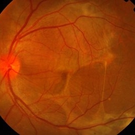
 Amelanotic Choroidal Melanoma
Apr 12 2019 by David L Kilpatrick, MD
Fundus photograph of a 69-year-old male with an amelanotic choroidal melanoma and corresponding exudative retinal detachment. Transvitreal biopsy was performed at the time of radioactive I-125 plaque placement. The genetic expression profile revealed a Class 1A, PRAME negative tumor.
Photographer: Retina Consultants of Alabama, P. C.
Imaging device: Optos
Condition/keywords: amelanotic melanoma
-
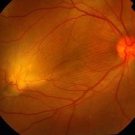
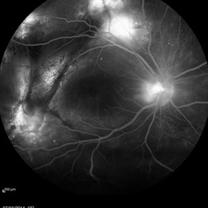
 Choroidal Melanoma
Jan 30 2019 by Karen Panzegrau
Ultra-wide field optos image of a 27-year-old male patient who presented with loss of vision for about 6-8 weeks. Previous choroidal nevus seen. Recommended annual monitoring. No exam for since 10/2014. Brachytherapy vs enucleation was discussed. Brachytherapy was decided as treatment. Full metastatic work up is being performed.
Photographer: Karen Panzegrau
Imaging device: Optos
Condition/keywords: choroidal nevus, exudative retinal detachment, malignant neoplasm of eye, Optos, ultra-wide field imaging
-
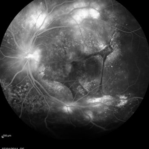
 Large, Dome-Shaped Peripheral Choroidal Melanoma - Widefield Color
Feb 13 2020 by Michael Seider, MD
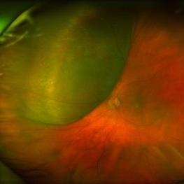
Large, dome-shaped peripheral choroidal melanoma of the left eye with inferior exudative retinal detachment. Note the lack of obvious orange pigment over the tumor and apparent drusen anteriorly. A lack of ophthalmoscopically obvious lipofuscin is not uncommon among larger choroidal melanomas. B-Scan ultrasonography (transverse, 10 o’clock) confirms a low-moderate internally reflective dome-shaped choroidal lesion with a small adjacent retinal detachment. Ultrasound biomicroscopy (radial, 10 o’clock) confirms no ciliary body involvement of the tumor.
-
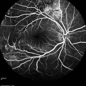
 Von Hippel-Lindau
Oct 13 2012 by Hamid Ahmadieh, MD
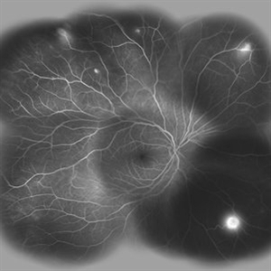
Wide field FA image of the right eye of a 25-year-old woman with retinal angiomatosis (Von Hippel-Lindau). Fundus of the right eye seemed to be normal in ophthalmoscopy.
Photographer: Soodabeh Fooladin, Negah Eye Center, Tehran
Imaging device: Heidelberg Spectralis
Condition/keywords: exudative retinal detachment, retinal angiomatous proliferation (RAP), Von Hippel-Lindau
 Loading…