Search results (2640 results)
-
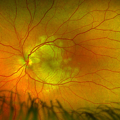
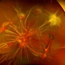
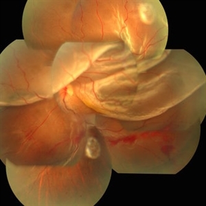 Globe Perforation With Retinal Detachment
Feb 7 2017 by Manish Nagpal, MD, FRCS (UK), FASRS
Patient presenting with globe perforation with a penetration seen at below the inferior arcade with some scattered hemorrhage and a retinal detachment.
Photographer: Rakesh Juneja
Condition/keywords: globe perforation
-
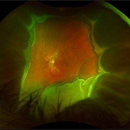
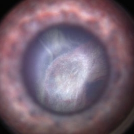
 Total Rhegmatogenous Retinal Detachment With Severe PVR
May 27 2015 by Darin R. Goldman, MD
63-year-old pseudophakic male with hand motion vision in the left eye due to a total retinal detachment with severe proliferative vitreoretinopathy.
Condition/keywords: proliferative vitreoretinopathy (PVR), retinal tear
-
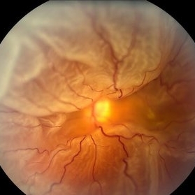
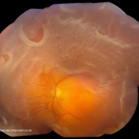
 Tractional Retinal Detachment
Dec 4 2019 by Janet Brazil
Fundus photograph of a 32-year-old female with severe end-stage diabetic tractional retinal detachment.
Photographer: Janet Atkinson, Eye Associates of New Mexico, Albuquerque, NM
Imaging device: Topcon TRC- 50EX
Condition/keywords: diabetes, proliferative diabetic retinopathy (PDR), tractional retinal detachment
-
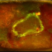
 Foreign Body SS
Feb 14 2015 by Shlomit Schaal, MD, PhD, MHCM
A four millimeter metal foreign body removed surgically using foreign body forceps. The macula is protected by PFCL heavy fluid. There is a traumatic retinal detachment inferiorly (top of photo, surgeon's view).
Photographer: Shlomit Schaal, MD, PhD
Condition/keywords: intraocular foreign body
-
 Massive Subretinal Hemorrhage With Near Total Retina Detachment
Nov 27 2013 by David W. Faber, MD
Fundus photo of an 71-year-old male with massive subretinal hemorrhage. Had been given 6 week Avastin treatments. Was put on coumadin for 6 weeks following knee surgery.
Photographer: Donna Knight, Rocky Mountain Retina Consultants, Salt Lake City, Utah
-
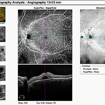 Acute Posterior Multifocal Placoid Pigment Epitheliopathy
Feb 20 2024 by Soobien Lee
12x12mm OCT Angiography of a 20-year-old caucasian female with viral prodrome and vision loss OS>OD secondary to Acute Posterior Multifocal Placoid Pigment Epitheliopathy (APPME). Imaging shows multifocal flow voids.
Photographer: Kim Seay, Elman Retina Group
Imaging device: 12x12mm OCT-Angiography
Condition/keywords: acute posterior multifocal placoid pigment epitheliopathy (APMPPE), bacillary layer detachment, OCT, OCT Angiography, Uveitis, white dot syndrome
-
 Giant Retinal Tear
Feb 20 2024 by Soobien Lee
Optos color fundus photograph of a 40-year-old caucasian male who is a UFC fighter with a total retinal detachment in his right eye secondary to a giant retinal tear from 10 o'clock to 2 o'clock.
Photographer: Trinity Wolf, Elman Retina Group
Imaging device: Optos Ultra-Widefield Imaging
Condition/keywords: giant retinal tear, optos, Retinal Detachment, Retinal tear with detachment, trauma
-
 Acute Posterior Multifocal Placoid Pigment Epitheliopathy
Feb 20 2024 by Soobien Lee
Optos color fundus photograph of a 20-year-old caucasian female with viral prodrome and vision loss OS>OD secondary to Acute Posterior Multifocal Placoid Pigment Epitheliopathy (APPME). Imaging of her left eye shows multiple bilateral creamy yellow-white placoid lesions at the level of RPE and choroid throughout the posterior pole.
Photographer: Ashley Metzger, Elman Retina Group
Imaging device: Optos Ultra-Widefield Imaging
Condition/keywords: acute posterior multifocal placoid pigment epitheliopathy (APMPPE), bacilliary layer detachment, Optos, uveitis, white dot syndrome
-
 Acute Posterior Multifocal Placoid Pigment Epitheliopathy
Feb 20 2024 by Soobien Lee
Optos fundus autofluorescence photograph of a 20-year-old caucasian female with viral prodrome and vision loss OS>OD secondary to Acute Posterior Multifocal Placoid Pigment Epitheliopathy (APPME). Imaging of her left eye shows hypoautofluorescent areas corresponding to multiple bilateral placoid lesions at the level of RPE and choroid throughout the posterior pole.
Photographer: Ashley Metzger, Elman Retina Group
Imaging device: Optos Ultra-Widefield Autoflurescence Imaging
Condition/keywords: acute posterior multifocal placoid pigment epitheliopathy (APMPPE), autofluorescence imaging, bacilliary layer detachment, Optos, OPTOS CALIFORNIA, uveitis, white dot syndrome
-
 Acute Posterior Multifocal Placoid Pigment Epitheliopathy
Feb 20 2024 by Soobien Lee
Fluorescein angiogram of a 20-year-old caucasian female with viral prodrome and vision loss OS>OD secondary to Acute Posterior Multifocal Placoid Pigment Epitheliopathy (APPME). Early blockage with late hyperfluorescent leakage can be seen on fluorescein angiography of the left eye.
Photographer: Ashley Metzger, Elman Retina Group
Imaging device: Optos Ultra-Widefield Fluorescein Angiography
Condition/keywords: acute posterior multifocal placoid pigment epitheliopathy (APMPPE), bacilliary layer detachment, FA, FA early phase, fluorescein angiogram (FA), Optos, uveitis, white dot syndrome
-
 Acute Posterior Multifocal Placoid Pigment Epitheliopathy
Feb 20 2024 by Soobien Lee
Fluorescein angiogram of a 20-year-old caucasian female with viral prodrome and vision loss OS>OD secondary to Acute Posterior Multifocal Placoid Pigment Epitheliopathy (APPME). Early blockage with late hyperfluorescent leakage can be seen on fluorescein angiography of the left eye.
Photographer: Ashley Metzger, Elman Retina Group
Imaging device: Optos Ultra-Widefield Fluorescein Angiography
Condition/keywords: acute posterior multifocal placoid pigment epitheliopathy (APMPPE), bacilliary layer detachment, FA, FA late phase, FA late phase leakage, fluorescein angiogram (FA), Optos, uveitis, white dot syndrome
-
 Acute Posterior Multifocal Placoid Pigment Epitheliopathy
Feb 20 2024 by Soobien Lee
A 20-year-old caucasian female with viral prodrome and vision loss OS>OD secondary to Acute Posterior Multifocal Placoid Pigment Epitheliopathy (APPME). OCT of the left macula shows bacillary layer detachment.
Photographer: Kim Seay, Elman Retina Group
Condition/keywords: acute posterior multifocal placoid pigment epitheliopathy (APMPPE), bacilliary layer detachment, OCT, Uveitis, white dot syndrome
-
 Disseminated Retinitis and Retinochoroiditis, Metastatic
May 16 2017 by Karen Panzegrau
Fundus photograph of 44-year-old male with plasmacytoma infiltation of the choroid confirmed by biopsy, associated with disseminated retinitis, and retinochoroiditis. Vision is LP. Patient treated with intravitreal methotrexate
Photographer: Karen Panzegrau
Imaging device: Optos
Condition/keywords: metastatic lesion, methotrexate, Optos, plasmacytoma, retinitis, retinochoroiditis, unilateral exudative retinal detachment
-
 360 Degree Retinal Detachment
Jun 29 2013 by Jason S. Calhoun
Total retinal detachment in the left eye.
Photographer: Jason S. Calhoun, Mayo Clinic Jacksonville, Florida
Imaging device: TOPCON TRC 50-EX
-
 360 Degree Retinectomy
Sep 11 2020 by Sham Talati, DOMS
A case of retinal detachment with PVR. Patient underwent pars plana vitrectomy with silicon oil injection with 360 degree retinectomy.
Photographer: Dr. Sham Talati,Retina Foundation,Ahmedabad
Imaging device: Nidek Mirante
Condition/keywords: proliferative vitreoretinopathy (PVR), retinectomy
-
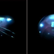
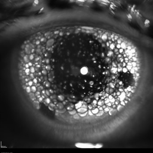 Oil Bubbles
Apr 27 2018 by Mark Lazcano
Infrared photograph of 56-year-old male with retinal detachment oil on lens.
Photographer: Mark Lazcano, University of Miami, Bascom Palmer Eye Institute
Imaging device: Heidelberg Spectralis
Condition/keywords: silicone oil
-
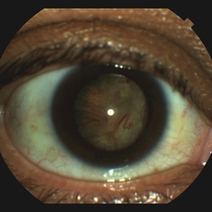
 Retinal Detachment
Apr 30 2020 by Giselle DeOliveira
External / Gonio Photograph of 13-month old male infant with retinopathy of prematurity, retinal detachment and Cohen syndrome.
Photographer: Giselle DeOliveira, University of Miami, Bascom Palmer Eye Institute
Imaging device: Retcam III
-
 Retinal Tear
Sep 16 2021 by Stefanie Palmer
Retinal Tear with a bridge vessel.
Photographer: Stefanie Palmer, CRA
Condition/keywords: detachment, tear
-
 Choroidal Melanoma
Nov 3 2022 by pedro fernandes souza neto
Transillumination of Enucleation specimen of Choroidal Melanoma: anterior chamber is closed. Total secondary retinal detachment with subretinal serous fluid and some subretinal hemorrhages are present.
Photographer: Eduardo Marback, Federal University of Bahia, Brazil
Condition/keywords: enucleation, melanoma
-
 Cohen Syndrome Retinal Detachment
Apr 30 2020 by Giselle DeOliveira
Gonio Photograph of 13-month infant male with retinal detachment, retinopathy of prematurity and Cohen Syndrome
Photographer: Giselle DeOliveira, University of Miami, Bascom Palmer Eye Institute
Imaging device: Retcam III
Condition/keywords: retinopathy of prematurity (ROP)
-
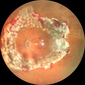
 Retinal Detachment With Multiple Retinal Tears
May 18 2017 by Kamal Kishore, MD, MBBS
77-year-old female presented with a report of gradual decreased vision over the span of one week. Vision finger count. Examination showed retinal detachment with multiple retinal tears and vitreous hemorrhage present.
Photographer: Lindsay Shepard, Illinois Retina and Eye Associates, Peru, IL
Imaging device: Topcon TRC- 50 EX
Condition/keywords: retinal tear
-
 Retinal Detachment with PVR (s/ SPR, PPV, MPV, 360 Retinectomy, PFO, PI, FAx, SO)
Aug 22 2019 by Merrick Avila
Ultra-wide field pseudocolor fundus photograph of a 64-year-old female with a treated retinal detachment with proliferative vitreoretinopathy. Patient has a history of complex retinal detachments that have been treated multiple times. On exam 8-22-19, there were large macular holes with LP vision. There was a long discussion about guarded nature of her condition and goals or trial for repair including globe sparing prevention of phthisis.
Photographer: Merrick Avila
Imaging device: Optos
Condition/keywords: diabetic retinopathy, hemorrhage, Optos, proliferative vitreoretinopathy (PVR), retinectomy, silicone oil
-
 Sickle Cell Retinopathy
Nov 5 2022 by Mateus Queiroz Corrêa, MD
19 -year-old young man with combined rhegmatogenous and tractional retinal detachment secondary to a proliferative sickle retinopathy ( stage V)
Photographer: Mateus Corrêa, Sorocaba Eye Bank Hospital
Imaging device: Optos California
Condition/keywords: Retinal detachment, sickle cell retinopathy
-
 Silicon Oil
Apr 27 2018 by Giselle DeOliveira
Infrared photo of 75-year-old male with retinal detachment.
Photographer: Giselle DeOliveira, University of Miami, Bascom Palmer Eye Institute
Imaging device: Heidelberg Spectralis
Condition/keywords: infrared image, silicone oil
-
 Tractional Retinal Detachment
Sep 27 2012 by Virgilio Morales-Canton, MD
OCT image of a 42-year-old male patient with a localized traction of the superior macula secondary to proliferative diabetic retinopathy.
Imaging device: Cirrus
Condition/keywords: tractional retinal detachment
 Loading…