 Initializing download.
-
 By Soobien Lee
Elman Retina Group
Co-author(s): Sidney Schechet, M.D. - Uploaded on Feb 20, 2024.
- Last modified by Joshua Friedman on Feb 20, 2024.
- Rating
- Appears in
- Miscellaneous
- Condition/keywords
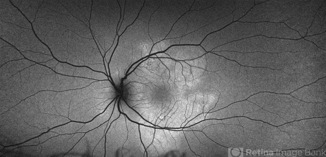
- acute posterior multifocal placoid pigment epitheliopathy (APMPPE), white dot syndrome, uveitis, bacilliary layer detachment, Optos, OPTOS CALIFORNIA, autofluorescence imaging
- Photographer
- Ashley Metzger, Elman Retina Group
- Imaging device
- Optos Ultra-Widefield Autoflurescence Imaging
- Description
- Optos fundus autofluorescence photograph of a 20-year-old caucasian female with viral prodrome and vision loss OS>OD secondary to Acute Posterior Multifocal Placoid Pigment Epitheliopathy (APPME). Imaging of her left eye shows hypoautofluorescent areas corresponding to multiple bilateral placoid lesions at the level of RPE and choroid throughout the posterior pole.


")


