Search results (43 results)
-
---thumb.JPG/image-square;max$300,300.ImageHandler) Lattice Degeneration
Lattice Degeneration
Jul 12 2013 by Jason S. Calhoun
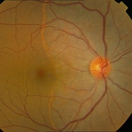
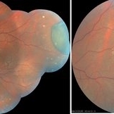
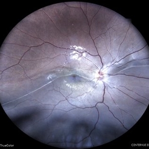
Composite of HD-OCT and fundus photo showing demarcation line of lattice degeneration inferiorly temporally at 4-o'clock in a young black female.
Photographer: Jason S. Calhoun, Department of Ophthalmology, Mayo Clinic Jacksonville, Florida
Condition/keywords: lattice degeneration
-
 Retinal detachment
Apr 12 2023 by Ahmed Abbas Hashmi, OD
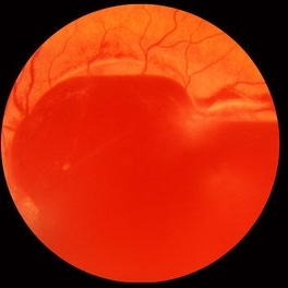
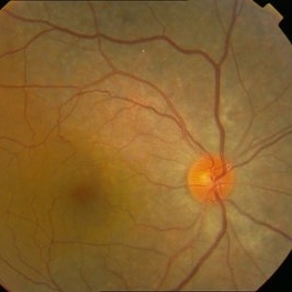
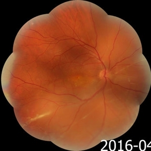
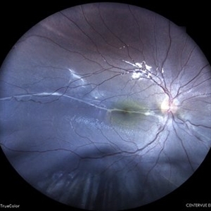
Color fundus photograph of the left eye of a 30-year-old man with asymptomatic inferior retinal detachment with pigmented demarcation line. Macula and Disc healthy.
Photographer: Ahmed Abbas Hashmi
Imaging device: Topcon TRC-NW8F
Condition/keywords: Pigmentary demarcation line, Retinal Detachment
-
 Chronic Inferior Retinal Detachment
Mar 1 2017 by Philip J. Polkinghorne, MD
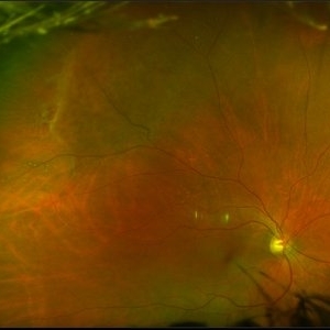
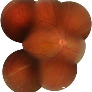
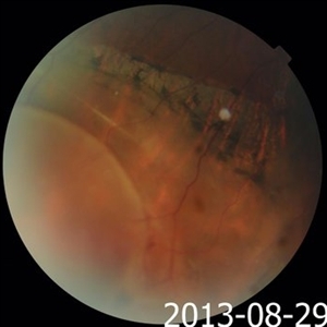
Color photograph of chronic retinal detachment with pigment demarcation line and atrophic holes visible. The vision was recorded at 20/20, and follow up is 3 years.
Photographer: Alex Fraser
Condition/keywords: atrophic retinal hole, demarcation line
-
 Acute Subhyaloid Hemorrhage CF
Oct 1 2012 by Jeffrey G. Gross, MD, FASRS
Acute subhyaloid hemorrhage CF.
Condition/keywords: demarcation line, subhyaloid hemorrhage
-
 Asymptomatic Rhegmatogenous Retinal Detachment
Sep 14 2012 by Sharon Fekrat, MD FACS FASRS
Fundus photograph of a 25-year-old emmetropic male graduate student with an inferotemporal phakic chronic asymptomatic rhegmatogenous retinal detachment with a demarcation line in the right eye. His sister who is an ophthalmology resident discovered this incidental finding. Vision 20/20.
Photographer: Brian Lutman CRA, Duke University Eye Center, Durham, NC
Condition/keywords: asymptomatic, demarcation line
-
 Retinal Detachment
May 9 2016 by Nichole Lewis
Retinal detachment with partial demarcation line and same day barrier laser treatment.
Photographer: Nichole Lewis
-
 Asymptomatic Chronic Retinal Detachment With Demarcation Line
Jun 11 2016 by Philip J. Polkinghorne, MD
A 65-year-old emmetrope with asymptomatic chronic retinal detachment with demarcation line.
Photographer: Alex Fraser, Greenlane Clinical Center, Auckland, New Zealand
Condition/keywords: chronic retinal detachment, fundus autofluorescence (FAF)
-
 AZOOR vs. AAOOR
Mar 19 2014 by Ali Tavallali, MD, FASRS
Color fundus photograph of a 47-year-old female with 20/20 VA of both eyes, note the demarcation line
Photographer: Neda Sheibani, Dr. Khodadoust Eye Hospital, Shiraz, Iran
Condition/keywords: acute zonal occult outer retinopathy (AZOOR)
-
 AZOOR vs. AAOOR
Mar 19 2014 by Ali Tavallali, MD, FASRS
Color fundus photograph of a 47-year-old female with 20/20 VA of both eyes, note the progression of demarcation line after 4 months
Photographer: Neda Sheibani, Dr. Khodadoust Eye Hospital, Shiraz, Iran
Condition/keywords: acute zonal occult outer retinopathy (AZOOR)
-
 AZOOR vs. AAOOR
Mar 19 2014 by Ali Tavallali, MD, FASRS
FAF of a 47-year-old female with 20/20 VA of both eyes, note the progression of demarcation line after 4 months
Photographer: Neda Sheibani, Dr. Khodadoust Eye Hospital, Shiraz, Iran
Condition/keywords: acute zonal occult outer retinopathy (AZOOR)
-
 Bullous Retinoschisis with Outer Retinal Holes
Jun 15 2020 by Olivia Rainey
Ultra-widefield pseudocolor fundus photograph of a 56-year-old female with bullous retinoschisis with outer retinal holes affecting her right eye. The physician noted superotemporal retinoschisis in her monoculcar functioning eye. There was no demarcation line and no inner or outer layer breaks at her first appointment in February of 2020. On 6/15/20 she had a new onset outer holes and SRF tracking inferiorly. The physician recommended observation, however if this continues to progress we have discussed indications for barrier laser.
Photographer: Olivia Rainey, OCT-C, COA
Imaging device: Optos California
Condition/keywords: bullous retinoschisis, Optos, outer layer breaks, outer layer hole, pseudocolor, subretinal fluid, superior retina, ultra-wide field imaging
-
 Choroidal Melanoma with Exudative Detachment
Apr 7 2025 by Virginia Gebhart
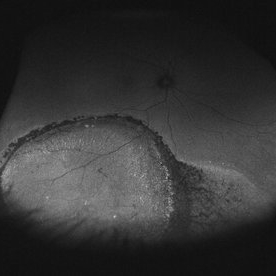
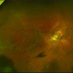
Autofluorescence image of 36 year old female showing demarcation line of fluid/detachment from new choroidal melanoma. Pt will be scheduled for brachytherapy pending CT scan results.
Photographer: Virginia Gebhart, Retina Consultants of Carolina
Imaging device: Optos California
Condition/keywords: Autoflourescence, autofluorescence imaging, choroidal melanoma, melanoma, retinal detachment
-
 Chronic retinal detachment changes
Apr 29 2022 by Otakar Dušek, M.D. Ph.D.
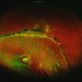
Colour fundus photo of 22-year-old woman with bulous retinal detachment number 5-9, old demarcation lines and inferotemporal periheral secondary retinal cyst.
Photographer: Otakar Dušek, Charles University, Prague
Imaging device: Zeiss Clarus
Condition/keywords: chronic retinal detachment, demarcation line, peripheral retinal cyst
-
 Chronic Retinal Detachment in a Young Myopic Patient
Nov 6 2019 by Kamal Kishore, MD, MBBS
Chronic retinal detachment in a 27-year-old myopic female showing spontaneous reattachment in inferotemporal quadrant, and demarcation line and subretinal gliosis in superotemporal quadrant.
Photographer: Stephanie Shaver
Imaging device: Topcon 50 EX with OIS Winstation
Condition/keywords: chronic retinal detachment, high myopia
-
 Chronic Retinal Detachment: Features Slide 1
Oct 22 2012 by Ronald C. Gentile, MD
Chronic retinal detachments can be associated with demarcation lines (tidemarks), subretinal bands or sheets, and retinal cysts. Fundus photo of a chronic inferior retinal detachment reveals multiple demarcation lines inferior to the center of the fovea as a result of an inferior temporal dialysis.
Photographer: The New York Eye & Ear Infirmary Department of Medical Imaging
Condition/keywords: chronic retinal detachment, demarcation line
-
 Chronic Retinal Detachment: Features Slide 2
Oct 22 2012 by Ronald C. Gentile, MD
Chronic retinal detachments can be associated with demarcation lines (tidemarks), subretinal bands or sheets, and retinal cysts. Fundus photo of a chronic retinal detachment reveals a branching subretinal band superior nasal to the macula with a portion extending to the inferior margin of the optic disc.
Photographer: The New York Eye & Ear Infirmary Department of Medical Imaging
Condition/keywords: chronic retinal detachment, subretinal bands
-
 Chronic Retinal Detachment: Features Slide 3
Oct 22 2012 by Ronald C. Gentile, MD
Chronic retinal detachments can be associated with demarcation lines (tidemarks), subretinal bands or sheets, and retinal cysts. Fundus photo of a chronic retinal detachment reveals a retinal cyst within the peripherally detached temporal retina.
Condition/keywords: chronic retinal detachment
-
 CHRPE
CHRPE
Jan 15 2021 by Priya Rasipuram Chandrasekaran, MBBS, DO, DNB, FRCS
This is the fundus photo and fundus photo montage of the left eye of a 25-year-old male showing flat, solitary, round, greyish pigmented lesion situated AT THE equator with a scalloped margin. Vessels overlying the lesion are normal and there is a clear demarcation line between this and normal retina. The margins are hypopigmented with few hypopigmented lacunae inside.
Condition/keywords: congenital hypertrophy of the retinal pigment epithelium (CHRPE)
-
 Guardian Angel
Dec 11 2024 by Virginia Gebhart
48 year old female 3 months s/p brachytherapy for choroidal melanoma. Persistent subretinal and increased subfoveal fluid. Will observe for now, will consider Ozurdex if no improvement. BCVA 20/80
Photographer: Virginia Gebhart, Retina Consultants of Carolina
Imaging device: Optos California
Condition/keywords: brachytherapy, demarcation line, fundus autofluorescence (FAF), serous detachment, subretinal fluid
-
 Longstanding Retinal Detachment Due to a Larg Retinal Tear
Dec 27 2016 by Hamid Ahmadieh, MD
Wide-field color fundus photograph of the right eye of a patient with longstanding retinal detachment. Demarcation lines are visible.
Photographer: Shabnam Poureh, Negah Eye Center, Tehran, Iran
Condition/keywords: color fundus photograph
-
 Longstanding Retinal Detachment Secondary to a Larg Retinal Tear
Dec 27 2016 by Hamid Ahmadieh, MD
Montaged color fundus photograph of the right eye of a patient with longstanding retinal detachment. Demarcation lines are visible.
Photographer: Shabnam Poureh, Negah Eye Center, Tehran, Iran
Condition/keywords: color fundus photograph, demarcation line
-
 Macula Sparring Tractional Retinal Detachment
Feb 9 2018 by Olivia Rainey
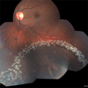
Ultra-wide field pseudocolor image of a 22-year-old male with a macula sparring tractional retinal detachment relating to retinopathy of prematuritiy affecting his right eye.
Photographer: Olivia Rainey
Imaging device: Optos
Condition/keywords: color fundus photograph, demarcation line, macula sparring, Optos, retinopathy of prematurity (ROP), tractional retinal detachment, ultra-wide field imaging
-
 Old Retinal Detachment
Apr 17 2024 by Akansha Sharma
Color fundus photograph of a 13 year old female with old retinal detachment at presentation as is demonstrated by a demarcation line.
Photographer: Dr. Akansha Sharma, Bharati Eye Hospital
Condition/keywords: delimited old retinal detachment, demarcation line, RD, Retinal Detachment
-
 Old Retinal Detachment
Jun 17 2024 by Akansha Sharma
Color fundus photograph of a 10 year old male with old retinal detachment with subretinal band.
Photographer: Dr. Akansha Sharma, Bharati Eye Hospital
Condition/keywords: demarcation line, OLD RD, subretinal bands
-
 Pigmented Demarcation Line and Retinal Macrocyst
Nov 14 2013 by Hamid Ahmadieh, MD
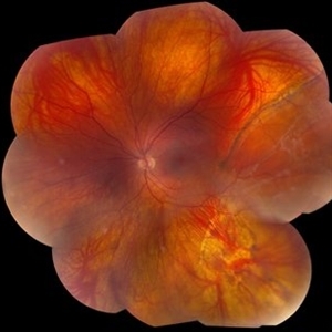
Color fundus photograph of the right eye of a 40-year-old man with longstanding retinal detachment showing a broad pigmented demarcation line and a retinal macrocyst as well as patches of retinal hemorrhages.
Photographer: Elham Salehi , Negah Eye Center, Tehran
Condition/keywords: demarcation line, fundus photograph, retinal macrocyst
 Loading…