Search results (544 results)
-
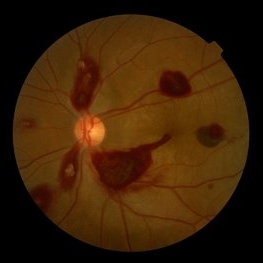
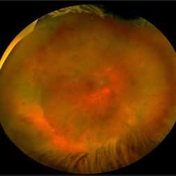
 Coats' Disease
Apr 27 2018 by Brenda Fallas
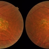
3-year-old boy with unilateral Coats' Disease fundus photo.
Photographer: Brenda Fallas, Bascom Palmer Eye Institute, Miami, FL
Imaging device: Retcam III 130 degree lens
Condition/keywords: Coats' disease, color fundus photograph, retinal telangiectasia
-
 Ocular Manifestation of Acute Leukemia
Sep 8 2012 by Hamid Ahmadieh, MD
Color fundus photograph of a 26-year-old man with acute leukemia.
Photographer: Hamid Ahmadieh, MD, Ophthalmic Research Center, Labbafinejad Medical Center, Shahid Beheshti University of Medical Sciences , Tehran
Imaging device: Topcon Fundus Camera
Condition/keywords: acute leukemia, white centered retinal hemorrhage (Roth Spot)
-
 Giant Retinal Tear
Feb 20 2024 by Soobien Lee
Optos color fundus photograph of a 40-year-old caucasian male who is a UFC fighter with a total retinal detachment in his right eye secondary to a giant retinal tear from 10 o'clock to 2 o'clock.
Photographer: Trinity Wolf, Elman Retina Group
Imaging device: Optos Ultra-Widefield Imaging
Condition/keywords: giant retinal tear, optos, Retinal Detachment, Retinal tear with detachment, trauma
-
 Acute Posterior Multifocal Placoid Pigment Epitheliopathy
Feb 20 2024 by Soobien Lee
Optos color fundus photograph of a 20-year-old caucasian female with viral prodrome and vision loss OS>OD secondary to Acute Posterior Multifocal Placoid Pigment Epitheliopathy (APPME). Imaging of her left eye shows multiple bilateral creamy yellow-white placoid lesions at the level of RPE and choroid throughout the posterior pole.
Photographer: Ashley Metzger, Elman Retina Group
Imaging device: Optos Ultra-Widefield Imaging
Condition/keywords: acute posterior multifocal placoid pigment epitheliopathy (APMPPE), bacilliary layer detachment, Optos, uveitis, white dot syndrome
-
 Torpedo Maculopathy
Feb 20 2024 by Soobien Lee
Optos color fundus photograph of a 35-year-old asymptomatic female with no ocular or medical history with stable and chronic appearing torpedo-shaped macula lesion in the left eye.
Photographer: Peter Sotirakos, Elman Retina Group
Imaging device: Optos Ultra-Widefield Imaging
Condition/keywords: macula, Optos, torpedo maculopathy
-
 Choroidal Melanoma with Exudative Retinal Detachment
Mar 2 2023 by Aditya S Kelkar, MS, FRCS, FASRS,FRCOphth
Color fundus photograph of the left eye of a 45 year old male showing choroidal melanoma with exudative retinal detachment.
Photographer: Dr. Pranali Surawase, National Institute of Ophthalmology, Pune, India.
Imaging device: Zeiss Clarus 500
Condition/keywords: choroidal mass, exudative retinal detachment, Retinal detachment
-
 Retinal Arterio-Venous Malformations
Apr 7 2017 by Deepak Bhojwani, MS
Multimodal imaging of a 16-year-old boy with retinal arterio-venous malformations(AVM). He also had cerebral AVM's on MRI-contrast studies suggesting Wyburn-Mason syndrome.
Photographer: DEEPAK BHOJWANI, RAGHUDEEP EYE HOSPITAL, AHMEDABAD.
Imaging device: Zeiss VISUCAM
Condition/keywords: color fundus photograph, FA early phase, optical coherence tomography (OCT), Wyburn-Mason
-
 Retinal Detachment with PVR (s/ SPR, PPV, MPV, 360 Retinectomy, PFO, PI, FAx, SO)
Aug 22 2019 by Merrick Avila
Ultra-wide field pseudocolor fundus photograph of a 64-year-old female with a treated retinal detachment with proliferative vitreoretinopathy. Patient has a history of complex retinal detachments that have been treated multiple times. On exam 8-22-19, there were large macular holes with LP vision. There was a long discussion about guarded nature of her condition and goals or trial for repair including globe sparing prevention of phthisis.
Photographer: Merrick Avila
Imaging device: Optos
Condition/keywords: diabetic retinopathy, hemorrhage, Optos, proliferative vitreoretinopathy (PVR), retinectomy, silicone oil
-
 Acute Idiopathic Occlusive Retinal Vasculitis
May 31 2014 by Hamid Ahmadieh, MD
Color fundus photograph of the right eye of a 28-year-old woman with sudden drop of vision due to acute occlusive retinal vasculitis leading to extensive nerve fiber layer infarction and retinal hemorrhages.
Photographer: Naghmeh Nozhat, Negah Eye Center, Tehran
Condition/keywords: color fundus photograph, cotton wool spots, retinal hemorrhage, retinal ischemia
-
 Central Areolar Choroidal Dystrophy
Jul 7 2015 by Hamid Ahmadieh, MD
Color fundus photograph of both eyes of a 58-year-old man with progressive loss of vision. VA OD is 20/60 and VA OS is 20/400.
Photographer: Soulmaz Shahmohammad, Negah Eye Center, Tehran, Iran
Imaging device: Topcon
Condition/keywords: central areolar choroidal dystrophy (CACD), color fundus photograph
-
 Coats' Disease Fundus
Jun 13 2019 by Feyene Art
This is an oil on canvas painting inspired by a fundus photograph posted by Ahmad B. Tarabishy from the Retina Specialists of Tampa. https://imagebank.asrs.org/file/29978/coats-disease-color-fundus-photograph
Photographer: Feyene Art
Imaging device: Oil on Canvas Painting
Condition/keywords: Coats' disease, color fundus photograph
-
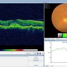
 Optic Disc Pit with Maculopathy
Feb 25 2021 by Niloofar Piri, MD
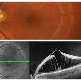
Color fundus photograph and SD OCT of a 6-year-old patient with optic disc pit associated with large retinoschisis involving the entire macula. SD OCT demonstrating large retinoschisis with ILM draping centrally giving it the appearance of pseudohole on the corresponding central area of color photo. Vision 20/80
Photographer: Lisa Breeding, St Louis University
Condition/keywords: maculopathy, optic disc
-
 Proliferative Diabetic Retinopathy with Pre-retinal Hemorrhage
Jan 16 2018 by Olivia Rainey
Ultra-wide field pseudo-color image of an 57-year-old male with a large pre-retinal hemorrhage secondary to proliferative diabetic retinopathy affecting his left eye.
Photographer: Olivia Rainey
Imaging device: Optos California
Condition/keywords: color fundus photograph, diabetic mellitus, hemorrhage, left eye, neovascularization (NV), Optos, proliferative diabetic retinopathy (PDR), pseudocolor, ultra-wide field imaging
-
 Traumatic Retinal Tear
Dec 5 2021 by Aditya S Kelkar, MS, FRCS, FASRS,FRCOphth
Color fundus photograph of a 34-year old man's left eye, 2 hours after a tennis ball injury, showing commotio retinae with Berlin's edema and cherry red spot in the fovea along with linear retinal tears in the temporal equatorial zone.
Photographer: Dr Sukanya Mondal. National Institute of Ophthalmology, Pune. India.
Imaging device: Zeiss Clarus 500
Condition/keywords: Berlin's edema, cherry red spot, commotio retinae, retinal tear
-
 Acute Idiopathic Occlusive Retinal Vasculitis
May 31 2014 by Hamid Ahmadieh, MD
Color fundus photograph of the left eye of a 28-year-old woman with acute drop of vision due to occlusive retinal vasculitis leading to extensive nerve fiber layer infarction and retinal hemorrhages.
Photographer: Naghmeh Nozhat, Negah Eye Center, Tehran
Condition/keywords: color fundus photograph, cotton wool spots, retinal hemorrhage, retinal ischemia
-
 Acute Retinal Necrosis secondary to Herpes Zoster Ophthalmicus
Jan 9 2018 by Olivia Rainey
Ultra-wide field Optos pseudocolor montage of an 40-year-old female presenting with acute retinal necrosis secondary to herpes zoster ophthalmicus affecting her right eye.
Photographer: Olivia Rainey
Imaging device: Optos California
Condition/keywords: acute retinal necrosis, color fundus photograph, Herpes zoster, montage, Optos, ultra-wide field imaging
-
 Advanced Proliferative Diabetic Retinopathy
Nov 4 2017 by Hamid Ahmadieh, MD
Merged color fundus photograph of the left eye of a 30-year-old woman with type1 diabetes since childhood. Note laser scars, severe fibrous proliferation, traction RD and macular dragging.
Photographer: Shabnam Poureh, Negah Eye Center, Tehran, Iran
Condition/keywords: color fundus photograph, diabetes, fibrous proliferation, proliferative diabetic retinopathy (PDR), severe traction
-
 Best Disease
Mar 9 2013 by Hamid Ahmadieh, MD
Color fundus photograph the left eye of a 49-year-old man with decreased VA due to advanced Best disease.
Photographer: Soodabeh Fooladin, Negah Eye Center, Tehran
Condition/keywords: Best disease
-
 Central Retinal Vein Occlusion: Case 1
Oct 12 2012 by Gregg T. Kokame, MD, MMM, FASRS
Color Fundus Photograph
Photographer: Jaclyn Pisano, Retina Consultants of Hawaii
Imaging device: Topcon 50IA / Eschalon
Condition/keywords: central retinal vein occlusion (CRVO)
-
 Choroidal Osteoma Plus CNV
Sep 2 2012 by Hamid Ahmadieh, MD
Color fundus photograph and OCT imaging of a 47-year-old man with a juxtafoveal CNV superimposed on a choroidal osteoma.
Photographer: Hamid Ahmadieh, Ophthalmic Research Center, Labbafinejad Medical Center
Imaging device: Topcon
Condition/keywords: choroidal neovascularization (CNV), choroidal osteoma, optical coherence tomography (OCT)
-
 Choroidal-Nevus
Feb 25 2023 by Aditya S Kelkar, MS, FRCS, FASRS,FRCOphth
Color fundus photograph of the left eye of a 55 year old male showing large choroidal nevus.
Photographer: Dr. Apoorva Jadhav, National Institute of Ophthalmology, Pune. India.
Imaging device: Zeiss Clarus 500
Condition/keywords: Choroidal nevus
-
 CNV due to AMPPE
Oct 16 2012 by Ratimir Lazic, MD, PhD
Color fundus photography of a 58-year-old male. White dots with juxtafoveolar subretinal fluid can be seen. BCVA of that eye is 0.35.
Photographer: Marko Lukic, MD
Imaging device: Zeis Visucam Lite 2
Condition/keywords: acute posterior multifocal placoid pigment epitheliopathy (APMPPE), choroidal neovascularization (CNV)
-
 Coat's Disease
Nov 17 2016 by Amir Manor, MD
Fundus photograph of a 25-year-old man with Coat's disease.
Photographer: Galit Yair-Pur
Condition/keywords: Coats' disease, color fundus photograph
-
 CRAO
CRAO
Oct 17 2014 by Avris Romario Diparaja Siahaan
A color fundus photograph of 35-year-old man that suffers central retinal artery occlusion in his right eye. He has underwent COA aspiration one day before.
Photographer: Dewi Tri Wulandari, Klinik Mata Nusantara
Imaging device: Topcon TRC 50 DX-IA
Condition/keywords: central retinal artery occlusion (CRAO), color fundus photograph
-
 Dislocated IOL
May 15 2018 by Morgan Benton
Ultra-wide field pseudocolor image of a 68-year-old male with a dislocated IOL after cataract surgery in the left eye. Patient was only able to count fingers at one foot and could pinhole to 20/60.
Photographer: Morgan Benton
Imaging device: Optos
Condition/keywords: color fundus photograph, dislocated intraocular lens (IOL), left eye, Optos, ultra-wide field imaging
 Loading…