Search results (415 results)
-
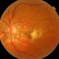
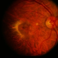
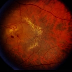
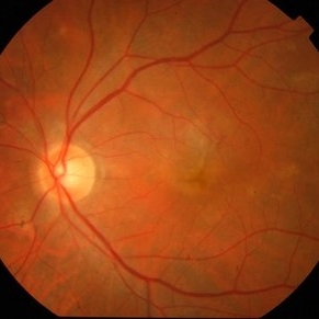 Active CNVM
Jul 11 2016 by Manish Nagpal, MD, FRCS (UK), FASRS
Colour photo showing an active CNVM.
Photographer: pooja barot
Condition/keywords: choroidal neovascular membrane (CNVM), optical coherence tomography (OCT)
-
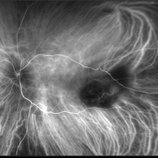
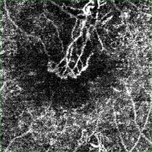 Active CNVM on Angio OCT
Jul 11 2016 by Manish Nagpal, MD, FRCS (UK), FASRS
Angio OCT picture showing neovascularization corresponding to the area of CNVM.
Photographer: pooja barot
Condition/keywords: choroidal neovascular membrane (CNVM), optical coherence tomography (OCT)
-
 Peripheral CNVM with Extensive Scarring
Oct 12 2019 by John S. King, MD
82-year-old white male with an acute loss of vision in the right eye was sent in to rule out a retinal detachment. Vision was 20/350; a dense VH was present, b-scan showed irregular areas of high reflectivity in the periphery that was c/w SRH. Peripherally, a few weeks later, there were areas that could be seen and were c/w peripheral CNVM (old and new). Anti-VEGF was administered. A month later vision was unchanged and patient wanted surgery to remove the VH. Pictured is one week since surgery; large peripheral scars are seen; diffuse areas of SR pigmentation is present; vitreous skirt present; and a few IRHs secondary to DR can be seen. He is currently 20/70 sc.
Photographer: Shelly Blair
Imaging device: Optos CA
Condition/keywords: choroidal neovascular membrane (CNVM), peripheral fundus lesion, vitreous blood
-
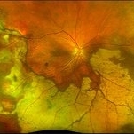
 Angioid Streaks
Jan 20 2021 by Nivesh Gupta
Fundus photograph of an 51-year-old female patient with angioid streaks with secondary choroidal neovascular membrane.
Photographer: Nivesh Gupta, Retina Fellow, Retina Foundation, Ahmedabad, India
Imaging device: NIDEK SLO MIRANTE
Condition/keywords: age-related macular degeneration (AMD), angioid streaks, choroidal neovascular membrane (CNVM)
-
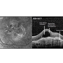 Outer Retinal Tubulation
Mar 27 2018 by Dhaivat Shah
Outer retinal tubulation (ORT) is a feature of photoreceptor rearrangement after chronic retinal damage due to refractory cme, long standing CNVM or old trauma. Photoreceptors lose adhesions to surrounding structures, resulting in outward folding and formation of new lateral contact between photoreceptors to form round structure. They generally remains stable over time. It is important to recognize ORT on OCT because it indicates a refractory state of the pathological condition and poor visual prognosis, and likely not to benefit from any treatment. Here is a case of 62-year-old female with history of 4 previous anti-VEGF injection in left eye for CNVM, with the recent OCT showing formation of ORT with subfoveal scarred membrane.
Photographer: Dr Dhaivat Shah
Condition/keywords: choroidal neovascular membrane (CNVM), outer retinal tubulation
-
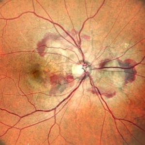
 RAP Lesions
Sep 29 2014 by Thomas A. Ciulla, MD, MBA, FASRS
Fluorescein angiogram of an 81-year-old man revealing several RAP lesions superior to fovea.
Photographer: Stuart Alfred CRA
Condition/keywords: choroidal neovascular membrane (CNVM), neovascular age-related macular degeneration (AMD), retinal angiomatous proliferation (RAP), wet age-related macular degeneration (wet AMD)
-
 Right Eye Color Photo With Hemorrhages in Case of CNVM With Angioid Streaks
Nov 29 2024 by Anand Temkar
A 45 year old male came with chief complaint of blurring vision in right eyes since past 4 days. His vision is 6/12 in right eye and 6/9 in left eye. His vision was 14 mmHg in right eye and 16 mmHg in left eye. He was diagnosed with Angioid Streaks in both eyes about a year ago, then he developed choroidal neovascularization in his left eye 8 months ago, for which he received AntiVEGF injections x 3. Left eye is a stable eye now. Patient presented with right eye choroidal neovascularization in a case of Angioid Streaks on recent follow up. We have advised him right eye AntiVEGF injections x 3. In this image, the right eye color photo shows bleed from CNVM in case of angioid streaks.
Photographer: Dr.Anand Temkar- Retina Foundation, Ahmedabad
Imaging device: Mirante
Condition/keywords: Angioid Streaks, choroidal neovascular membrane (CNVM)
-
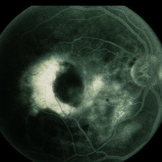
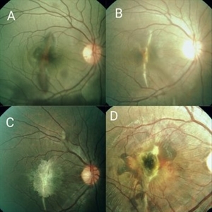 Serial Photographs of a Young Boy With Choroidal Rupture Progressing to Secondary CNVM
Sep 21 2017 by S. Natarajan, MD, FASRS, FRCS (GLASGOW) , FICO, D.Sc, FELA
A 16-year-old boy had a blunt injury to the right eye from a cricket ball. Fig A shows Berlins' edema with choroidal rupture which progresses to develop a secondary CNVM (Fig. D) over 1 year.
Photographer: Miss Ashwini borde
Imaging device: FF 450 Plus IR Zeiss
Condition/keywords: Berlin's edema, choroidal neovascular membrane (CNVM), choroidal rupture
-
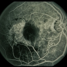
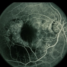
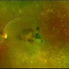 Subretinal Hemorrhage with Chorioretinal Macular Scars
Sep 28 2022 by Denica Rodriguez
Ultra-widefield pseudocolor fundus photograph of a 59 year old female with Subretinal Hemorrhage with Chorioretinal Macular Scars affecting her left eye. The physician presumes the etiology is CNV from adjacent scarring/choroidal rupture. Patient has history of ocular trauma with cricket ball at age 10-12 years old. She suspects that she might have suffered choroidal rupture, which has resulted in secondary CNV and hemorrhage that we are seeing today. She recommends treatment with Eylea sample injection in a series of 4 at a 4-5 week interval. The patient's vision at the time of her appointment was Dcc20/40-2 PHNI.
Photographer: Denica Rodriguez, COA
Imaging device: Optos California
Condition/keywords: antiVEGF therapy, chorioretinal scar, choroidal neovascular membrane (CNVM), fundus photography, left eye, macular scar, Optos, peripheral drusen, pseudocolor, secondary CNV, subretinal hemorrhage, ULTRA WIDE FIELD, ultra-wide field imaging
-
 Angioid Streaks with Associated Disc Drusen and CNV
Jan 6 2020 by Sarah Oelrich
Angioid streaks with associated disc drusen and CNV.
Photographer: Sarah Oelrich CRA COT OCT-C
Imaging device: Topcon, Heidelberg
Condition/keywords: angioid streaks, choroidal neovascular membrane (CNVM), disc drusen
-
 ICG: Choroidal Aspergilloma With Secondary Choroidal Neovascularization and Exudative Retinal Detachment
Mar 21 2019 by Scott D Walter, MD, MSc, FASRS
Multimodal imaging of a transplant patient with disseminated Aspergillosis and vision loss in her left eye.
Condition/keywords: choroidal neovascular membrane (CNVM), choroidal neovascularization (CNV), exudative detachment, focal chorioretinitis, fungal endophthalmitis, granulomatous choroiditis
-
 10 Days Post Subretinal TPA
Jan 9 2019 by John S. King, MD
76-year-old white male with history of treat/extend with Eylea OD for a PPCNVM; also monocular due to large scar in fellow eye. Two months since last injection, had acute decrease in vision OD and was seen that day. Vision CF; moderate SRH involving the fovea. Discussed monotherapy with anti-VEGF vs displacement, and elected for PPV, srTPA, AFx, SF6. Total of 0.2 cc of 25 microgram/0.1 ml of srTPA administered from two different areas in the temporal macula. 10 days post-op vision is improving; 20/200 J7; displacement of heme (photo)
Photographer: Kay Dalby
Imaging device: Topcon 50
Condition/keywords: choroidal neovascular membrane (CNVM), peripapillary hemorrhage, presumed ocular histoplasmosis syndrome (POHS)
-
 A rare case of a 45-year-old male with choroidal neovascular membrane in Familial Dominant Drusen (Doyne Honeycomb Drusen) in both eyes treated with intravitreal injections.
Nov 30 2022 by SHRADDHA ASHOK CHANDORKAR, DNB DO FVRS
A 45-year-old man presented with diminution of vision in both eyes with metamorphopsia, which was painless and gradually progressive in nature. BCVA at presentation were 6/40 and 6/36 for the right and left eye respectively. Anterior segment examination of both eyes was unremarkable. IOP were within normal limits. Fundus examination showed bilateral numerous yellowish white round closely spaced lesions extending radially from the vascular arcades till the periphery associated with an elevated grayish macular choroidal neovascular membrane (CNV) with multiple drusen in the macular area and posterior pole. Impression was Familial Dominant Drusen (Doyne Honeycomb Drusen) associated with CNVM, both eyes. Color fundus photograph and autofluorescence showed Familial Dominant Drusen with CNVM. Subsequently , the patient underwent periodic intravitreal injections of Ranibizumab in both eyes under guarded visual prognosis, for which he tolerated well.
Photographer: NATIONAL INSTITUTE OF OPHTHALMOLOGY, PUNE
Imaging device: ZEISS CLARUS
Condition/keywords: choroidal neovascular membrane (CNVM), Doyne's Honeycomb, FAMILIAL DOMINANT DRUSEN, IMIM (Online Mendelian Inheritance in Man), intravitreal injection, Malattia Leventinese
-
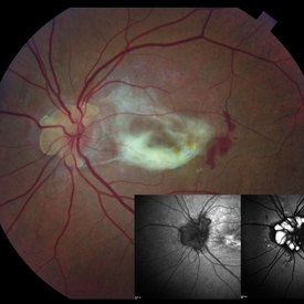
 Active CNVM
Jul 12 2023 by Harsh Vardhan Singh, MS
55-year male with left eye sub-retinal hemorrhage due to Active CNVM, Colour fundus photograph of left eye subretinal hemorrhage due to Active CNVM
Photographer: Harsh Vardhan Singh
Condition/keywords: choroidal neovascular membrane (CNVM), CNVM, subretinal hemorrhage
-
 Active CNVM
Jul 12 2023 by Harsh Vardhan Singh, MS
55-year male with left eye sub-retinal hemorrhage due to Active CNVM, Colour fundus photograph of left eye subretinal hemorrhage due to Active CNVM; Red-free image of left eye sub-retinal hemorrhage due to Active CNVM
Photographer: Harsh Vardhan Singh
Condition/keywords: choroidal neovascular membrane (CNVM), CNVM, subretinal hemorrhage
-
 Angioid Streaks
Jan 20 2021 by Nivesh Gupta
Fundus photograph of an 51-year-old female patient with angioid streaks with secondary choroidal neovascular membrane.
Photographer: Nivesh Gupta, Retina Fellow, Retina Foundation, Ahmedabad, India
Imaging device: NIDEK SLO MIRANTE
Condition/keywords: age-related macular degeneration (AMD), angioid streaks, choroidal neovascular membrane (CNVM)
-
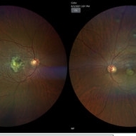
 Angioid streaks with choroidal neovascular membrane
Aug 24 2022 by Ruchir Mehta, DO, DNB, FRCS
Color fundus and red free fundus pictures of a 38 year old male with angioid streaks and choroidal neovascular membrane in the right eye
Photographer: Ruchir Mehta, Mehta Superspeciality Eye Hospital, Jamnagar, Gujarat, India
Imaging device: Zeiss Visucam 500
Condition/keywords: Angioid Streaks, choroidal neovascular membrane (CNVM)
-
 Angioid Streaks with scarred CNVM
Mar 28 2022 by Rutul R Patel, MD Ophthalmology
Case of 50 year female with Bilateral Angioid streaks and Left eye scarred CNVM
Photographer: Vidhi Bavishi
Imaging device: Topcon Maestro
Condition/keywords: angioid streaks, choroidal neovascular membrane (CNVM)
-
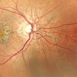
 ARMD / CNVM / PED With RPE Tear
Nov 7 2014 by David Callanan, MD
59-year-old male, ARMD / CNVM / PED with RPE Tear.
Condition/keywords: choroidal neovascular membrane (CNVM), pigment epithelial detachment (PED), retinal pigment epithelium (RPE) tear
-
 ARMD / CNVM / PED With RPE Tear
Nov 7 2014 by David Callanan, MD
59-year-old male, ARMD / CNVM / PED with RPE Tear.
Condition/keywords: choroidal neovascular membrane (CNVM), pigment epithelial detachment (PED), retinal pigment epithelium (RPE) tear
-
 ARMD / CNVM / PED With RPE Tear
Nov 7 2014 by David Callanan, MD
59-year-old male, ARMD / CNVM / PED with RPE Tear.
Condition/keywords: choroidal neovascular membrane (CNVM), pigment epithelial detachment (PED), retinal pigment epithelium (RPE) tear
-
 ARMD / CNVM / PED With RPE Tear
Nov 7 2014 by David Callanan, MD
59-year-old male, ARMD / CNVM / PED with RPE Tear.
Condition/keywords: choroidal neovascular membrane (CNVM), pigment epithelial detachment (PED), retinal pigment epithelium (RPE) tear
-
 ARMD / CNVM / PED With RPE Tear
Nov 7 2014 by David Callanan, MD
59-year-old male, ARMD / CNVM / PED with RPE Tear.
Condition/keywords: choroidal neovascular membrane (CNVM), pigment epithelial detachment (PED), retinal pigment epithelium (RPE) tear
-
 ARMD / PED / CNVM
Nov 3 2014 by David Callanan, MD
81-year-old male, ARMD / PED / CNVM.
Condition/keywords: choroidal neovascular membrane (CNVM), pigment epithelial detachment (PED)
-
 ARMD / PED / CNVM
Nov 3 2014 by David Callanan, MD
81-year-old male, ARMD / PED / CNVM.
Condition/keywords: choroidal neovascular membrane (CNVM), pigment epithelial detachment (PED)
 Loading…