Search results (257 results)
-
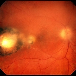
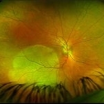
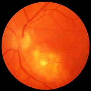
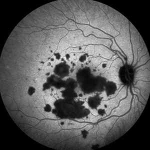 Relentless Placoid Chorioretinitis
Jan 9 2019 by Janet Brazil
ICG photo of a 24-year-old male with Relentless placoid chorioretinitis
Photographer: Janet Atkinson, Eye Associates of New Mexico
Imaging device: Heidelberg HRA Spectralis
Condition/keywords: placoid retinal lesions
-
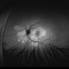 Acute Syphilitic Posterior Placoid Chorioretinitis
Oct 16 2024 by César Adrián Gómez Valdivia, MD
Fundus autofluorescence image of an acute syphilitic posterior placoid chorioretinitis found in a HIV positive 28 YO male patient with suspected neurosyphilis. A beautiful butterfly autofluorescence pattern can be appreciated.
Photographer: @eyemissu2
Imaging device: California ICG OPTOS
Condition/keywords: acute syphilitic posterior placoid chorioretinitis, chorioretinitis, syphilis
-
 Acute Syphilitic Posterior Placoid Chorioretinitis
Sep 3 2016 by ADRIANO FERREIRA
66-year-old woman with acute visual acuity loss.
Photographer: Claudio Zett Lobo, UNIFESP
Condition/keywords: acute syphilitic posterior placoid chorioretinitis
-
 Chorioretinitis with Overlying Vitreous Stranding/Vitritis
Mar 23 2023 by Isaac Agranoff
Fundus photograph of a 37-year-old woman presenting with chorioretinitis with overlying vitreous stranding/vitritis that has remained unchanged for multiple years. Patient presented with irritation and blurred vision and her vision was 20/40 OD. The OCT revealed evidence of low-grade inflammation and the recommend treatment was anti-inflammatory eye drops at this time and to obtain second opinion with another physician in the office.
Photographer: Isaac Agranoff, Technician
Imaging device: Optos California
Condition/keywords: chorioretinal scar, chorioretinitis, inflammation, Optos, ultra-wide field imaging, vitritis
-
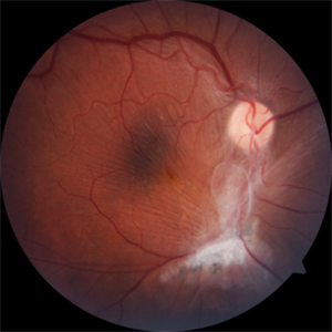
 Idiopathic Uveal Effusion Syndrome
Aug 22 2024 by Jordyn Beckman
61 year old male with Idiopathic Uveal Effusion Syndrome with starry night appearance on fluorescein. 3 weeks s/p single external drainage retinotomy and 9 weeks of oral pred with recurrent choroidal effusions. Has since returned to surgery for secondary drainage retinotomy; subretinal fluid remain persistent.
Photographer: Jordyn Beckman
Imaging device: Optos California
Condition/keywords: chorioretinitis, Choroidal, exudative detachment, window defect
-
 Retinocoroiditis Inactiva Por Toxoplasmosis
Apr 28 2025 by Paulina Araujo
Fundus photography demonstrates a 2-disc-diameter chorioretinal scar in the superior temporal arcade, consistent with inactive toxoplasmic retinochoroiditis. The lesion exhibits pigmented borders and central atrophy, with adjacent splinter hemorrhages and vascular sheathing. No vitreous inflammation or active satellite lesions are present.
Photographer: Paulina D.Araujo Martínez, Asociación para Evitar la Ceguera en México I.A.P., Hospital Dr Luis Sánchez Bulnes.
Condition/keywords: toxoplasmosis chorioretinitis
-
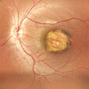
 Toxo Lesion
Dec 29 2016 by Manish Nagpal, MD, FRCS (UK), FASRS
Healing toxo chorioretinitis lesion post a course of anti-toxo regime.
Photographer: Avijit Vishnoi
Condition/keywords: toxoplasmosis chorioretinitis
-
 Toxoplasma Retinochoroiditis
Feb 25 2013 by Henry J. Kaplan, MD
Toxoplasmosis, right eye: reactivation of congenital toxoplasmosis as an active retinitis lesion with overlying vitritis adjacent to an old scar.
Condition/keywords: toxoplasmosis chorioretinitis, toxoplasmosis reactivation
-
 Acute Syphilitic Posterior Placoid Chorioretinitis
Oct 20 2024 by César Adrián Gómez Valdivia, MD
Fundus autofluorescence image of an acute syphilitic posterior placoid chorioretinitis found in a HIV positive 28 YO male patient with suspected neurosyphilis. A beautiful butterfly autofluorescence pattern can be appreciated.
Photographer: @eyemissu2
Imaging device: California ICG OPTOS
Condition/keywords: acute syphilitic posterior placoid chorioretinitis
-
 Acute Syphilitic Posterior Placoid Chorioretinitis
May 4 2021 by RAFAEL REIS PEREIRA, MD
A 31-year-old patient with a complaint of photophobia and low visual acuity OD in the previous three weeks. BCVA was 20/60 and 20/20 The fundus examination revealed a placoid white lesion in the posterior pole and vitreous cells in the right eye. The left eye was unremarkable. Fluorescein angiography reveals hyperfluorescent plaque with distinctive “leopard spots” hypofluorescence.
Imaging device: Opto California
Condition/keywords: acute syphilitic posterior placoid chorioretinitis
-
 Acute syphilitic posterior placoid chorioretinitis
Apr 24 2022 by Aniruddha K Agarwal, MD
Green-light fundus autofluorescence (FAF) of the right eye from a 55-year-old man with risk factors for sexually trasnmitted diseases who presented to the retina clinic for a central scotoma. Funduscopy revealed a placoid lesion in the posterior pole. FAF highlights a hyperautofluorescent placoid lesion involving the macula with granular hyperfluorescence. The patient tested positive for syphilis and received intravenous penicillin treatment.
Photographer: Esther CIANCAS, MD, PhD, Gema CRESPO-RODRÍGUEZ, RN
Imaging device: Zeiss Clarus fundus camera
Condition/keywords: chorioretinitis, IUSG, syphilis, uveitis
-
 Acute Syphilitic Posterior Placoid Chorioretinitis (ASPPC)
May 12 2021 by Joseph D Boss, MD
Ultra-widefield fundus photograph of a 36-year-old male with acute syphilitic posterior placoid chorioretinitis. Subsequent testing reviewed a positive RPR 1:256 and positive syphilis antibody.
Photographer: Joseph Boss, MD; Retina Specialists of Michigan
Condition/keywords: acute syphilitic posterior placoid chorioretinitis, syphilitis uveitis
-
 Childhood Acquired Ocular Toxoplasmosis
Sep 13 2023 by Deepak Bhojwani, MS
Fundus image of a 16 year old boy diaagnosed with Ocular Toxoplasmosis since the age of 10 years showing the classic toxo chorioretinitis scar on the posterior pole. Luckily the scar is loacted juxtatemporal to fovea on OCT and so the boy has good vision of 20/30.
Photographer: DR DEEPAK BHOJWANI
Imaging device: OPTCAL COHERENCE TOMOGRAPHY
Condition/keywords: posterior uveitis, toxo chorioretinitis
-
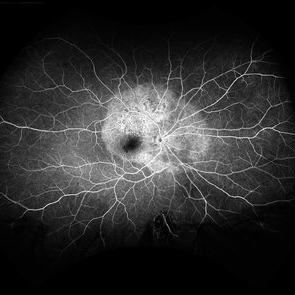
 Disseminated Chorioretinitis With Unknown Etiology
Apr 5 2018 by Kim Barrett
Ultra-wide field fluorescein angiogram of a 31-year-old female with intermittent pain in her left eye. Her condition has been managed in Liberia until recently when she moved to the United States. She suffers from multiple modalities including central retinal artery occlusion, posterior synechiae of the iris, interstitial keratitis, disseminated chorioretinitis, as well as HIV. An infectious cause is high on the differential in light of her HIV status. DDx: hypertensive crisis, an embolism (? IV drug use), coagulopathy, trauma, infectious. Blood work was normal. Her current vision is 20/30 right eye and 20/400 left eye.
Photographer: Kim Barrett, COA
Imaging device: Optos
Condition/keywords: central retinal artery occlusion (CRAO), chorioretinal scar, ciliary artery sparring, disseminated chorioretinitis, HIV, left eye, optic atrophy, staining
-
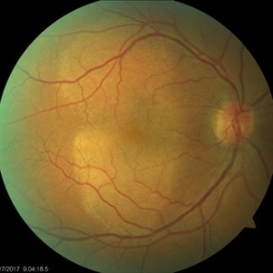
 Macular Traction Related to Toxoplasma Chorioretinitis
Jan 7 2021 by Lucas Zago Ribeiro, MD
Fundus image of a 50-year-old woman with macular traction and epiretinal membrane after toxoplasma chorioretinitis.
Photographer: Lucas Zago Ribeiro, UNIFESP / EPM, Brazil
Condition/keywords: epiretinal membrane (ERM), toxoplasmosis
-
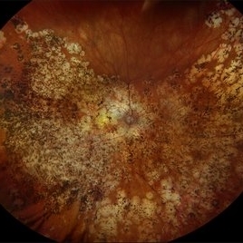 Multifocal Choroiditis with Panuveitis
Oct 16 2025 by Virginia Gebhart
39 year old female diagnosed with MCP in 2009. Extensive RPE changes and hypertrophy, arterial attenuation and pale nerve. Currently no active inflammation.
Photographer: Virginia Gebhart, Retina Consultants of Carolina
Imaging device: Optos California
Condition/keywords: corticosteroid-induced glaucoma, hypertrophy, multifocal chorioretinitis (MCP), PALE DISC
-
 Posterior Placoid Chorioretinopathy
Dec 19 2020 by John S. King, MD
44-year-old white female seen over the weekend complaining of a "spot" in her vision centrally OD for three days. She was referred over by another eye doctor who was concerned about a possible retinal detachment vs ARN in that eye. Her past medical history includes adrenal insufficiency for which she takes a low dose of hydrocortisone, thyroxine (post thyroidectomy), Plaquenil (inflammatory arthritis). She is divorced with one partner and denies any IVDU. Va 20/200 OD and 20/20 OS, IOP 12 OU, pupils mydriatic post gtts (light desaturation OD). There was 1+ A/C cell OD, O/W unremarkable anterior segment OU; in the posterior segment OD there was 1+ vitritis with a diffusely swollen optic disc and a large yellowish placoid lesion in the macula with yellowish border and extended out past the arcades inferiorly, as well as another lesion smaller in the IN periphery, and two possible smaller spots SN (See Photo above). There was a trace vitreous cell OS with a large, granular placoid lesion nasally. The OCT showed mild subfoveal fluid with nodular areas in the RPE and some overlying irregular architecture of the outer retina. Syphilis was a concern at this point. She denied any hand or foot rash, and said that she was recently working on the house, and her hands were dried out. There did appear to be a rash on the hand, and later learned that she had a rash on the soles of her feet. She was sent to ED for a work-up and her syphilis IgG was positive and VDRL 1:128, and negative for HIV. She was started on a course IV Penicillin (40mg PO steroid two days after tx started). She has responded well. A few days after treatment her visual acuity has improved to 20/60 OD; there was no anterior segment inflammation OU, and decreased vitreous cell OU. Disc edema was improved. The large placoid lesion in the macula of the right eye was slightly enlarged, but more granular in appearance without a distinct yellowish border, and the smaller lesions SN had dissipated. OCT showed resolution of the subfoveal fluid and an improved appearance of the outer retina and RPE layer.
Imaging device: Optos CA
Condition/keywords: acute syphilitic posterior placoid chorioretinitis, syphilis
-
 Syphilis CR
Sep 1 2017 by Annal D Meleth, MD, MS
Syphilis CR
Photographer: Kenneth Thompson
Condition/keywords: acute syphilitic posterior placoid chorioretinitis, syphilis
-
 Syphilitic Maculopathy
Aug 27 2012 by Logan Milad Haak, MD
51 year-old HIV+ man presenting with "Gray screen over vision" RPR and FTA-Abs were positive.
Photographer: Illinois Retina Associates
Condition/keywords: acute syphilitic posterior placoid chorioretinitis
-
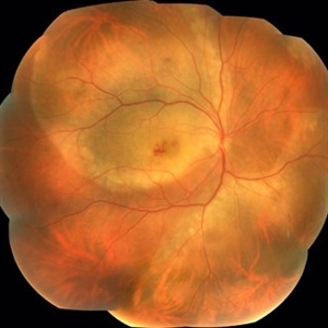
 Toxo Lesion on Macula
Jul 25 2019 by Manish Nagpal, MD, FRCS (UK), FASRS
Fundus photo of old toxo lesion on macula.
Photographer: Gayathri Mohan, Retina Foundation
Imaging device: Nidek Mirante SLO
Condition/keywords: toxoplasmosis, toxoplasmosis chorioretinitis
-
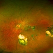
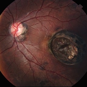
 Toxoplasma chorioretinitis 1
Jan 11 2013 by Alex P. Hunyor, MD
Toxoplasmosis 1 - chorioretinal scar from previous toxoplasma chorioretinitis. See image 2 - recurrent todo adjacent to this scar
Condition/keywords: inactive toxoplasmosis, ocular toxoplasmosis, toxoplasmosis, toxoplasmosis retinitis
-
 Toxoplasmosis
Jun 3 2017 by Gabriel Costa Andrade, PhD
Fundus photograph of an 14-year-old boy with multiple chorioretinal scars secondary to toxoplasmosis.
Photographer: Gabriel Costa de Andrade
Imaging device: Optos® California
Condition/keywords: congenital toxoplasmosis, ocular toxoplasmosis, toxoplasmosis chorioretinitis, toxoplasmosis uveitis
-
 Toxoplasmosis Chorioretinitis
Oct 10 2012 by Jeffrey G. Gross, MD, FASRS
Toxoplasmosis chorioretinitis, 20/70, 5 weeks after treatment, APD
Condition/keywords: 20/70, afferent pupillary defect (APD), toxoplasmosis chorioretinitis
-
 Wagon-Wheel Lesion
Jun 5 2025 by César Adrián Gómez Valdivia, MD
Wagon-wheel lesion found in a 12 YO male patient diagnosed with congenital toxoplasmosis. Findings were bilateral.
Photographer: @eyemissu2
Imaging device: TOPCON TRC-50DX
Condition/keywords: toxoplasmosis chorioretinitis, Wagon-wheel lesion
-
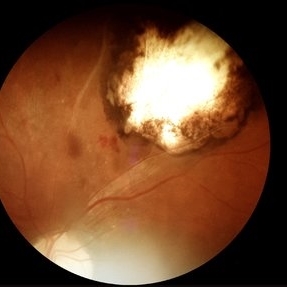
 Acute Syphilitic Posterior Placoid Chorioretinitis
Aug 23 2012 by Gerardo Garcia-Aguirre, MD
Fundus photograph of a 42 year-old male with positive VDRL and FTA-ABS, with a yellowish placoid lesion in the posterior pole.
Photographer: Ricardo Montoya, Asociación para Evitar la Ceguera en México
Condition/keywords: acute syphilitic posterior placoid chorioretinitis, syphilis
 Loading…