Search results (257 results)
-
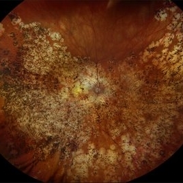
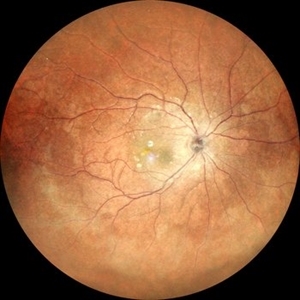
 Toxoplasmosis Chorioretinitis
Dec 16 2025 by Kimberly Wakester
Optomap RGB image of a 22-year-old man with Toxoplasmosis Chorioretinitis in both eyes. Recommended yearly examinations.
Photographer: Kimberly Wakester, COA, OCT-C
Imaging device: Optos California
Condition/keywords: toxoplasmosis chorioretinitis
-
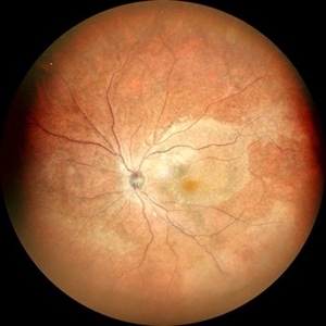
 Chorioretinitis
Dec 16 2025 by Kimberly Wakester
Optomap RGB of a 37 year-old woman with Chorioretinitis. The chorioretinitis remains largely stable in both eyes on exam and compared to prior photos. Clinical and diagnostic findings in both eyes continue to be most consistent with punctate inner choroidopathy (PIC) /Multifocal choroiditis and panuveitis (MCP). Will continue follow up care every 6 months with dilated exam and repeat testing.
Photographer: Kimberly Wakester, COA, OCT-C
Imaging device: Optos California
Condition/keywords: chorioretinitis, multifocal chorioretinitis (MCP), punctate inner choroidopathy (PIC)
-
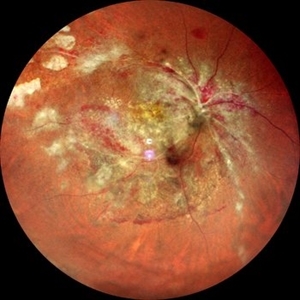
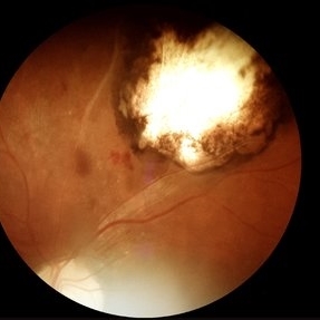
 Acute Syphilitic Posterior Placoid Chorioretinitis
Nov 1 2025 by Julián Villarreal, MD
A 48 year old male presented with Acute syphilitic posterior placoid chorioretinitis (ASPPC) a large, roundish, yellowish, placoid lesion occurring at level of the retinal pigment epithelium (RPE) at the macular/paramacular area. Further testing showed a reactive VDRL and a positive FTA-ABS.
Photographer: Julián Villarreal MD
Imaging device: Mirante
Condition/keywords: Preretinal precipitates
-
 Chorioretinitis
Oct 30 2025 by Kimberly Wakester
Optomap RGB of an 37-year-old-woman with stable Chorioretinitis in the right eye. Patient is to return in 6 months for dilated exam and repeat diagnostic testing.
Photographer: Kimberly Wakester, COA, OCT-C
Imaging device: Optos California
Condition/keywords: chorioretinitis
-
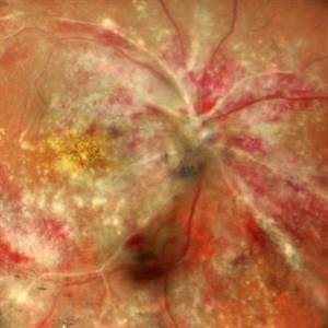
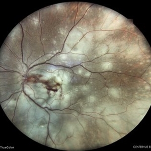
 Multifocal Choroiditis with Panuveitis
Oct 16 2025 by Virginia Gebhart
39 year old female diagnosed with MCP in 2009. Extensive RPE changes and hypertrophy, arterial attenuation and pale nerve. Currently no active inflammation.
Photographer: Virginia Gebhart, Retina Consultants of Carolina
Imaging device: Optos California
Condition/keywords: corticosteroid-induced glaucoma, hypertrophy, multifocal chorioretinitis (MCP), PALE DISC
-
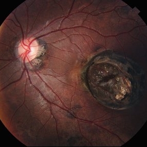
 Kyrieleis Plaques
Aug 18 2025 by Helder Vasconcelos
Kyrieleis Plaques and vitritis in a patient with posterior uveitis secondary to presumed ocular toxoplasmosis.
Photographer: Helder Vasconcelos
Imaging device: Smartphone Fundoscopy
Condition/keywords: kyrieleis plaques, toxoplasmosis chorioretinitis
-
 CMV Retinitis: Turning Retina into Abstract Art Since Immunosuppression
Aug 4 2025 by rohan jain
We report a case of 34 years old HIV positive male who presented with Diminution of vision in OD since 1 month .Examination of OD showed hazy media due to vitritis, diffuse yellowish-whitish retinal necrosis and retinal hemorrhages around the disc and attenuated retinal vessels.
Photographer: Dr. ROHAN JAIN
Imaging device: mirante
Condition/keywords: CMV chorioretinitis, CMV retinitis, cytomegalovirus (CMV), Cytomegalovirus Retinitis
-
 CMV Retinitis: Turning Retina into Abstract Art Since Immunosuppression
Aug 4 2025 by rohan jain
We report a case of 34 years old HIV positive male who presented with Diminution of vision in OD since 1 month. Examination of OD showed hazy media due to vitritis, diffuse yellowish-whitish retinal necrosis and retinal hemorrhages around the disc and attenuated retinal vessels.
Photographer: Dr. ROHAN JAIN
Imaging device: mirante
Condition/keywords: CMV chorioretinitis, CMV retinitis, cytomegalovirus (CMV), Cytomegalovirus Retinitis
-
 Wagon-Wheel Lesion
Jun 5 2025 by César Adrián Gómez Valdivia, MD
Wagon-wheel lesion found in a 12 YO male patient diagnosed with congenital toxoplasmosis. Findings were bilateral.
Photographer: @eyemissu2
Imaging device: TOPCON TRC-50DX
Condition/keywords: toxoplasmosis chorioretinitis, Wagon-wheel lesion
-
 Retinocoroiditis Inactiva Por Toxoplasmosis
Apr 28 2025 by Paulina Araujo
Fundus photography demonstrates a 2-disc-diameter chorioretinal scar in the superior temporal arcade, consistent with inactive toxoplasmic retinochoroiditis. The lesion exhibits pigmented borders and central atrophy, with adjacent splinter hemorrhages and vascular sheathing. No vitreous inflammation or active satellite lesions are present.
Photographer: Paulina D.Araujo Martínez, Asociación para Evitar la Ceguera en México I.A.P., Hospital Dr Luis Sánchez Bulnes.
Condition/keywords: toxoplasmosis chorioretinitis
-
 Posterior Placoid Chorioretinitis
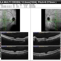
Mar 9 2025 by Oscar Francisco Miranda, MD
A 36-year-old male with bilateral visual loss of 3 months' duration, with no relevant medical history on inquiry. A round-shaped lesion with well-defined borders and a yellowish-white color is observed in the macula of both eyes, accompanied by vitreous cellularity. The macular OCT shows a dentate RPE. The VDRL, FTA-ABS, and HIV tests were positive.
Photographer: Oscar Francisco Miranda-Gómez
Imaging device: Heidelberg Spectralis
Condition/keywords: acute syphilitic posterior placoid chorioretinitis, OCT, Ocular syphilis
-
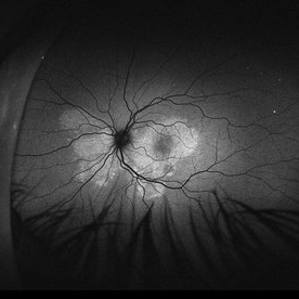
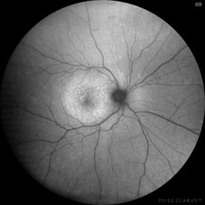 Posterior Placoid Chorioretinitis
Mar 9 2025 by Oscar Francisco Miranda, MD
A 36-year-old male with bilateral visual loss of 3 months' duration, with no relevant medical history on inquiry. A round-shaped lesion with well-defined borders and a yellowish-white color is observed in the macula of both eyes, accompanied by vitreous cellularity. The macular OCT shows a dentate RPE. The VDRL, FTA-ABS, and HIV tests were positive.
Photographer: Oscar Francisco Miranda-Gómez
Imaging device: Autofluorescence Zeiss Clarus 700
Condition/keywords: acute posterior placoid chorioretinitis, Autofluorescence, ocular syphilis
-
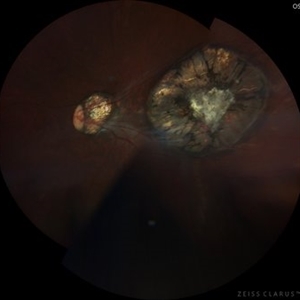
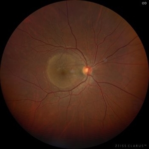 Posterior Placoid Chorioretinitis
Mar 9 2025 by Oscar Francisco Miranda, MD
A 36-year-old male with bilateral visual loss of 3 months' duration, with no relevant medical history on inquiry. A round-shaped lesion with well-defined borders and a yellowish-white color is observed in the macula of both eyes, accompanied by vitreous cellularity. The macular OCT shows a dentate RPE. The VDRL, FTA-ABS, and HIV tests were positive.
Photographer: Oscar Francisco Miranda-Gómez
Imaging device: Zeiss Clarus 700
Condition/keywords: acute posterior placoid chorioretinitis, Ocular syphilis
-
 Acute Syphilitic Posterior Placoid Chorioretinitis
Oct 20 2024 by César Adrián Gómez Valdivia, MD
Fundus autofluorescence image of an acute syphilitic posterior placoid chorioretinitis found in a HIV positive 28 YO male patient with suspected neurosyphilis. A beautiful butterfly autofluorescence pattern can be appreciated.
Photographer: @eyemissu2
Imaging device: California ICG OPTOS
Condition/keywords: acute syphilitic posterior placoid chorioretinitis
-
 Acute Syphilitic Posterior Placoid Chorioretinitis
Oct 16 2024 by César Adrián Gómez Valdivia, MD
Fundus autofluorescence image of an acute syphilitic posterior placoid chorioretinitis found in a HIV positive 28 YO male patient with suspected neurosyphilis. A beautiful butterfly autofluorescence pattern can be appreciated.
Photographer: @eyemissu2
Imaging device: California ICG OPTOS
Condition/keywords: acute syphilitic posterior placoid chorioretinitis, chorioretinitis, syphilis
-
 Idiopathic Uveal Effusion Syndrome
Aug 22 2024 by Jordyn Beckman
61 year old male with Idiopathic Uveal Effusion Syndrome with starry night appearance on fluorescein. 3 weeks s/p single external drainage retinotomy and 9 weeks of oral pred with recurrent choroidal effusions. Has since returned to surgery for secondary drainage retinotomy; subretinal fluid remain persistent.
Photographer: Jordyn Beckman
Imaging device: Optos California
Condition/keywords: chorioretinitis, Choroidal, exudative detachment, window defect
-
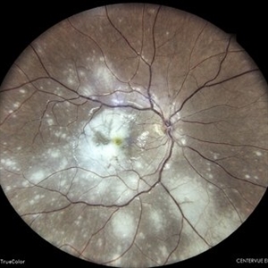
 Focal Chorioretinitis
Jul 11 2024 by Virginia Gebhart
67 year old female with punched-out CR scars. Hx of laser 3x for apparent peripapillary CNV. ESR, CRP, toxo, IgG/IgM all "normal." Bartonella, quant gold, and FTA-ABS ordered given possibility of neuroretinitis. Vision CF
Photographer: Virginia Gebhart
Imaging device: Optos California
Condition/keywords: FA, fluorescein angiogram (FA), FLUORESCEIN ANGIOGRAPHY, focal chorioretinitis, optic neuritis
-
 Multifocal Chorioretinitis
Apr 9 2024 by Akansha Sharma
Color fundus photograph of a 34 year old male patient with multifocal chorioretinitis with subretinal bleed.
Photographer: Dr. Akansha Sharma, Bharati Eye Hospital
Condition/keywords: chorioretinal inflammations, chorioretinitis, subretinal hemorrhage
-
 Multifocal Chorioretinitis
Apr 9 2024 by Akansha Sharma
Color fundus photograph of a 34 year old male patient with multifocal chorioretinitis.
Photographer: Dr. Akansha Sharma, Bharati Eye Hospital
Condition/keywords: chorioretinal inflammations, chorioretinitis
-
 Toxoplasmosis Disease
Mar 30 2024 by Karen Flores Guevara
Fundus photograph of a 7-year-old-child with a macular scar observed over time for growth.
Photographer: Diana Elizabeth Arellano Acosta MD Pediatric Retina,Asociación para Evitar la Ceguera en México IAP. México
Condition/keywords: toxoplasmosis chorioretinitis
-
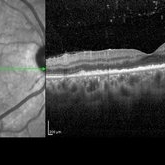
 Syphilitic Posterior Uveitis
Mar 22 2024 by Anjana Mirajkar, MS Ophthalmology
An OCT image BE of 36 year old female showing RPE granularity and IS/OS irregularity in a case of syphilitic posterior placoid chorioretinitis
Photographer: Dr. Anjana Mirajkar -Retina Foundation, Ahmedabad
Condition/keywords: acute posterior placoid chorioretinitis, acute syphilitic posterior placoid chorioretinitis
-
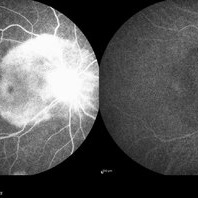 Syphilitic Posterior Uveitis
Mar 22 2024 by Anjana Mirajkar, MS Ophthalmology
FA image of RE of a 36 year old female showing hyper-fluorescence (staining) from early to late phases of the angiogram in a case syphilitic posterior placoid chorioretinitis. ICG image depicts hypo-cyanence from early to late phases.
Photographer: Dr. Anjana Mirajkar -Retina Foundation, Ahmedabad
Imaging device: Heidelberg
Condition/keywords: acute syphilitic posterior placoid chorioretinitis
-
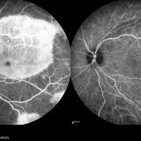 Syphilitic Posterior Uveitis
Mar 22 2024 by Anjana Mirajkar, MS Ophthalmology
FA image of LE of a 36 year old female showing hyper-fluorescence (staining) from early to late phases of the angiogram in a case syphilitic posterior placoid chorioretinitis. ICG image depicts hypo-cyanence from early to late phases.
Photographer: Dr. Anjana Mirajkar -Retina Foundation, Ahmedabad
Condition/keywords: acute syphilitic posterior placoid chorioretinitis
-
 Syphilitic Posterior Uveitis
Mar 22 2024 by Anjana Mirajkar, MS Ophthalmology
A color photo image of RE of a 36 year old female showing hypopigmented lesions at the posterior pole(ground glass appearance) in a case of syphilitic posterior placoid chorioretinitis
Photographer: Dr. Anjana Mirajkar -Retina Foundation, Ahmedabad
Imaging device: Mirante-Nidek
Condition/keywords: posterior uveitis
-
 Syphilitic Posterior Uveitis
Mar 22 2024 by Anjana Mirajkar, MS Ophthalmology
A color photo image of LE of a 36 year old female showing hypopigmented lesions at the posterior pole(ground glass appearance) in a case of syphilitic posterior placoid chorioretinitis
Photographer: Dr. Anjana Mirajkar -Retina Foundation, Ahmedabad
Imaging device: Mirante-Nidek
Condition/keywords: syphilitic posterior uveitis
 Loading…