Search results (129 results)
-
 Acute Posterior Multifocal Placoid Pigment Epitheliopathy
Feb 20 2024 by Soobien Lee
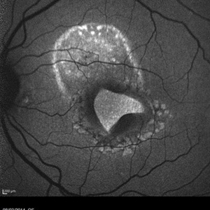
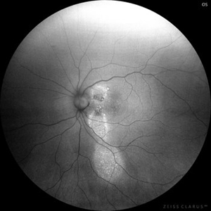
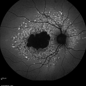
Optos fundus autofluorescence photograph of a 20-year-old caucasian female with viral prodrome and vision loss OS>OD secondary to Acute Posterior Multifocal Placoid Pigment Epitheliopathy (APPME). Imaging of her left eye shows hypoautofluorescent areas corresponding to multiple bilateral placoid lesions at the level of RPE and choroid throughout the posterior pole.
Photographer: Ashley Metzger, Elman Retina Group
Imaging device: Optos Ultra-Widefield Autoflurescence Imaging
Condition/keywords: acute posterior multifocal placoid pigment epitheliopathy (APMPPE), autofluorescence imaging, bacilliary layer detachment, Optos, OPTOS CALIFORNIA, uveitis, white dot syndrome
-
 Puzzle Retinitis
Jan 20 2021 by Jamin S. Brown, MD
Puzzle artifact after imaging on a smaller field of view with blue light autofluorescence.
Photographer: Stefanie Palmer CRA, Retina Vitreous Surgeons of CNY
Condition/keywords: autofluorescence imaging, normal eye
-
 Torpedo Maculopathy
Feb 20 2024 by Soobien Lee
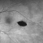
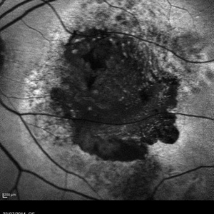
Optos fundus autofluorescence photograph of a 35-year-old asymptomatic female with no ocular or medical history with stable and chronic appearing torpedo-shaped macula lesion in the left eye.
Photographer: Peter Sotirakos, Elman Retina Group
Imaging device: Optos Ultra-Widefield Autoflurescence Imaging
Condition/keywords: autofluorescence imaging, genetics, macula, maculopathy, Optos, torpedo maculopathy
-
 Branch Retinal Artery Occlusion With Calcium Embolus at the Disc - Fundus Autofluorescence Imaging (FAF)
Apr 7 2018 by Rameez N Hussain, MD
Acute branch retinal artery occlusion with a calcium embolus at the disc which is hyper autofluorescent in fundus autofluorescence imaging (FAF) -resembles an LED light source ('LED sign').
Photographer: DR RAMEEZ N HUSSAIN
Imaging device: Heidelberg Spectralis
Condition/keywords: branch retinal artery occlusion (BRAO), embolus, fundus autofluorescence (FAF), retinal edema
-
 Branch Retinal Artery Occlusion With Calcium Embolus at the Disc - Fundus Autofluorescence Imaging (FAF)
Apr 7 2018 by Rameez N Hussain, MD
Acute branch retinal artery occlusion with a calcium embolus at the disc which is hyper autofluorescent in fundus autofluorescence Imaging (FAF) -resembles an LED light source ('LED sign').
Photographer: DR RAMEEZ N HUSSAIN
Imaging device: Heidelberg Spectralis
Condition/keywords: branch retinal artery occlusion (BRAO), embolus, fundus autofluorescence (FAF), retinal edema
-
 Central Serous Retinopathy
Mar 19 2024 by Corey Grant
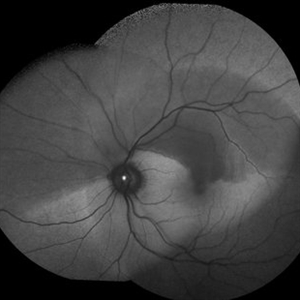
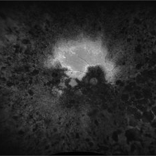
Ultra Wide-Field Fundus Autofluorescence Imaging of a 37 year old female with Central Serous Retinopathy affecting her right eye. Patient Visual Acuity was 20/20 in both eyes. Patient reported black spots in her vision onset three years ago, with associating flashes of light. Patient reports history of cortisone back injections a few years ago and denies Flonase use. The physician stated that there is hyperautofluorescence in the area of gutter of Sub-Retinal Fluid which likely happened from CSR.
Photographer: Corey Grant, OSC
Imaging device: OPTOS CALIFORNIA RGB
Condition/keywords: Central Serous Chorioretinopathy (CSR), central serous retinopathy (CSR), fundus autofluorescence (FAF), Guttering, hyperautofluorescence, inferior retina, OPTOS, Retina, Right Eye, subretinal fluid, ULTRA WIDE FIELD
-
 Choroidal Osteoma
Mar 29 2013 by Henry J. Kaplan, MD
Autofluorescence in choroidal osteoma.
Condition/keywords: autofluorescence imaging, choroidal osteoma
-
 Macular Tear
May 14 2014 by Avris Romario Diparaja Siahaan
Blue autofluorescence (BAF) a 40-year-old man with macular tear (had a photocoagulation laser).
Photographer: Avris Romario Diparaja Siahaan
Imaging device: Heidelberg HRA + OCT Spectralis
Condition/keywords: autofluorescence imaging, macular hole
-
 AMD
AMD
Jul 26 2014 by Avris Romario Diparaja Siahaan
An autofluorescence image of a 78-year-old-man with an age-related macular degeneration on his both eyes.
Photographer: Avris Romario Diparaja Siahaan, Klinik Mata Nusantara
Imaging device: Heidelberg Spectralis
Condition/keywords: age-related macular degeneration (AMD), autofluorescence imaging
-
 Case 2 Retinitis Pigmentosa BAF IRAF OD
May 14 2014 by Avris Romario Diparaja Siahaan
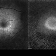
Fundus image a 57-year-old man with retinitis pigmentosa on both eyes. These image were taken with blue auto fluorescein mode (BAF) and infrared auto fluorescence (IRAF).
Photographer: Avris Romario Diparaja Siahaan
Imaging device: Heidelberg HRA + OCT Spectralis
Condition/keywords: autofluorescence imaging, fundus photograph, infrared image, retinitis pigmentosa
-
 Case 2 Retinitis Pigmentosa BAF IRAF OS
May 14 2014 by Avris Romario Diparaja Siahaan
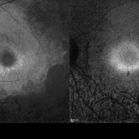
Fundus image a 57-year-old man with retinitis pigmentosa on both eyes. These image were taken with blue auto fluorescein mode (BAF) and infrared auto fluorescence (IRAF).
Photographer: Avris Romario Diparaja Siahaan
Imaging device: Heidelberg HRA + OCT Spectralis
Condition/keywords: autofluorescence imaging, fundus photograph, infrared image, retinitis pigmentosa
-
 Chronic CSR - Dancing doll
Nov 20 2023 by Harsh Vardhan Singh, MS
37-year male with chronic CSR
Photographer: Harsh Vardhan Singh
Imaging device: Zeiss clarus 700
Condition/keywords: autofluorescence imaging, Central Serous Chorioretinopathy (CSR), fundus autofluorescence (FAF), idiopathic central serous choroidopathy (ICSC)
-
 Chronic Multifocal Central Serous Chorio-Retinopathy
Jan 30 2019 by Aditya S Kelkar, MS, FRCS, FASRS,FRCOphth
A 47-year-old male presented with left eye diminision of vision since 2 months. Left eye fundus autofluorescence image shows multiple hypofluorescent areas with numerous discrete small hyperfluroscent dots suggestive of inactive chronic multifocal central serous chorio-retinopathy.
Photographer: Dr. Abhishek Pandit
Condition/keywords: autofluorescence imaging, multifocal central serous chorioretinopathy (CSCR)
-
 Comet Tail Sign In Chronic Central Serous Chorioretinopathy
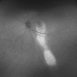
Jul 23 2023 by Aditya S Kelkar, MS, FRCS, FASRS,FRCOphth
Fundus Autofluorescence imaging of an 57-year-old woman with Chronic Central Serous Chorioretinopathy demonstrating a Comet Tail Sign.
Photographer: Dr. Ajinkya Rawale. National institute of Ophthalmology, Pune, India.
Imaging device: OPTOS DAYTONA
Condition/keywords: chronic central serous chorioretinopathy (CSCR)
-
---thumb.JPG/image-square;max$300,300.ImageHandler) FAF of Macular Degeneration
FAF of Macular Degeneration
Jul 12 2013 by Jason S. Calhoun
Autofluresence or FAF photo of bilateral age related macular degeneration in both eyes.
Photographer: Jason S. Calhoun, Department of Ophthalmology, Mayo Clinic Jacksonville, Florida
Condition/keywords: autofluorescence imaging
-
 Geographic Atrophy - Case 1: Photo 1 of 6
Oct 4 2012 by Gregg T. Kokame, MD, MMM, FASRS
NIRAF (near infrared autofluorescence) Image of patient with Geographic Atrophy
Photographer: Jaclyn Pisano, Retina Consultants of Hawaii
Imaging device: Heidelberg Spectralis
Condition/keywords: autofluorescence imaging, geographic atrophy, near infrared autofluorescence (NIRAF)
-
 Geographic Atrophy - Case 1: Photo 2 of 6
Oct 4 2012 by Gregg T. Kokame, MD, MMM, FASRS
Blue-peak Autofuorescence Image of patient with Geographic Atrophy
Photographer: Jaclyn Pisano, Retina Consultants of Hawaii
Imaging device: Heidelberg Spectralis
Condition/keywords: autofluorescence imaging, geographic atrophy
-
 Hemangioma of Retina (FAF)
Mar 5 2025 by Virginia Gebhart
Fundus autofluorescence of 64 year old male with choroidal hemangioma in the macula and STA. Persistent IRF and new cuff of SRF compared to previous photos. BCVA CF@face. Pt has had PDT in the past with no significant improvement. Will observe closely
Photographer: Virginia Gebhart, Retina Consultants of Carolina
Imaging device: Optos California
Condition/keywords: autofluorescence imaging, hemangioma, inferior subretinal fluid
-
 Idiopathic Choroidal Folds
Aug 22 2017 by Carolyn Daley
This is an autofluorescence image of a 77-year-old male with idiopathic choroidal folds in his right eye.
Photographer: Carolyn Daley
Imaging device: Heidelberg Spectralis
Condition/keywords: autofluorescence imaging, choroidal folds
-
 Late Stage Stargardt's Disease
Mar 13 2013 by Hamid Ahmadieh, MD
Autofluorescence imaging of the right eye of a 46-year-old man with decreased VA due to advanced Stargardt's disease.
Photographer: Nayereh Hadipoor, Negah Eye Center, Tehran
Imaging device: Heidelberg Spectralis
Condition/keywords: autofluorescence imaging, Stargardt disease
-
 Optic Disc Drusen Autofluorescence
Jul 16 2023 by Aditya S Kelkar, MS, FRCS, FASRS,FRCOphth
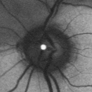
Fundus autofluorescence imaging of a 22-year-old male with optic disc drusen seen as hyperautofluorescent spot.
Photographer: Dr. Harsh Jain
Condition/keywords: Autofluorescence imaging of Optic Disc Drusen
-
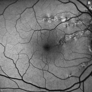
 Radiation Retinopathy FAF
Apr 26 2019 by Carissa Hurdstrom
Autofluorescence radiation retinopathy
Photographer: Carissa Hurdstrom
Imaging device: Optos
Condition/keywords: autofluorescence imaging, radiation retinopathy
-
 Retinitis Pigmentosa
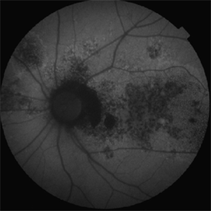
Apr 17 2025 by Virginia Gebhart
Fundus autofluorescence of 75 year old female with Retinitis Pigmentosa. Pt diagnosed at age 53. Diffuse RPE atrophy with minimal central sparing present in both eyes. Stable and unchanged compared to previous exams. BCVA 20/200 OD, NLP OS
Photographer: Virginia Gebhart, Retina Consultants of Carolina
Imaging device: Optos California
Condition/keywords: autofluorescence imaging, bone spicule, retinitis pigmentosa, RP
-
 Retinitis Pigmentosa
Nov 7 2023 by Jolee Rodriguez
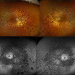
Bilateral fundus photography and fundus autofluorescence imaging of a 62-year-old male with Retinitis Pigmentosa. Patient reported visual field defects and dark adapting issues. Patient's vision at the time images were taken were sc20/20 of the right eye and sc20/25 of the left eye. Dr. Sutherland determined that based on the patient's lack of family history, the most likely route of inheritance is autosomal recessive.
Photographer: Jolee Rodriguez
Imaging device: Optos California RGB
Condition/keywords: autofluorescence imaging, fundus photography, hereditary retinal dystrophy, Optos, OPTOS CALIFORNIA RGB, retinitis pigmentosa, ultra-wide field imaging, Ultra-wide field retinal imaging, ultra-widefield image
-
 Serpiginous Choroiditis - Fundus Autofluorescence
Sep 20 2014 by Rameez N Hussain, MD
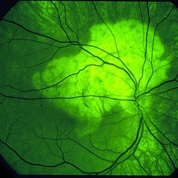
Fundus autofluorescence of serpiginous choroiditis showing decreased autofluorescence area corresponding to the inactive lesion (RPE atrophy) and increased autofluorescence area corresponding to active lesion.
Photographer: Dr.Rameez N Hussain, MD, Central Imaging Center, Vitreo Retinal Services, Giridhar Eye Institute, Cochin, India
Imaging device: Heidelberg Blue Peak Autofluorescence imaging.
Condition/keywords: serpiginous choroiditis
 Loading…