Search results (18 results)
-
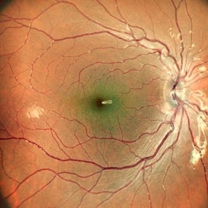
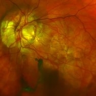
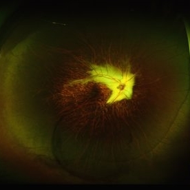
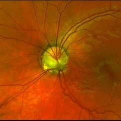 Optic Nerve Pit Left Eye
Feb 15 2021 by Kim Barrett
A 14-year-old male presented with vision loss and VF defect. Patient was treated for presumed amblyopia with patching since age 4. He has had neurologic care for post traumatic skull fracture and brain bleed in 2012. Patient has a superior hemifield defect OS on HVF. IOP's WNL. There are vessels emanating from the optic pit OS. Patient is at risk of serous detachment. Current VA 20/20-2+2
Photographer: Kim Barrett C.O.A. Retina Specialist of Michigan, Grand Rapids, MI
Imaging device: Optos California
Condition/keywords: amblyopia, hemifield, Humphrey visual field, nerve, optic nerve pit, visual field defect
-
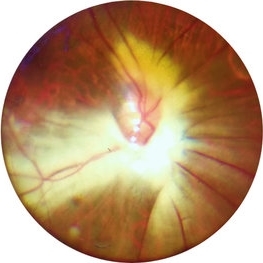
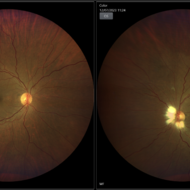
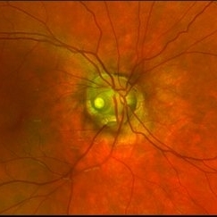 Optic Nerve Pit Right Eye
Feb 15 2021 by Kim Barrett
A 14-year-old male presented with vision loss and VF defect. Patient was treated for presumed amblyopia with patching since age 4. He has had neurologic care for post traumatic skull fracture and brain bleed in 2012. IOP's WNL. OD is without retinoschisis or subretinal fluid. Patient is at risk of serous detachment. Current VA OD 20/200+1 PH 20/80.
Photographer: Kim Barrett C.O.A. Retina Specialist of Michigan, Grand Rapids, MI
Imaging device: Optos California
Condition/keywords: amblyopia, hemifield, Humphrey visual field, nerve, optic nerve pit, visual field defect
-
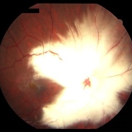
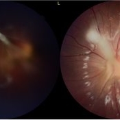
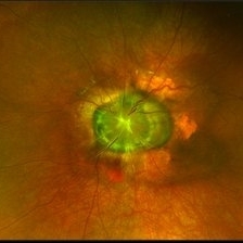 Morning Glory Syndrome
Jan 6 2020 by Olivia Rainey
Ultra-wide field pseudocolor image of a 23-month-old male with morning glory syndrome affecting his left eye. Patient presented with esotropia affecting his left eye and strabismic amblyopia affecting both eyes. He could fix and follow on exam and his medical history was unremarkable.
Photographer: Olivia Rainey
Imaging device: Optos California
Condition/keywords: esotropia, left eye, macular, Morning Glory Syndrome, Optos, strabismic amblyopia, ultra-wide field imaging
-
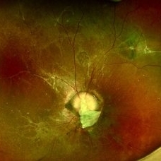
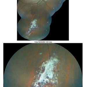 Retinal Cavernous Hemangioma
Apr 23 2021 by Aparna Ghodake
Fundus photograph of a 14-year-old girl having amblyopia was incidentally found to have numerous dark blood filled saccular aneurysms embedded in white fibroglial tissue giving it a 'bunch of grapes' appearance characteristic of retinal cavernous hemangioma.
Photographer: Dr. Aparna Ghodake, Sri Sankaradeva netralaya, Guwahati, Assam, India
Imaging device: Zeiss visucam 500
Condition/keywords: cluster of grapes, fundus photograph
-
 AMBLYOPIA
Oct 19 2022 by Akansha Sharma
COLOUR FUNDUS PHOTOGRAPH OF A 12 YEAR OLD MALE CHILD WITH HIGH REFRACTIVE ERROR WITH AMBLYOPIA
Photographer: Dr. Akansha Sharma-Retina Foundation, Ahmedabad
Condition/keywords: amblyopia
-
 Combined Hamartoma of the Retina and Retinal Pigment Epithelium
Oct 7 2019 by Sophia El Hamichi, MD
A 7-year-old female followed for combined hamartoma of the retina and the retinal pigment epithelium with amblyopia
Photographer: Abby Orcutt-Hayes, Murray Ocular Oncology and Retina
Condition/keywords: combined hamartoma, montage
-
 Extensive Myelinated Nerve Fibres
May 20 2021 by Anmol Naik
A 21-year-old Indian male presented with incidentally discovered subnormal vision in the left eye. On examination, he had esotropia with high myopia of -14 dioptres. Fundus examination revealed extensively myelinated nerve fibres around the optic disc extending along the arcade but sparing the fovea. The association of myelinated nerve fibres with high myopia and amblyopia is well documented but the causal association between these is unproven. Early detection of refractive error and aggressive therapy to prevent amblyopia has been reported with some success.
Photographer: Anmol Naik, Nakshatra Superspeciality Eye Hospital, Pune, India.
Imaging device: Zeine slit-lamp mounted Fundus imaging system
Condition/keywords: amblyopia, high myopia, myelinated nerve fibers
-
 Fluorescein Angiogram of Combined Hamartoma of the Retina and Retinal Pigment Epithelium
Oct 7 2019 by Sophia El Hamichi, MD
A 7-year-old female followed for combined hamartoma of the retina and the retinal pigment epithelium with amblyopia.
Photographer: Abby Orcutt-Hayes, Murray Ocular Oncology and Retina
Condition/keywords: combined hamartoma, fluorescein angiogram (FA), montage
-
 MNF with ERM with LMH
Mar 24 2020 by Sugnesh Parmar
32-year-old male with unilateral high myopia with amblyopia on routine fundus examination was having large MNF with ERM with LMH
Photographer: Dr. Sugnesh Parmar, Radheshyam retina hospital, Bhavnagar, Gujarat, India
Condition/keywords: high myopia, lamellar macular hole
-
 Morning Glory Anomaly With Retinal Detachment Managed With Amniotic Membrane Graft
Oct 15 2024 by Hemanth Murthy, MBBS, MD, FASRS
10 year-old boy presented with noticed blurring of vision. He had total retinal detachment with complicated cataract. He underwent lensectomy with 240 band and vitrectomy with silicone oil. The retina failed to settle due to minute breaks in the inferior part of the disc. Repeat surgery with AMG was done to cover the inferior part of disc. The retina settled under silicone oil. Silicone oil was removed and he is presently undergoing amblyopia treatment. Vision is 2/60 with +14.5 diopter lens.
Photographer: Mr Veda Vyas
Condition/keywords: amniotic membrane graft, Morning Glory Anomaly
-
 Morning-Glory-Syndrome
Dec 22 2017 by James B. Soque, CRA, OCT-C, COA, FOPS
68-year-old WM with Morning Glory Syndrome with PVD OS with Staphyloma surrounding optic nerve and extending into the macula. Also, esotropia OS from V1 nerve paresis from birth, with amblyopia.
Photographer: James B Soque, CRA OCT-C COA FOPS
Imaging device: Optos Daytona
Condition/keywords: color photo, esotropia, fundus photograph, Optomap, Optos, peripheral vascular disease (PVD), posterior vitreous detachment, staphyloma, ultra-wide field imaging, wide angle imaging
-
 Myelinated Nerve Fiber
May 5 2021 by Priya Rasipuram Chandrasekaran, MBBS, DO, DNB, FRCS
A 31-year-old male presented with a decreased vision of 20/125 N24 with -6.50 DS/-3.50 cyl 90 in the left eye. Fundus examination revealed peripapillary MNF progressing superiorly, obscuring disc and vessels and sparing the macula. OCT of ONH showed hyper reflective NFL and an abrupt ending of RPE and inner retinal layers (IRL) with underlying shadowing at the beginning of hyper reflectivity. The absence of photoreceptor integrity line (PIL) in the macula is believed to cause refractory amblyopia in such patients.
Condition/keywords: myelinated nerve fibers
-
 Myelinated Nerve Fibers
Apr 18 2025 by DR Rohit Gupta
The **myelinated nerve fibers of the optic disc** (also known as **medullated nerve fibers**) are retinal nerve fibers that retain their myelin sheath as they pass through the optic nerve head. Normally, retinal nerve fibers are unmyelinated to allow for light transparency, but in some cases, myelination extends anteriorly into the retina, appearing as a striking white, feathery patch on the optic disc or peripapillary retina. ### **Key Features:** 1. **Appearance:** - Dense, white, striated patches with feathery edges. - Typically located at the superior or inferior pole of the optic disc. - May obscure retinal vessels underneath. 2. **Clinical Significance:** - Usually **benign** and asymptomatic. - **Congenital** (present at birth or early childhood). - Rarely associated with **visual field defects** (e.g., scotomas corresponding to the area of myelination). - Occasionally linked with **high myopia** or **amblyopia** if extensive. 3. **Pathophysiology:** - Failure of oligodendrocytes or Schwann cells to stop myelination at the lamina cribrosa. - Normally, myelination stops at the optic nerve head, but in this condition, it extends into the retina. 4. **Diagnosis:** - **Fundoscopy:** Classic white, feathery appearance. - **Optical Coherence Tomography (OCT):** Shows thickened retinal nerve fiber layer (RNFL). - **Visual Field Testing:** May detect defects if large. 5. **Differential Diagnosis:** - Optic disc edema - Cotton wool spots - Retinoblastoma (rarely, but must be ruled out in children) 6. **Management:** - No treatment required if asymptomatic. - Monitor for amblyopia in children. - Rare cases with significant visual impairment may need further evaluation. ### **Fun Fact:** Myelinated nerve fibers are seen in **~0.5-1%** of the population and are usually an incidental finding.
Photographer: Dr Rohit gupta
Imaging device: Samsung S21
Condition/keywords: Medulated Nerve fibre, Medullated Nerve fibres, myelinated nerve fibers, Myelinated Nerve Fibres, optic disc drusen
-
 Myelinated Nerve Fibres in left eye with old tributary vein occlusion in left eye
Jul 18 2023 by Harsh Vardhan Singh, MS
55 year female with left eye amblyopia & high myopia with MNF and Right eye showed signs of old macular branch retinal vein occlusion
Photographer: Harsh Vardhan Singh, AIIMS, Guwahati
Imaging device: Zeiss Clarus 700
Condition/keywords: BRVO, macular branch retinal vein occlusion (BRVO), Medullated Nerve fibres, MNF, Myelinated Nerve Fibres, TRVO
-
 Posterior-PFV
Jul 27 2024 by Gokcen Deniz Gulpinar Ikiz
7 Year old girl presented with blurred vision on the left eye, with intermittent esotopia. She had been followed conservatively for intermittent esotropia on the left eye, recently advised for patching of the right eye. The vision is 1.0 on the right eye and 0.4 (Snellen) on the left eye. Anterior segment is natural bilaterally, except 20 PD esotropia on the left eye, with alternation and fixation. Refraction was +0.25 +0.25 x180 and +1.00-1.50 x60 on the right and left eyes respectively. Dilated fundus examination was natural on the right eye. However, there was a fibrotic stalk originating from the optic nerve head extending to the vitreous, terminating in the middle of the vitreous cavity, in a spider web configuration. Which also causes nasal dragging of the macula, leading to partial shallow detachment of the fovea nasally. Vitrectomy is advised for the left eye, with lens preserving approach, to preserve the current functional potential and the anatomy of the globe in long term.
Photographer: Gokcen Deniz Gulpinar Ikiz, Special Eye Clinic
Condition/keywords: amblyopia, posterior PFV, vitrectomy
-
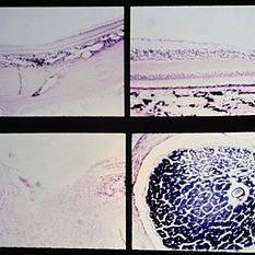 Slide 9-89
Feb 26 2019 by Lancaster Course in Ophthalmology
Nutritional amblyopia. The nerve fiber and ganglion cell layers are absent in the macular area (upper views). The temporal side of the optic nerve head (lower left) is partially atrophy, with marked reduction in the size of the nerve fiber bundles and secondary gliosis.
Condition/keywords: amblyopia, atrophy, gliosis, macular
-
 Straatsma Syndrome
Nov 17 2020 by Linda A Cernichiaro- Espinosa, MD
5-year-old male with leukocoria, high myopia and myelinated nerve fibers (Straatsma Syndrome). OCT showed a foveal depression and intact photoreceptors. Amblyopia management was started.
Photographer: Guillermo Salcedo-Villanueva, MD
Imaging device: Zeiss Clarus
Condition/keywords: high myopia, leukocoria, myelinated nerve fibers, pediatric retina
-
 Straatsma Syndrome
Aug 29 2024 by César Adrián Gómez Valdivia, MD
Fundus photograph of a 11 year-old female patient with unilateral myelinated retinal nerve fibers, axial myopia, amblyopia and strabismus.
Photographer: @eyemissu2
Imaging device: California ICG OPTOS
Condition/keywords: Straatsma Syndrome
 Loading…